Abstract
Clostridium botulinum produces heat-resistant endospores that may germinate and outgrow into neurotoxic cultures in foods. Sporulation is regulated by the transcription factor Spo0A and the alternative sigma factors SigF, SigE, SigG, and SigK in most spore formers studied to date. We constructed mutants of sigF, sigE, and sigG in C. botulinum ATCC 3502 and used quantitative reverse transcriptase PCR and electron microscopy to assess their expression of the sporulation pathway on transcriptional and morphological levels. In all three mutants the expression of spo0A was disrupted. The sigF and sigE mutants failed to induce sigG and sigK beyond exponential-phase levels and halted sporulation during asymmetric cell division. In the sigG mutant, peak transcription of sigE was delayed and sigK levels remained lower than that in the parent strain. The sigG mutant forespore was engulfed by the mother cell and possessed a spore coat but no peptidoglycan cortex. The findings suggest that SigF and SigE of C. botulinum ATCC 3502 are essential for early sporulation and late-stage induction of sigK, whereas SigG is essential for spore cortex formation but not for coat formation, as opposed to previous observations in B. subtilis sigG mutants. Our findings add to a growing body of evidence that regulation of sporulation in C. botulinum ATCC 3502, and among the clostridia, differs from the B. subtilis model.
INTRODUCTION
Through production of resistant endospores, the Gram-positive anaerobe Clostridium botulinum can persist under extreme environmental conditions. The spores may survive pasteurization and further germinate and outgrow into neurotoxic cultures in anaerobically stored foods, which poses a serious public health risk (1–3). C. botulinum spores may also germinate in the gut, posing a health risk for small babies and those adults with disturbed intestinal microbial populations due to heavy antimicrobial treatments (4, 5).
Despite the high significance of C. botulinum spores, not much is known about the molecular mechanisms behind spore formation in this organism. Sporulation has, however, been well characterized in the Gram-positive aerobic model organism, Bacillus subtilis (6–10). The morphological stages (0 to VII) of sporulation are much the same in all spore formers. The key regulators include Spo0A, the essential sporulation master regulator, and the sigma-70 family alternative sigma factors SigF, SigE, SigG, and SigK, which govern the sporulation process in B. subtilis and have homologues in C. botulinum (11) and many other clostridia (12–14).
In B. subtilis, sporulation is triggered by nutrient limitation and initiated upon activation of Spo0A by a phosphorelay system (15, 16). Phosphorylated Spo0A (Spo0A∼P) then induces the downstream sigma cascade regulating the sporulation process. Sporulation stage 0 is denoted by cell elongation. At this point, Spo0A∼P is active and positively regulates the spoIIA operon containing sigF and the spoIIG locus containing sigE and sigG while causing a negative feedback loop and repressing spo0A expression. Chromatin filaments form at stage I; however, this is visually indistinct from stage 0 (17). At stage II, the cell divides into the forespore and mother cell, where SigF and SigE are activated, respectively. In Bacillus, SigF is part of a feed-forward loop initiated by Spo0A activation (18). SigF activation, in turn, leads to sigE transcription in another feed-forward loop (19). SigG is activated in the forespore at stage III as the mother cell engulfs the forespore (20). Upon engulfment, peptidoglycan spore cortex formation begins. In B. subtilis, at stage IV, SigK is activated in the mother cell and regulates spore coat formation by expression of the coat and germination-related genes (21).
Although the key morphological stages of sporulation in Bacillus and clostridia are much the same, several structural and functional differences on the molecular level have been identified. First, the phosphorelay system activating Spo0A in Bacillus appears to be absent from all clostridial strains (14, 22). Thus, how Spo0A is activated in C. botulinum remains to be elucidated. Second, the sigma factor cascade controlling sporulation gene expression in clostridia seems to possess several specific characteristics. While SigK in B. subtilis is exclusively involved in late-stage sporulation, studies in C. perfringens (23), C. botulinum (24), and C. acetobutylicum (25) have demonstrated roles for SigK in both early and late stages of sporulation. Moreover, many clostridial strains lack the sigK-intervening (skin) element found in B. subtilis (26), suggesting this sigma factor is regulated differently. A notable exception among the clostridia is C. difficile, which possesses a skin element, but this element has been proposed to play a different role than that of the skin element of B. subtilis (27, 28). Apart from sporulation, SigK of C. botulinum also has been associated with stress response (29). In addition to SigK, SigG also appeared to possess various roles, as related mutants in the clostridia were reported to advance further in sporulation than those in B. subtilis (28, 30–32). Finally, on a population level, sporulation among the clostridia and, as a result, in C. botulinum generally is asynchronous (33, 34), whereas in B. subtilis synchronized sporulation can be induced (6).
In this study, we determined the essential role of SigF, SigE, and SigG in the sporulation of C. botulinum ATCC 3502. Strains with insertionally disrupted sigF, sigE, and sigG were evaluated for spore formation and imaged by electron microscopy. Reverse transcription-quantitative PCR (RT-qPCR) analysis of spo0A, sigF, sigE, sigG, and sigK was performed in the ATCC 3502 parent and mutant strains to determine the transcriptional relationship between these sigma factors during spore formation.
MATERIALS AND METHODS
Strains, plasmids, and culture.
Bacterial strains, intron insertion sequences, and plasmids are listed in Table 1. The sense-oriented mutant strains (for all experiments) were designated the sigF522s::CT, sigE441s::CT, and sigG546s::CT mutants, and the antisense-oriented mutants (sporulation assay and spore staining) were designated the sigF487a::CT, sigE385a::CT, and sigG457a::CT mutants, according to Kuehne and Minton (35). The C. botulinum ATCC 3502 parent and the sigma factor mutants were stored at −70°C in anaerobic Protect microorganism preservation system beads (Technical Service Consultants Ltd., Lancashire, United Kingdom). C. botulinum was grown in anaerobic tryptone-peptone-glucose-yeast extract (TPGY) broth (5% tryptone, 0.5% peptone, 2% yeast extract [Difco, BD Diagnostic Systems, Sparks, MD], 0.4% glucose [VWR International, Leuven, Belgium], 0.1% sodium thioglycolate [Merck, Darmstadt, Germany]) or on anaerobic TPGY plates (1.5% agar). TPGY was supplemented with 15 μg/ml thiamphenicol, 250 μg/ml d-cycloserine, and 5 μg/ml erythromycin where appropriate. Cultures were incubated at 37°C in an anaerobic cabinet (MG1000 Anaerobic Work Station; Don Whitley Scientific Ltd., Shipley, United Kingdom) with an atmosphere of 85% N2, 10% CO2, and 5% H2. Escherichia coli cultures were grown aerobically at 37°C in Luria-Bertani (LB) broth (Difco) supplemented with 25 μg/ml chloramphenicol and 30 μg/ml kanamycin (Sigma-Aldrich, St. Louis, MO). Overnight cultures from frozen stocks were grown in TPGY broth at 37°C (supplemented with erythromycin for mutant strains) and streaked on TPGY plates. Cultures then were individually inoculated into 10 ml of TPGY broth and cultured overnight at 37°C before use. All experiments were performed in triplicate.
TABLE 1.
Strains and plasmids
| Strain or plasmid | Characteristic | Sequencea | Source/reference |
|---|---|---|---|
| Strains | |||
| C. botulinum ATCC 3502 | Parent strain | ATCC, Manassas, VA | |
| C. botulinum ATCC 3502 sigF522s::CT | ClosTron insertional mutant of sigF in sense orientation | ATACATCAAGATGATGGAGCACCAGTTTTG*CTTATTGATAAAATA | This study |
| C. botulinum ATCC 3502 sigF487a::CT | ClosTron insertional mutant of sigF in antisense orientation | CTGGTGCTCCATCATCTTGATGTATAACAT*CATATAAATATTGGG | This study |
| C. botulinum ATCC 3502 sigE441s::CT | ClosTron insertional mutant of sigE in sense orientation | TTAAATATTGATTGGGATGGAAATGAGCTA*TTACTTTCAGATATA | This study |
| C. botulinum ATCC 3502 sigE385a::CT | ClosTron insertional mutant of sigE in antisense orientation | TTAAAGGTTCATAAAAGGATATTTCTGCTT*TTACTTTACTATTTC | This study |
| C. botulinum ATCC 3502 sigG546s::CT | ClosTron insertional mutant of sigG in sense orientation | ATATATCATGATGGTGGTGACGCCATATTT*GTTATGGATCAAATA | This study |
| C. botulinum ATCC 3502 sigG457a::CT | ClosTron insertional mutant of sigG in antisense orientation | TAGCGTCTAAAGCAAACACTACCTCTTCTC*TTGGTAGTTCTAATT | This study |
| E. coli 5-alpha | Chemically competent cloning strain | Invitrogen, Carlsbad, CA | |
| E. coli CA434 | Conjugation donor strain containing R702 plasmid | 39 | |
| Plasmids | |||
| pMTL007C-E2 | Clostridia mutagenesis vector | 37 | |
| pMTL007C-E2::Cbo-sigF522s::CT | pMTL007C-E2 targeting sigF in sense orientation | This study; DNA 2.0 | |
| pMTL007C-E2::Cbo-sigF487a::CT | pMTL007C-E2 targeting sigF in antisense orientation | This study; DNA 2.0 | |
| pMTL007C-E2::Cbo-sigE441s::CT | pMTL007C-E2 targeting sigE in sense orientation | This study; DNA 2.0 | |
| pMTL007C-E2::Cbo-sigE385a::CT | pMTL007C-E2 targeting sigE in antisense orientation | This study; DNA 2.0 | |
| pMTL007C-E2::Cbo-sigG546s::CT | pMTL007C-E2 targeting sigG in sense orientation | This study; DNA 2.0 | |
| pMTL007C-E2::Cbo-sigG457a::CT | pMTL007C-E2 targeting sigG in antisense orientation | This study; DNA 2.0 |
ClosTron insertion sites are represented by asterisks.
Construction of the ClosTron insertional mutants.
A mobile group II intron from Lactococcus lactis (Ll.ltrB) (36) was inserted into sigF (cbo3087), sigE (cbo2533), and sigG (cbo2532) in the sense and antisense orientations using the ClosTron plasmid pMTL007C-E2 (37). The insertion sites were selected using the Perutka algorithm (38), made available online by the University of Nottingham, United Kingdom, on the ClosTron website. The retargeted ClosTron plasmids for generation of mutations were constructed by DNA 2.0 (Menlo Park, CA, USA). These plasmids were chemically transformed into E. coli CA434 (39) and grown overnight in LB supplemented with kanamycin and chloramphenicol to select for the R702 conjugation plasmid and the ClosTron plasmid, respectively. The ClosTron plasmids then were individually introduced into C. botulinum ATCC 3502 via conjugation. One ml of the E. coli CA434 donor strain containing the ClosTron plasmid was centrifuged at 5,000 rpm for 2 min and washed with phosphate-buffered saline (PBS). The pellet was resuspended in 1 ml of the C. botulinum parent strain overnight culture and spotted onto a blood agar plate. This was incubated under anaerobic conditions at 37°C for 6 h. Growth after 6 h was collected into 1 ml of PBS solution. Aliquots of 100 μl were spread on TPGY plates supplemented with thiamphenicol for ClosTron plasmid selection and with cycloserine to purify the parent strain and incubated anaerobically at 37°C. Pure C. botulinum transconjugants then were transferred onto TPGY plates supplemented with erythromycin to select for integrants. Confirmation of successful integration into the target sites of all genes was performed by PCR using primers (Table 2) flanking the insertion site and a primer targeted to the intron. A Southern blot was performed according to Palonen et al. (40) to confirm a single integration event in each mutant strain. Genomic DNA was extracted from the parent and mutant strains using a modified Marmur method (41). HindIII was used to digest the DNA, and a digoxigenin-labeled probe was added. This probe was specific for the erythromycin resistance gene in the ClosTron insert.
TABLE 2.
Primers used in this study
| Name and use | Efficiencya | Sequence (5′–3′) |
|---|---|---|
| Mutant confirmation | ||
| EBS universal | NA | CGAAATTAGAAACTTGCGTTCAGTAAAC |
| sigFF | NA | GCTACAGAATCATCGAGTTCC |
| sigFR | NA | CTTGACACCTGTACTTGACTTATTC |
| sigEF | NA | GGAACTATAGGACTAATTAAGGCAG |
| sigER | NA | CCCAGCATATCTGCTACTTC |
| sigGF | NA | CTGGTAAGGATTAGGAACGG |
| sigGR | NA | CCTATTTCATCGGCTACTTCC |
| RT-qPCR | ||
| 16S rrnF | 1.99 | AGCGGTGAAATGCGTAGAGA |
| 16S rrnR | GTCTTAATTTATCAGCCACCATTG | |
| spo0AF | 2.00 | TATAATACTACAGCGAGCAGAGTG |
| spo0AR | GTCTTAATTTATCAGCCACCATTG | |
| sigFqF | 1.99 | GGATGCATCGGTTTAGTAAAGGC |
| sigFqR | CATAATGTAATTTTTTTGCCGCTG | |
| sigEqF | 1.9 | AGCAACCTATGCTTCAAGATGT |
| sigEqR | AGCTCATTTCCATCCCAATC | |
| sigGqF | 1.94 | ATGGTGGTGACGCCATATTT |
| sigGqR | TTCATCGGCTACTTCCATTTG | |
| sigKqF2 | 1.94 | ACTTATGGGATGTACTAGGAAGTG |
| sigKqR2 | TTCTTCTTCATCACTTAGAGGCTT |
Efficiency was calculated for RT-qPCR primers only. NA, not applicable.
RNA isolation and cDNA synthesis.
The ATCC 3502 parent and the sigF, sigE, and sigG mutant strains with the ClosTron insert in sense orientation were inoculated from overnight cultures into 100 ml of fresh TPGY at a 1:100 dilution. Growth was monitored by optical density at 600 nm (OD600). One-ml samples were collected 5, 11, 15, and 24 h after inoculation, representing the exponential, late-exponential, transition, and stationary phases of growth, respectively. The samples were mixed with 200 μl of ice-cold stop solution containing phenol (95%) and ethanol (99.5%) at a ratio of 1:9 and stored on ice for 30 min. The samples were centrifuged, and the supernatant was discarded. The pellets were stored at −70°C until RNA isolation. The frozen cell pellets were suspended in 250 μl of a lysis buffer containing lysozyme (50 mg/ml; Sigma-Aldrich), mutanolysin (1,000 U/ml; Sigma-Aldrich), and Tris-EDTA (10 mM Tris-HCl, 1 mM EDTA, pH 8.0) and incubated at 37°C for 30 min. RNA isolation and purification were performed using the RNeasy minikit (Qiagen, Hilden, Germany) with a DNase treatment (Qiagen). RNA was eluted twice with RNase-free water, and a second DNase treatment was performed according to the Ambion DNA-free kit (Thermo Fisher Scientific, Waltham, MA). RNA concentration was measured at 260 nm with a NanoDrop 1000 spectrophotometer (Thermo Scientific, Waltham, MA), and purity was examined by measuring the A260/A280 ratio. Integrity of the RNA was examined using an Agilent 2100 Bioanalyzer (Agilent Technologies, Santa Clara, CA, USA). A total of 500 ng of RNA was used for each reverse transcription (RT) reaction. The DyNAmo cDNA synthesis kit (Thermo Scientific) with random hexamer primers was used as instructed. RT was performed in duplicate for each RNA sample. The cDNA samples were stored at −20°C before use as templates in qPCR. Two PCR replicates were analyzed for each cDNA replicate per run. RT controls from pooled samples of RNA (5 samples per pool) without reverse transcriptase were included in each qPCR run to control DNA contamination of the RNA. In addition, nontemplate controls were included in each run.
qPCR.
The Maxima SYBR green qPCR master mix kit (Thermo Scientific) was used for qPCR. Primers were designed for the target genes spo0A, sigF, sigE, sigG, and sigK and for the reference gene 16S rrn (42) (Table 2) using Primer3 (43, 44). Each reaction mixture contained 12.5 μl of master mix (Thermo Scientific), 0.3 μM each primer, 4.0 μl of diluted cDNA template (10−6-fold for 16S rrn, 10−4-fold for sigF, and 10−3-fold for spo0A, sigE, sigG, and sigK), and nuclease-free water up to a final volume of 25 μl. The qPCR runs (Rotor Gene 3000 real-time thermal cycler; Qiagen) included an enzyme activation step at 95°C for 10 min, followed by 40 cycles of denaturation at 95°C for 15 s, annealing and extension at 60°C for 60 s, and a final extension at 60°C for 1 min. At the end of each run, a melting curve analysis from 60°C to 99°C at 0.5°C per 5 s was performed. Standard curves based on dilution series of pooled cDNA (independent of the RT control RNA pool) were constructed to determine the efficiency of each primer pair. The reaction efficiencies (Table 2) were determined from the slopes of the standard curves using Rotor Gene software. Cq values were calculated using Rotor Gene software. The Cq (quantification cycle) values of the target genes spo0A (cbo1872), sigF, sigE, sigG, and sigK (cbo2541) were normalized to Cq values of 16S rrn. To compare relative gene expression over time, Cq values were calibrated to the values at the 5-h exponential-phase time point. To compare parent and mutant strain expression levels for each gene over time, additional calibration to the parent strain at the 5-h time point also was performed. The calculations were performed according to the equation R = [(1 + Egene)ΔCq, gene (calibrator − sample)]/[(1 + E16Srrn)ΔCq, 16Srrn (calibrator − sample)] outlined by Pfaffl (45), where R is the expression ratio, Egene is the reaction efficiency of the target gene primers, E16Srrn is the efficiency of the 16S rrn primers, ΔCq, gene is the deviation of the target gene Cq between the calibration time point (5 h) and the time point under investigation, ΔCq, 16Srrn is the Cq deviation of 16S rrn between the calibration time point and the time point under investigation, and calibrator − sample is the Cq value at 5 h minus the Cq value at the sampling time point in question. Significance of the differences in gene transcripts between the parent and mutant strains was tested using a two-tailed Student's t test on the expression data normalized by log2 transformation in PASW 18 (Predictive Analytics SoftWare Statistics 18; IBM).
Sporulation assay and spore staining.
The C. botulinum ATCC 3502 parent and the sigF, sigE, and sigG mutants with sense- and antisense-oriented ClosTron insertions were incubated in TPGY broth at 37°C under anaerobic conditions for 7 days (24, 46). One-ml aliquots of each culture were heated at 80°C for 15 min to kill vegetative cells. Growth from heat-treated samples determined the presence of heat-resistant spores. The parent and mutant strains also were grown on TPGY plates for 7 days and stained on a glass slide with malachite green to detect spores (47).
Transmission electron microscopy (TEM).
The preparation of the cells and spores for electron microscopy was based on the protocol of Stevenson et al. (48). The parent and mutant strains with sense-oriented ClosTron insertions were incubated in TPGY broth for 7 days, and 2-ml aliquots were centrifuged at 3,000 × g for 10 min. The supernatant was removed, and the pellet was resuspended in 2 ml of a solution containing 2.5% glutaraldehyde and 18.5% formaldehyde and incubated at room temperature for 2 h for optimal spore fixation. The suspension was centrifuged at 3,000 × g for 10 min, and a 500-μl layer of distilled water was pipetted over the pellet for storage at 4°C prior to embedding. Cells were embedded in Epon 812 epoxy resin (TAAB Materials, Berks, United Kingdom) for ultrathin sectioning (60 to 80 nm) with a diamond knife. Ultrathin sections were collected onto carbon mesh grids and stained with uranyl acetate (0.5%) and Pb-citrate (1%) for 30 min. The sections were observed using an FEI Tecnai 12 transmission electron microscope (FEI, Hillsboro, OR, USA) at the Electron Microscopy unit at the University of Helsinki.
RESULTS
Confirmation of mutations.
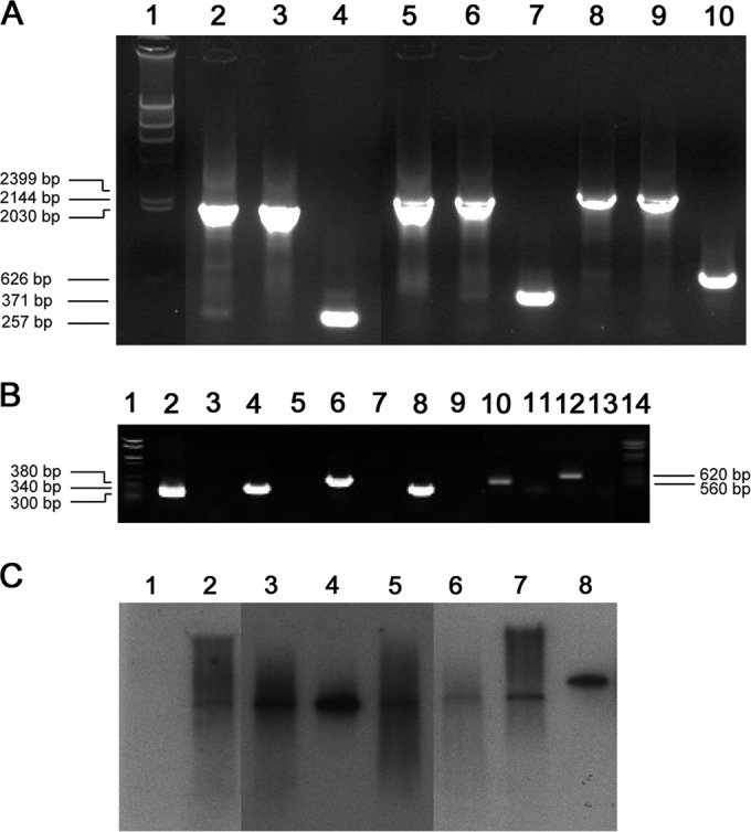
Insertion of the group II intron into the intended site in sigF, sigE, or sigG was confirmed by PCR screening of erythromycin-resistant colonies. Primers flanking the insertion sites yielded PCR products with sizes corresponding to 2,030 bp for the sigF mutants, 2,144 bp for the sigE mutants, and 2,399 bp for the sigG mutants, while the parent strain gave noticeably smaller products corresponding to 257, 371, and 626 bp in size, respectively (Fig. 1A). Correct orientation of each insertion was confirmed by PCR combining a forward flanking primer with the EBS universal primer specific for the insert itself. These yielded the expected products of 300 bp and 340 bp for the sigF sense and antisense mutations, respectively, 380 bp and 340 bp for the sigE sense and antisense mutations, respectively, and 560 bp and 620 bp for the sigG sense and antisense mutations, respectively (Fig. 1B). Finally, a Southern blot using a probe specific for the insert, with the ClosTron plasmid as a positive control and DNA of the parent strain as a negative control, demonstrated a single insertion in each mutant strain (Fig. 1C).
FIG 1.

(A) PCR confirmation of ClosTron intron insertion into target genes of Clostridium botulinum ATCC 3502. Lane 1, molecular weight marker (MWM) IV (Roche, Basel, Switzerland); lane 2, sigF sense mutant; lane 3, sigF antisense mutant; lane 4, C. botulinum ATCC 3502 (sigFF and sigFR pimers); lane 5, sigE sense mutant; lane 6, sigE antisense mutant; lane 7, ATCC 3502 (sigEF and sigER primers); lane 8, sigG sense mutant; lane 9, sigG antisense mutant; lane 10, ATCC 3502 (sigGF and sigGR primers). (B) PCR confirmation of ClosTron intron insertion in correct orientation. Lane 1, MWM VI; lane 2, sigF sense mutant; lane 3, ATCC 3502 (EBS and sigFF primers); lane 4, sigF antisense mutant; lane 5, ATCC 3502 (EBS and sigFR primers); lane 6, sigE sense mutant; lane 7, ATCC 3502 (EBS and sigEF primers); lane 8, sigE antisense mutant; lane 9, ATCC 3502 (EBS and sigER primers); lane 10, sigG sense mutant; lane 11, ATCC 3502 (EBS and sigGF primers); lane 12, sigG antisense mutant; lane 13, ATCC 3502 (EBS and sigGR primers); lane 14, MWM VI. (C) Southern blotting on HindIII-digested genomic DNA with probe specific for intron confirming a single integration event in each mutant strain. Shown is a composite image of a single gel where lanes 1 and 2 as well as 6 to 8 were exposed for 20 s and lanes 3 to 5 were exposed for 10 s. Lane 1, ATCC 3502; lane 2, sigF sense mutant; lane 3, sigF antisense mutant; lane 4, sigE sense mutant; lane 5, sigE antisense mutant; lane 6, sigG sense mutant; lane 7, sigG antisense mutant; lane 8, ClosTron plasmid.
RT-qPCR analysis of spo0A, sigF, sigE, sigG, and sigK.
The C. botulinum ATCC 3502 parent and sigF, sigE, and sigG mutant strains showed similar growth patterns according to OD600 measurements at 37°C over 24 h. RNA was isolated at 5, 11, 15, and 24 h, representing exponential, late-exponential, transition-phase, and stationary-phase growth. The relative expression levels of each target gene were calculated by normalization to 16S rrn expression. 16S rrn was stably expressed during the study, and its Cq values had an overall coefficient of variation (CV) of 3.8% (42, 49), whereas the CVs of the target genes ranged from 8.3 to 11.8%. Gene expression data were calibrated to the 5-h time point to show gene expression changes over time within each strain (Fig. 2 and 3). Additional calibration to parent strain gene expression at 5 h allowed comparison between the parent and mutant strains at each time point (Fig. 4).
FIG 2.

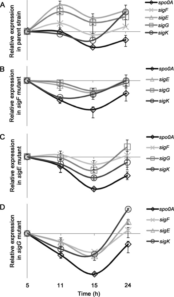
Gene expression trends of Clostridium botulinum ATCC 3502 parent strain (A), sigF mutant (B), sigE mutant (C), and sigG mutant (D) with sense-oriented ClosTron insertions showing target gene spo0A, sigF, sigE, sigG, and sigK expression levels at 5, 11, 15, and 24 h of growth. Transcript levels of each gene were normalized to 16S rrn transcript levels and calibrated to levels at 5 h. As the target genes could not be calibrated to each other, values on the y axis were omitted, as relative expression ratios between target genes within each strain cannot be quantifiably compared.
FIG 3.
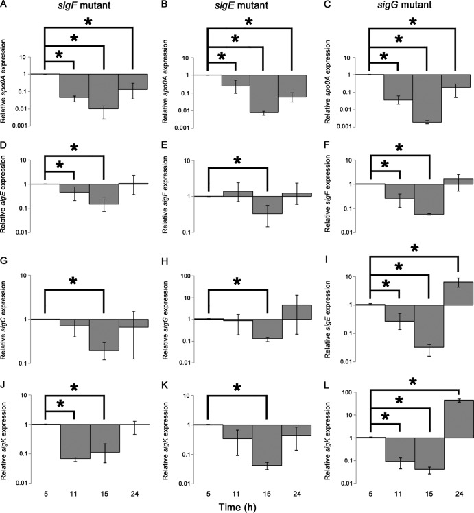
Relative gene expression ratios in the sigF mutant (spo0A [A], sigE [D], sigG [G], and sigK [J]), sigE mutant (spo0A [B], sigF [E], sigG [H], and sigK [K]), and sigG mutant (spo0A [C], sigF [F], sigE [I], and sigK [L]) strains with sense-oriented ClosTron insertions over 24 h of growth. Transcript levels of each gene were normalized to 16S rrn transcript levels and calibrated to those at 5 h. Significant (P < 0.05) changes in relative expression between the 5-h and subsequent time points are marked with an asterisk. Error bars represent maximum and minimum expression values from three replicates.
FIG 4.
Relative gene expression ratios in the Clostridium botulinum ATCC 3502 parent and sigF, sigE, and sigG mutant strains with sense-oriented ClosTron insertions over 24 h of growth for spo0A (A and B), sigF (C and D), sigE (E and F), sigG (G and H), and sigK (I and J). Bar charts (A, C, E, G, and I) show differences in relative transcript levels between parent and mutant strains at individual time points (*, P < 0.05). Line charts (B, D, F, H, and J) show the trend in gene expression for each gene expressed in each strain. Expression ratios were normalized to 16S rrn expression and calibrated to the parent strain expression levels at the 5-h time point to compare mutant and parent expression ratios. Error bars represent maximum and minimum expression values from three replicates.
In the parent strain (Fig. 2A), spo0A was expressed at 5 and 11 h and decreased thereafter. Expression of sigF, sigE, and sigG was induced at 11 h in the parent strain. Expression of sigE and sigG followed a similar pattern and remained high throughout the experiment. The expression pattern of sigK in the parent strain appeared biphasic and showed highest expression at 5 h and 24 h (Fig. 2A). These results are in line with those observed in a previous study (42).
In the sigF mutant, spo0A expression levels collapsed after 5 h (Fig. 2B) and remained significantly (P < 0.05) lower than levels at 5 h (Fig. 3A). Moreover, relative spo0A expression levels in the sigF mutant were significantly lower than those in the parent strain at 5, 11, and 15 h (Fig. 4A). The sigF mutant failed to induce its sigE and sigG expression (Fig. 2B, 3D, G, and H, and 4E and G) but showed induction of sigK similar to that of the parent strain, although the level of sigK expression was significantly lower than that in the parent strain at 5 h and 24 h and did not follow the same biphasic pattern as that of the parent strain (Fig. 4I and J). As in the parent strain, sigE and sigG expression trends were identical and appeared linked.
In the sigE mutant, the relative spo0A expression levels collapsed after 5 h (Fig. 2C and 3B) and were significantly lower than those in the parent strain at 11 and 15 h (Fig. 4A). No induction above the initial 5-h levels was observed for sigF or sigK; however, sigG expression was slightly induced above the 5-h levels at 24 h (Fig. 2C and 3E, H, and K). Expression levels of sigF and sigG were significantly lower in the sigE mutant than in the parent strain at 11 h (Fig. 4C and G), and that of sigK was significantly lower than that of the parent strain at 24 h (Fig. 4I); thus, it did not possess the same biphasic expression pattern as that observed in the parent strain (Fig. 4J). However, expression of sigK in the exponential phase appeared unaffected.
Similar to all other mutants, the sigG mutant decreased its spo0A expression after 5 h (Fig. 2D and 3C) to levels significantly lower than those in the parent strain (Fig. 4A). Expression levels of sigF and sigE were not induced above the initial 5-h levels for 15 h (Fig. 2D); however, at 24 h sigE was induced 7-fold in relation to the 5-h levels (Fig. 3I), with the final levels being similar to those in the parent strain (Fig. 4E). In the sigG mutant, sigK was expressed in a biphasic pattern similar to that of the parent strain (Fig. 2D; 4J), with a 44-fold induction at 24 h (Fig. 3L). However, the relative 24-h sigK transcript level was significantly lower than that in the parent strain (Fig. 4I).
Sporulation assays and TEM.
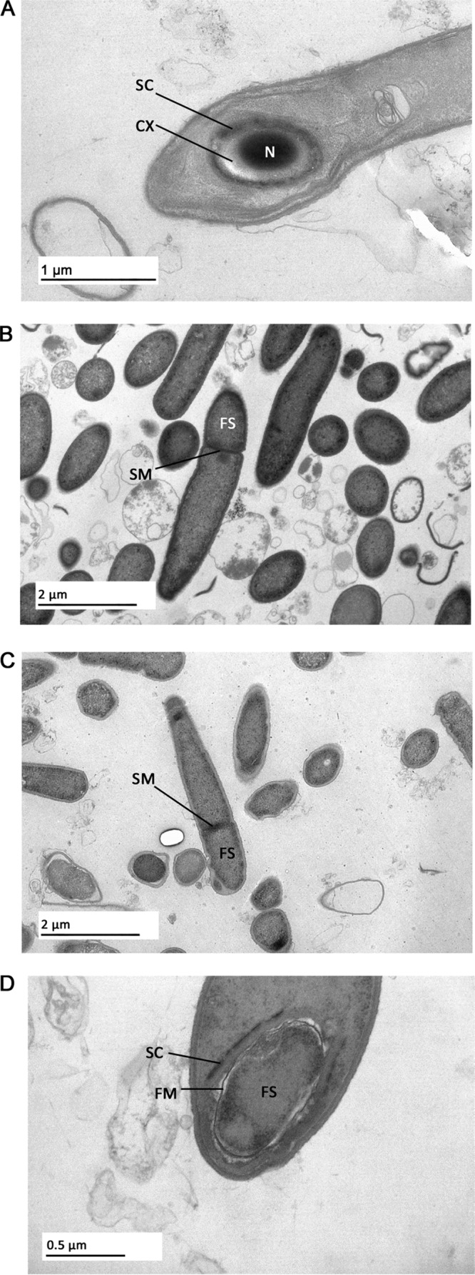
The C. botulinum ATCC 3502 parent and sigF, sigE, and sigG mutant strains were allowed to sporulate for 7 days at 37°C. The heat treatment assay detected no viable spores in any of the mutant strains, and spores were not observed in TEM images of the mutants. The parent strain successfully formed viable spores that were detectable after heat treatment. These spores stained positive with malachite green and were visibly mature under TEM (Fig. 5A). The sigF and sigE mutants stained negative with malachite green. TEM imaging revealed sporulation halted during stage II in the sigF (Fig. 5B) and sigE (Fig. 5C) mutants. The sigG mutants stained positive for spores with malachite green, suggesting the presence of a spore coat. TEM showed the sigG mutant had a spore coat-like structure surrounding the engulfed forespore (Fig. 5D). No peptidoglycan cortex was observed in this mutant.
FIG 5.

Transmission electron micrographs of the Clostridium botulinum ATCC 3502 parent (A) and sigF (B), sigE (C), and sigG (D) mutant strains with sense-oriented ClosTron insertions grown in TPGY broth for 7 days at 37°C. Indicated are the nucleoid area (N), spore coat (SC), spore cortex (CX), forespore membrane (FM), forespore (FS), and potential septation membranes (SM).
DISCUSSION
Here, we demonstrate the essential role of SigF, SigE, and SigG in the sporulation of C. botulinum ATCC 3502. We constructed insertional knockout mutants for each of the three sigma factor genes using the ClosTron system (37). The relative expression levels of the main transcriptional regulatory genes involved in sporulation, spo0A, sigF, sigE, sigG, and sigK, were examined in mutants with sense-oriented ClosTron insertions by RT-qPCR. This showed that the expression of the sporulation regulators was disrupted in the mutants compared to that in the parent strain. Sporulation was characterized in the three mutants and the parent strain in a sporulation assay, by spore staining, and by electron microscopy. All three mutants failed to produce heat-resistant spores and halted sporulation prior to complete spore formation. In order to verify that disruption of sigF, sigE, and sigG was responsible for the nonsporulating phenotype, ClosTron insertional mutants also were designed for integration in the opposite (antisense) orientation. The antisense mutants also displayed nonsporulating phenotypes, demonstrating that the nonsporulating phenotype likely was due to disruption of sigF, sigE, and sigG in C. botulinum ATCC 3502 and not biased by polar effects.
The RT-qPCR analysis revealed that in the sigF and sigE mutant strains, expression of the sporulation pathway genes appeared to be disrupted at an early stage. In these strains, expression of spo0A appeared to collapse at an earlier stage than in the parent strain, which expressed its spo0A until the transition growth phase before shutdown. It is not known if and how SigF and SigE, or their disruption, affect spo0A, but the results provide an interesting hypothesis for further testing. Previous observations suggest that the first peak of the biphasic expression pattern of sigK, already observed in the mid-exponential growth phase of the wild-type cultures, possess a link to spo0A expression in C. botulinum (24) and in C. acetobutylicum (25). Thus, the low spo0A expression observed in the sigF mutant may also be due to reduced sigK levels in the mid-exponential growth phase. However, expression of sigK appeared normal in the sigE mutant, so the collapsed spo0A levels cannot be explained by reduced sigK activity in this strain. Thus, regulation of spo0A in clostridia remains an important question for future research.
In the sigF and sigE mutants, expression of sigF, sigE, sigG, and sigK was not induced above the exponential-phase levels. In contrast, the parent strain induced its sigF, sigE, and sigG in late-exponential to stationary growth, concurrent with previous studies on sigma factor gene expression in clostridia (28, 42, 50). These data suggest that sporulation in the sigF and sigE mutant was disrupted at an early stage. Electron micrographs of these mutants showed formation of an asymmetric membrane at one or both poles, consistent with sporulation disrupted at stage II. Similar observations were made in sigF and sigE mutants of C. difficile (28, 32) and of C. perfringens (23, 51) and in sigE mutants of C. acetobutylicum (31). However, disruption of sigF in C. acetobutylicum halted sporulation prior to asymmetric cell division, which is earlier than it occurs in the other clostridia (52). Mutants of B. subtilis sigF and sigE display an abortive and sometimes abortively disporic phenotype where membrane septation occurs at both poles of the cell. These cells are unable to progress beyond stage II (6). Thus, SigF and SigE of C. botulinum ATCC 3502 appear to be essential in early-stage sporulation, particularly in progression of sporulation beyond stage II, similar to their role in B. subtilis and in other clostridia.
As with the sigF and sigE mutants, expression of the sporulation pathway appeared to be delayed in the sigG mutant. Similarly, the spo0A expression levels had already decreased in the late-exponential growth phase, again probably due to low sigK expression in exponential growth. In turn, the apparently delayed induction of sigF and sigE in the sigG mutant until the stationary phase of growth could follow the low levels of spo0A. However, a high induction of sigK occurred during the stationary phase in the sigG mutant, following the biphasic pattern observed in the parent strain, while no such reinduction of sigK was observed in the sigF and sigE mutants. These findings suggest that both SigF and SigE, but not SigG, are necessary for the reinduction of sigK expression in the stationary phase of growth.
Interestingly, the sigG mutants stained positive with malachite green despite not forming viable spores, indicating the presence of a spore coat. This suggested sporulation halted in a late stage. Electron microscopy revealed that the sigG mutant possessed a thin spore coat sufficient to retain the malachite green. The sigG mutant cells had progressed to stage III of sporulation, as the forespore was clearly engulfed by the mother cell. However, no peptidoglycan cortex signifying progression to stage IV was visible. Similarly, sigG mutants of C. acetobutylicum formed spore coats postengulfment (31), and those of C. difficile also showed spore coat formation (28, 32). These findings are in contrast to observations of sigG mutants of B. subtilis where no spore coat formation was observed postengulfment (30). In B. subtilis, SigG activates SigK, which regulates spore coat genes in the late stages of sporulation (9). It has been suggested that SigK activation is independent of SigG in C. difficile and C. acetobutylicum (31, 53). Active SigK accounts for spore coat formation in sigG mutants of C. acetobutylicum and C. difficile (28, 31, 32), which possessed phenotypes similar to those of our sigG mutant phenotype. Thus, SigG may not be responsible for SigK activation in C. botulinum. Our results indicate that SigG plays an essential role in regulating spore cortex formation, but not coat formation, in C. botulinum.
In summary, our findings suggest that the sporulation sigma factors E, F, and G are essential for viable spore formation in C. botulinum ATCC 3502. Our observations imply that the order of transcription induction of the genes encoding these sigma factors begins with sigK and spo0A in the exponential phase, followed by sigF and sigE together with sigG induction during late-exponential growth, with subsequent reinduction of sigK in the stationary growth phase (24, 42). In all three mutant strains, expression of spo0A was disrupted. It is unclear how spo0A is downregulated and if the downstream sigma factors in the sporulation pathway can influence the decision to enter sporulation via Spo0A. Additionally, sigE and sigG followed identical expression patterns in the parent and sigF mutant strains, suggesting a transcriptional link between them at the spoIIG locus. SigF and SigE may be required for induction of sigG in C. botulinum ATCC 3502. SigG of C. botulinum ATCC 3502 may play a major role in spore cortex formation rather than coat formation, in contrast to observations for B. subtilis sigG mutants (30). Our sigG mutant possessed a spore coat without a thick peptidoglycan cortex, similar to sigG mutants in other clostridia (28, 31, 32). Expression of sigK during stationary growth appears to rely on functional SigF and SigE, although SigG does not appear to be necessary. The biphasic nature of sigK expression in the parent strain and the phenotype of our sigG mutant suggest a dual role for SigK within the sporulation pathway of C. botulinum ATCC 3502, consistent with previous findings in C. botulinum ATCC 3502 and in other clostridia (23–25, 42, 50). Our findings add to a growing body of evidence that regulation of sporulation in C. botulinum ATCC 3502, and among the clostridia, differs from the B. subtilis model.
ACKNOWLEDGMENTS
The work was performed in the Finnish Centre of Excellence in Microbial Food Safety Research and was supported by the Academy of Finland (grants 257602 and 141140), the European Community's Seventh Framework Program FP7/2007-2013 (grant 237942 “CLOSTNET”), and the Finnish Foundation of Veterinary Research.
Nigel Minton and the University of Nottingham are thanked for the use of the ClosTron technology. Mervi Lindman of the electron microscopy unit at the University of Helsinki and Hanna Korpunen, Kirsi Ristkari, and Esa Penttinen are thanked for their technical assistance.
Footnotes
Published ahead of print 13 June 2014
REFERENCES
- 1.Peck MW, Stringer SC. 2005. The safety of pasteurised in-pack chilled meat products with respect to the foodborne botulism hazard. Meat Sci. 70:461–475. 10.1016/j.meatsci.2004.07.019 [DOI] [PubMed] [Google Scholar]
- 2.Peck MW. 2006. Clostridium botulinum and the safety of minimally heated, chilled foods: an emerging issue? J. Appl. Microbiol. 101:556–570. 10.1111/j.1365-2672.2006.02987.x [DOI] [PubMed] [Google Scholar]
- 3.Lindström M, Vuorela M, Hinderink K, Korkeala H, Dahlsten E, Raahenmaa M, Kuusi M. 2006. Botulism associated with vacuum-packed smoked whitefish in Finland, June-July 2006. Euro Surveill. 11:E060720.3 http://www.eurosurveillance.org/ViewArticle.aspx?ArticleId=3004 [DOI] [PubMed] [Google Scholar]
- 4.Pickett J, Berg B, Chaplin E, Brunstetter-Shafer MA. 1976. Syndrome of botulism in infancy: clinical and electrophysiologic study. N. Engl. J. Med. 295:770–772. 10.1056/NEJM197609302951407 [DOI] [PubMed] [Google Scholar]
- 5.McCroskey LM, Hatheway CL. 1988. Laboratory findings in four cases of adult botulism suggest colonization of the intestinal tract. J. Clin. Microbiol. 26:1052–1054 [DOI] [PMC free article] [PubMed] [Google Scholar]
- 6.Piggot PJ, Coote JG. 1976. Genetic aspects of bacterial endospore formation. Bacteriol. Rev. 40:908–962 [DOI] [PMC free article] [PubMed] [Google Scholar]
- 7.Errington J. 2003. Regulation of endospore formation in Bacillus subtilis. Nat. Rev. Microbiol. 1:117–126. 10.1038/nrmicro750 [DOI] [PubMed] [Google Scholar]
- 8.Eichenberger P, Fujita M, Jensen ST, Conlon EM, Rudner DZ, Wang ST, Ferguson C, Haga K, Sato T, Liu JS, Losick R. 2004. The program of gene transcription for a single differentiating cell type during sporulation in Bacillus subtilis. PLoS Biol. 2:e328. 10.1371/journal.pbio.0020328 [DOI] [PMC free article] [PubMed] [Google Scholar]
- 9.Hilbert DW, Piggot PJ. 2004. Compartmentalization of gene expression during Bacillus subtilis spore formation. Microbiol. Mol. Biol. Rev. 68:234–262. 10.1128/MMBR.68.2.234-262.2004 [DOI] [PMC free article] [PubMed] [Google Scholar]
- 10.Higgins D, Dworkin J. 2012. Recent progress in Bacillus subtilis sporulation. FEMS Microbiol. Rev. 36:131–148. 10.1111/j.1574-6976.2011.00310.x [DOI] [PMC free article] [PubMed] [Google Scholar]
- 11.Sebaihia M, Peck MW, Minton NP, Thomson NR, Holden MT, Mitchell WJ, Carter AT, Bentley SD, Mason DR, Crossman L, Paul CJ, Ivens A, Wells-Bennik MH, Davis IJ, Cerdeño-Tárraga AM, Churcher C, Quail MA, Chillingworth T, Feltwell T, Fraser A, Goodhead I, Hance Z, Jagels K, Larke N, Maddison M, Moule S, Mungall K, Morbertczak H, Rabbinowitsch E, Sanders M, Simmonds M, White B, Whithead S, Parkhill J. 2007. Genome sequence of a proteolytic (group I) Clostridium botulinum strain Hall A and comparative analysis of the clostridial genomes. Genome Res. 17:1082–1092. 10.1101/gr.6282807 [DOI] [PMC free article] [PubMed] [Google Scholar]
- 12.Sauer U, Santangelo JD, Treuner A, Buchholz M, Dürre P. 1995. Sigma factor and sporulation genes in Clostridium. FEMS Microbiol. Rev. 17:331–340. 10.1111/j.1574-6976.1995.tb00216.x [DOI] [PubMed] [Google Scholar]
- 13.Paget MS, Helmann JD. 2003. The σ70 family of sigma factors. Genome Biol. 4:203. 10.1186/gb-2003-4-1-203 [DOI] [PMC free article] [PubMed] [Google Scholar]
- 14.Paredes CJ, Alsaker KV, Papoutsakis ET. 2005. A comparative genomic view of clostridial sporulation and physiology. Nat. Rev. Microbiol. 3:969–978. 10.1038/nrmicro1288 [DOI] [PubMed] [Google Scholar]
- 15.Burbulys D, Trach KA, Hoch JA. 1991. Initiation of sporulation in B. subtilis is controlled by a multicomponent phosphorelay. Cell 64:545–552. 10.1016/0092-8674(91)90238-T [DOI] [PubMed] [Google Scholar]
- 16.Fujita M, Sadaie Y. 1998. Feedback loops involving Spo0A and AbrB in in vitro transcription of the genes involved in the initiation of sporulation in Bacillus subtilis. J. Biochem. 124:98–104. 10.1093/oxfordjournals.jbchem.a022103 [DOI] [PubMed] [Google Scholar]
- 17.Waites WM, Kay D, Dawes IW, Wood DA, Warren SC, Mandelstam J. 1970. Sporulation in Bacillus subtilis. Correlation of biochemical events with morphological changes in asporogeneous mutants. Biochem. J. 118:667–676 [DOI] [PMC free article] [PubMed] [Google Scholar]
- 18.Wang ST, Setlow B, Conlon EM, Lyon JL, Imamura D, Sato T, Setlow P, Losick R, Eichenberger P. 2006. The forespore line of gene expression in Bacillus subtilis. J. Mol. Biol. 358:16–37. 10.1016/j.jmb.2006.01.059 [DOI] [PubMed] [Google Scholar]
- 19.Narula J, Devi SN, Fujita M, Igoshin OA. 2012. Ultrasensitivity of the Bacillus subtilis sporulation decision. Proc. Natl. Acad. Sci. U. S. A. 109:E3513–E3522. 10.1073/pnas.1213974109 [DOI] [PMC free article] [PubMed] [Google Scholar]
- 20.Sun DX, Stragier P, Setlow P. 1989. Identification of a new σ-factor involved in compartmentalized gene expression during sporulation of Bacillus subtilis. Genes Dev. 3:141–149. 10.1101/gad.3.2.141 [DOI] [PubMed] [Google Scholar]
- 21.Kodama T, Matsubayashi T, Yanagihara T, Komoto H, Ara K, Ozaki K, Kuwana R, Imamura D, Takamatsu H, Watabe K, Sekiguchi J. 2011. A novel small protein of Bacillus subtilis involved in spore germination and spore coat assembly. Biosci. Biotechnol. Biochem. 75:1119–1128. 10.1271/bbb.110029 [DOI] [PubMed] [Google Scholar]
- 22.Wörner K, Szurmant H, Chiang C, Hoch JA. 2006. Phosphorylation and functional analysis of the sporulation initiation factor Spo0A from Clostridium botulinum. Mol. Microbiol. 59:1000–1012. 10.1111/j.1365-2958.2005.04988.x [DOI] [PubMed] [Google Scholar]
- 23.Harry KH, Zhou R, Kroos L, Melville SB. 2009. Sporulation and enterotoxin (CPE) synthesis are controlled by the sporulation-specific sigma factors SigE and SigK in Clostridium perfringens. J. Bacteriol. 191:2728–2742. 10.1128/JB.01839-08 [DOI] [PMC free article] [PubMed] [Google Scholar]
- 24.Kirk DG, Dahlsten E, Zhang Z, Korkeala H, Lindström M. 2012. Involvement of Clostridium botulinum ATCC 3502 sigma factor K in early-stage sporulation. Appl. Environ. Microbiol. 78:4590–4596. 10.1128/AEM.00304-12 [DOI] [PMC free article] [PubMed] [Google Scholar]
- 25.Al-Hinai MA, Jones SW, Papoutsakis ET. 2014. σK of Clostridium acetobutylicum is the first known sporulation-specific sigma factor with two developmentally separated roles, one early and one late in sporulation. J. Bacteriol. 196:287–299. 10.1128/JB.01103-13 [DOI] [PMC free article] [PubMed] [Google Scholar]
- 26.Oke V, Losick R. 1993. Multilevel regulation of the sporulation transcription factor σK in Bacillus subtilis. J. Bacteriol. 175:7341–7347 [DOI] [PMC free article] [PubMed] [Google Scholar]
- 27.Haraldsen JD, Sonenshein AL. 2003. Efficient sporulation in Clostridium difficile requires disruption of the σK gene. Mol. Microbiol. 48:811–821. 10.1046/j.1365-2958.2003.03471.x [DOI] [PubMed] [Google Scholar]
- 28.Pereira FC, Saujet L, Tomé AR, Serrano M, Monot M, Couture-Tosi E, Martin-Verstraete I, Dupuy B, Henriques AO. 2013. The spore differentiation pathway in the enteric pathogen Clostridium difficile. PLoS Genet. 9:e1003782. 10.1371/journal.pgen.1003782 [DOI] [PMC free article] [PubMed] [Google Scholar]
- 29.Dahlsten E, Kirk D, Lindström M, Korkeala H. 2013. Alternative sigma factor SigK has a role in stress tolerance of group I Clostridium botulinum ATCC 3502. Appl. Environ. Microbiol. 79:3867–3869. 10.1128/AEM.04036-12 [DOI] [PMC free article] [PubMed] [Google Scholar]
- 30.Cutting S, Oke V, Driks A, Losick R, Lu S, Kroos L. 1990. A forespore checkpoint for mother cell gene expression during development in B. subtilis. Cell 62:239–250. 10.1016/0092-8674(90)90362-I [DOI] [PubMed] [Google Scholar]
- 31.Tracy BP, Jones SW, Papoutsakis ET. 2011. Inactivation of σE and σG in Clostridium acetobutylicum illuminates their roles in clostridial-cell-form biogenesis, granulose synthesis, solventogenesis, and spore morphogenesis. J. Bacteriol. 193:1414–1426. 10.1128/JB.01380-10 [DOI] [PMC free article] [PubMed] [Google Scholar]
- 32.Fimlaid KA, Bond JP, Schutz KC, Putnam EE, Leung JM, Lawley TD, Shen A. 2013. Global analysis of the sporulation pathway in Clostridium difficile. PLoS Genet. 9:e1003660. 10.1371/journal.pgen.1003660 [DOI] [PMC free article] [PubMed] [Google Scholar]
- 33.Brown WL, Ordal ZJ, Halvorson HO. 1957. Production and cleaning of spores of putrefactive anaerobe 3679. Appl. Microbiol. 5:156–159 [DOI] [PMC free article] [PubMed] [Google Scholar]
- 34.Perkins WE, Tsuji K. 1962. Sporulation of Clostridium botulinum. II. Effect of arginine and its degradation products on sporulation in a synthetic medium. J. Bacteriol. 84:86–94 [DOI] [PMC free article] [PubMed] [Google Scholar]
- 35.Kuehne SA, Minton NP. 2012. ClosTron-mediated engineering of Clostridium. Bioengineered 3:247–254. 10.4161/bioe.21004 [DOI] [PMC free article] [PubMed] [Google Scholar]
- 36.Karberg M, Guo H, Zhong J, Coon R, Perutka J, Lambowitz AM. 2001. Group II introns as controllable gene targeting vectors for genetic manipulation of bacteria. Nat. Biotechnol. 19:1162–1167. 10.1038/nbt1201-1162 [DOI] [PubMed] [Google Scholar]
- 37.Heap JT, Kuehne SA, Ehsaan M, Cartman ST, Cooksley CM, Scott JC, Minton NP. 2010. The ClosTron: mutagenesis in Clostridium refined and streamlined. J. Microbiol. Methods 80:49–55. 10.1016/j.mimet.2009.10.018 [DOI] [PubMed] [Google Scholar]
- 38.Perutka J, Wang W, Goerlitz D, Lambowitz AM. 2004. Use of computer-designed group II introns to disrupt Escherichia coli DExH/D-box protein and DNA helicase genes. J. Mol. Biol. 336:421–439. 10.1016/j.jmb.2003.12.009 [DOI] [PubMed] [Google Scholar]
- 39.Purdy D, O'Keeffe TA, Elmore M, Herbert M, McLeod A, Bokori-Brown M, Ostrowski A, Minton NP. 2002. Conjugative transfer of clostridial shuttle vectors from Escherichia coli to Clostridium difficile through circumvention of the restriction barrier. Mol. Microbiol. 46:439–452. 10.1046/j.1365-2958.2002.03134.x [DOI] [PubMed] [Google Scholar]
- 40.Palonen E, Lindström M, Karttunen R, Somervuo P, Korkeala H. 2011. Expression of signal transduction system encoding genes of Yersinia pseudotuberculosis IP32953 at 28°C and 3°C. PLoS One 6:e25063. 10.1371/journal.pone.0025063 [DOI] [PMC free article] [PubMed] [Google Scholar]
- 41.Hyytiä E, Björkroth J, Hielm S, Korkeala H. 1999. Characterisation of Clostridium botulinum groups I and II by randomly amplified polymorphic DNA analysis and repetitive element sequence-based PCR. Int. J. Food Microbiol. 48:179–189. 10.1016/S0168-1605(99)00050-1 [DOI] [PubMed] [Google Scholar]
- 42.Kirk DG, Palonen E, Korkeala H, Lindström M. 2014. Evaluation of normalization reference genes for RT-qPCR analysis of spo0A and four sporulation sigma factor genes in Clostridium botulinum group I strain ATCC 3502. Anaerobe 26:14–19. 10.1016/j.anaerobe.2013.12.003 [DOI] [PubMed] [Google Scholar]
- 43.Koressaar T, Remm M. 2007. Enhancements and modifications of primer design program Primer3. Bioinformatics 23:1289–1291. 10.1093/bioinformatics/btm091 [DOI] [PubMed] [Google Scholar]
- 44.Untergasser A, Cutcutache I, Koressaar T, Ye J, Faircloth BC, Remm M, Rozen SG. 2012. Primer3—new capabilities and interfaces. Nucleic Acids Res. 40:e115. 10.1093/nar/gks596 [DOI] [PMC free article] [PubMed] [Google Scholar]
- 45.Pfaffl MW. 2001. A new mathematical model for relative quantification in real-time RT-PCR. Nucleic Acids Res. 29:2002–2007. 10.1093/nar/29.9.e45 [DOI] [PMC free article] [PubMed] [Google Scholar]
- 46.Burns DA, Heap JT, Minton NP. 2010. The diverse sporulation characteristics of Clostridium difficile clinical isolates are not associated with type. Anaerobe 16:618–622. 10.1016/j.anaerobe.2010.10.001 [DOI] [PubMed] [Google Scholar]
- 47.Lechtman MD, Bartholomew JW, Phillips A, Russo M. 1965. Rapid methods of staining bacterial spores at room temperature. J. Bacteriol. 89:848–854 [DOI] [PMC free article] [PubMed] [Google Scholar]
- 48.Stevenson KE, Vaughn RH, Crisan EV. 1972. Fixation of mature spores of Clostridium botulinum. J. Bacteriol. 109:1295–1297 [DOI] [PMC free article] [PubMed] [Google Scholar]
- 49.de Jonge HJ, Fehrmann RS, de Bont ES, Hofstra RM, Gerbens F, Kamps WA, de Vries EG, van der Zee AG, te Meerman GJ, ter Elst A. 2007. Evidence based selection of housekeeping genes. PLoS One 19:e898. 10.1371/journal.pone.0000898 [DOI] [PMC free article] [PubMed] [Google Scholar]
- 50.Jones SW, Paredes CJ, Tracy B, Cheng N, Sillers R, Senger RS, Papoutsakis ET. 2008. The transcriptional program underlying the physiology of clostridial sporulation. Genome Biol. 9:R114. 10.1186/gb-2008-9-7-r114 [DOI] [PMC free article] [PubMed] [Google Scholar]
- 51.Li J, McClane BA. 2010. Evaluating the involvement of alternative sigma factors SigF and SigG in Clostridium perfringens sporulation and enterotoxin synthesis. Infect. Immun. 78:4286–4293. 10.1128/IAI.00528-10 [DOI] [PMC free article] [PubMed] [Google Scholar]
- 52.Jones SW, Tracy BP, Gaida SM, Papoutsakis ET. 2011. Inactivation of σF in Clostridium acetobutylicum ATCC 824 blocks sporulation prior to asymmetric division and abolishes σE and σG protein expression but does not block solvent formation. J. Bacteriol. 193:2429–2440. 10.1128/JB.00088-11 [DOI] [PMC free article] [PubMed] [Google Scholar]
- 53.Saujet L, Pereira FC, Serrano M, Soutourina O, Monot M, Shelyakin PV, Gelfand MS, Dupuy B, Henriques AO, Martin-Verstraete I. 2013. Genome-wide analysis of cell type-specific gene transcription during spore formation in Clostridium difficile. PLoS Genet. 9:e1003756. 10.1371/journal.pgen.1003756 [DOI] [PMC free article] [PubMed] [Google Scholar]


