

Abstract
AIM
To determine the prevalence of ophthalmopathy in Hashimoto's patients and to make a comparison in subgroups of patients
METHODS
The study involved 110 Hashimoto's thyroiditis patients and 50 control subjects attending to the endocrinology department of the hospital. Subgroup classification of patients was made as euthyroid, subclinic and clinic in Hashimato's thyroiditis. All patients were evaluated by a single experienced ophthalmologist for the prevalence and characteristics of eye signs.
RESULTS
The overall prevalences of eye changes were 22.7% (25 patients) in patients and 4% (2 persons) in control subjects respectively (P=0.002). In patients the most common symptom was retrobulbar eye pain with or without any eye movement. Thirteen patients had significant upper eyelid retraction (11.8%). Six patients had eye muscle dysfunction as reduced eye movements in up gaze. In control patients one person had proptosis and another had lid retraction. The clinical activity score and classification of the ophthalmopathy did not show any significant differences among subgroups.
CONCLUSION
The eye signs were mostly mild (22.7%) and the most common eye sign was the presence of upper eyelid retraction (11.8%). Additionally six patients had eye muscle dysfunction as reduced eye movements in up gaze. Therefore we recommend to make a routine ophthalmic examination in Hashimoto's thyroiditis patients in order not to omit the associated ophthalmopathy.
Keywords: Graves' ophthalmopathy, Hashimoto's thyroiditis, upper eyelid retraction, thyroid-associated ophthalmopathy
INTRODUCTION
Thyroid-associated ophthalmopathy (TAO) is an autoimmune disorder of the extraocular muscles and surrounding orbital connective tissue which is generally associated with Graves' disease (GD)[1],[2]. Typical signs include upper eyelid retraction (UER), periorbital oedema, proptosis, and impairment of eye motility[3]. The pathophysiology of TAO remains unclear but sensitized T-cells and autoantibodies against thyroid-stimulating hormone receptor (TSH-r) located on thyroid cells, orbit muscles and fibroblasts are targeted in patients developing ophthalmopathy [4]. Although TAO generally occurs in patients with hyperthyroidism due to Graves' disease, it may accompany to Hashimoto's disease as well[5]. However, TSH-r hypothesis cannot explain TAO in Hashimoto's thyroiditis, because patients with Hashimoto's thyroiditis are usually negative for TSH-r[6]. Although a few cases have been reported, the spectrum of eye signs and their prevalences in Hashimoto's thyroiditis are not well documented[7]–[9]. The aim of the present study is to determine the prevalence of eye signs in patients with Hashimoto's thyroiditis.
SUBJECTS AND METHODS
The study involved 110 Hashimoto's thyroiditis patients and 50 control subjects attending to the endocrinology department of the hospital. The local ethics committee's approval was received for the study, and informed consent of the participating subjects was obtained. The diagnosis of Hashimoto's thyroiditis was based on standard clinical criteria and confirmed by thyroid function testing, thyroid antibody tests. Subgroup classification of patients was made as euthyroid, subclinic and clinic Hashimato's thyroiditis[10]. We also studied 50 age- and sex-matched healthy subjects with no personal or family history of any thyroid disease, goiter, ophthalmopathy or other autoimmun induced disorders among clinic staff. The laboratory findings of patients and controls, including plasma-free thyroxine (fT4) and thyroid stimulating hormone (TSH) levels and titers of thyroid peroxidase (TPO) and thyroglobulin (Tg) antibodies of the subgroups are summarized in Table 1 and 2.
Table 1. The laboratory findings in patients and control subjects.
| Groups | n | fT4 (ng/dL) |
TSH ( µIU/mL) |
||||
| Min | Max | Median | Min | Max | Median | ||
| Patients | 110 | 0.06 | 2.30 | 1.17 | 0.2 | 99 | 3.5 |
| Control | 50 | 0.90 | 1.60 | 1.2 | 0.3 | 4.0 | 1.3 |
fT4: Free thyroxine (normal range:0.7-1.7 ng/dL); TSH: Thyroid stimulating hormone (normal range: 0.2-4.2 µIU/mL)
Table 2. The laboratory findings in subgroup of Hashimoto's thyroiditis patients.
| Presence of HT | n | fT4 (ng/dL) |
TSH ( µIU/mL) |
Anti TG (IU/mL) |
AntiTPO (IU/mL) |
||||||||
| Min | Max | Median | Min | Max | Median | Min | Max | Median | Min | Max | Median | ||
| Euthyroid | 66 | 0.78 | 1.70 | 1.27 | 0.02 | 4.20 | 2.67 | 5 | 4000 | 145 | 5 | 1000 | 228 |
| Subclinic | 22 | 0.70 | 1.67 | 1.14 | 4.5 | 19.6 | 8.5 | 19 | 4000 | 281 | 10 | 625 | 280 |
| Clinic | 22 | 0.06 | 0.9 | 0.8 | 4.30 | 99 | 11.9 | 1 | 1416 | 245 | 3 | 729 | 316 |
fT4: Free thyroxine (normal range:0.7-1.7 ng/dl); TSH: Thyroid stimulating hormone (normal range: 0.2-4.2 µIU/ml); AntiTg: Antithyroglobulin (normal range 0-115 IU/ml); AntiTPO: Antithyroid peroxidase (normal range 0-35 IU/ml).
All patients were evaluated by a single experienced ophthalmologist for the presence of ophthalmopathy. The diagnosis of ophthalmopathy was based mainly on the clinical state (eyelid retraction, periorbital swelling, diplopia and others)[3]. The grade, severity, and activity of the cases were classified according to the NOSPECS classification and the clinical activity score (CAS)[11],[12]. The lid retraction was assessed by measuring the upper eyelid margin-reflex distance, which is the distance between the centre of the pupillary light reflex and upper eye lid margin in primary gaze position. A measurement of 3-5 mm is considered as normal and greater than 5 mm was considered as UER[13]. According to Hertel measurements, difference of >2 mm between two eyes or proptosis of >17 mm was accepted as significant proptosis for the Turkish population studied[14].
Statistical analysis was performed using the Statistical Package for the Social Sciences version 13.0 (SPSS, Chicago, IL, USA). Datas were expressed as median levels. Paerson Chi square test was used for the comparison of categoric variables between two groups and Mann-Whitney U test was used for the comparison of continious variables between two groups. Finally, a P <0.05 was considered as statistically significant.
RESULTS
Characteristics of Hashimoto's thyroiditis patients and control groups are shown in Table 3. Sex and age prevalence was similar between two groups. We have examined 110 diagnosed patients and 50 healthy control subjects for eye and upper eyelid abnormalities. The overall prevalences of eye changes were 22.7% (25 patients) in patients and 4% (2 persons) in control subjects respectively. This difference reached statistical significance (P=0.002). In patients the most common symptom was retrobulbar eye pain with or without any eye movement. In control patients, one person had proptosis and another had lid retraction. According to the NOSPECS classification, one patient (0.9%) was class 1, 12 were (10.9%) class 2, 2 were (1.8%) class 3, 6 were (5.5%) class 4, whereas no patients were class 5 or 6 (Table 4). The disease was inactive (CAS score 0) in 85 patients; mildly active (CAS 1–3) in 23 patients; and active (CAS≥4) in 2 patients (Table 5). The CAS and NOSPECS did not show any significant differences among subgroups (Tables 6, 7). Thirteen patients had significant UER (11.8%). Six patients had eye muscle dysfunction as reduced eye movements in up gaze. All of these patients also had UER.
Table 3. Characteristics of patients and control groups.
| Parameter | Patients (n=110) | Control (n=50) | P |
| Age (mean±SD) | 41±11 | 43±13 | 0.41 |
| Sex (M/F) | 7/103 | 3/47 | 0.93 |
| Eye sign (%) | 22.7 | 4 | 0.002 |
| UER (%) | 11.8 | 1 | 0.03 |
| Exophthalmos | 6.4 | 1 | 0.24 |
HT: Hashimoto's thyroiditis; UER: Upper eyelid retraction.
Table 4. NOSPECS classification of Hashimoto's thyroiditis patients.
| Class | Patients (n=110) | % |
| 0 | 89 | 80.9 |
| 1 | 1 | 0.9 |
| 2 | 12 | 10.9 |
| 3 | 2 | 1.8 |
| 4 | 6 | 5.5 |
Table 5. Clinical activity score in Hashimoto's thyroiditis patients.
| CAS | Patients (n=110) | % |
| 0 | 85 | 77.3 |
| 1 | 6 | 5.5 |
| 2 | 8 | 7.3 |
| 3 | 9 | 8.2 |
| 4 | 2 | 1.8 |
CAS: Clinical activity score.
Table 6. Clinical activity score among subgroup of Hashimoto's thyroiditis patients.
| Clinical stage of the disease | CAS (%) |
P | ||||
| 0 | 1 | 2 | 3 | 4 | ||
| Euthyroid | 78.8 | 4.5 | 7.6 | 7.6 | 1.5 | 0.63 |
| Subclinic | 72.7 | 9.1 | 13.6 | 4.5 | 0 | |
| Clinic | 77.3 | 4.5 | 0 | 13.6 | 4.5 | |
CAS: Clinic activity score.
Table 7. NOSPECS classification in subgroup of Hashimoto's thyroiditis patients.
| Clinical stage of the disease | NOSPECS (%) |
P | ||||
| 0 | 1 | 2 | 3 | 4 | ||
| Euthyroid | 81.8 | 1.5 | 9.1 | 3.0 | 4.5 | 0.92 |
| Subclinic | 81.8 | 0 | 13.6 | 0 | 4.5 | |
| Clinic | 77.3 | 0 | 13.6 | 0 | 9.1 | |
DISCUSSION
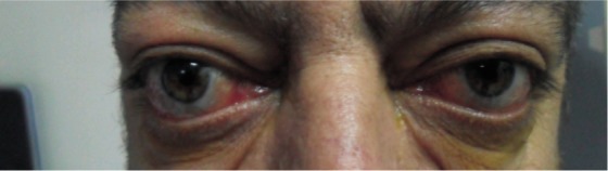
Thyroid-associated orbitopathy, is a set of symptoms, caused by an autoimmune process, typical for Graves' disease and rarely accompanies to Hashimoto's thyroiditis[15]. Immunologic crossreactivity of sensitized T lymphocytes and/or autoantibodies against thyroid and orbit may trigger the inflammatory process[16]. In both of Graves' and Hashimoto's disease, antithyroglobulin and anti-thyroid peroxidase antibodies are detected. Therefore a transformation of Graves' disease into Hashimoto's thyroiditis disease and the reverse is also known[6]. Typical signs are upper lid retraction, proptosis, periorbital oedema and restrictive myopathy[3]. Although numerous studies have been reported about the prevalence of Graves' ophthalmopathy, similar studies about the prevalence of Hashimato's ophthalmopathy are sparse. The first study about the prevalence of ophthalmopathy in Hashimoto's thyroiditis was reported by Tjiang et al[13], he studied 91 recently diagnosed patients with Hashimoto's thyroiditis and reported that the overall prevalence of any eye signs was 34% and about one third of patients had UER. Besides, two patients had eye muscle dysfunction and about a third of the patients had severe inflammatory changes such as periorbital swelling, chemosis, conjunctival injection, and proptosis (Figure 1). In the present study we studied the prevalence of ophthalmopathy in 110 Hashimoto's thyroiditis for eye signs and compared these with healthy control subjects. We showed that the prevalence of eye signs were 22.7% in patients, which were less than Tjiang's study, and 4% in control subjects and the difference reached a statistical significance (P=0.002). In patients, 11.8% had UER, 6.4% had proptosis, 5.5% had ocular myopathy as shown on Table 3. We also determined the possible effect of the clinic stage on ophthalmopathy, neverthless the CAS and NOSPECS classification did not show any significant difference among these subgroups. One of the important characteristics of our study which differs it from Tjiang's study is that we studied with larger number of patients and compared with healthy subjects that could enhance the power of the study. The levator palpebrae superioris and extraocular muscle involvement in Hashimoto's thyroiditis is common and has been emphasized in some studies. Grzesiuk et al[8] reported a 36-year-old man with Hashimoto's thyroiditis who had enlargement of the medial and inferior rectus muscles bilaterally, with eye pain, conjunctival redness, swelling of the eye lid and exophthalmos. Hiraga et al[7] reported a 76 years old woman with Hashimoto's thyroiditis suffering from diplopia due to limitations of upper gaze in the right eye. She had enlargement of inferior rectus muscle unilaterally. In an another study reporting two cases with Hashimoto's thyroiditis, the CAS activity score was found high and magnetic resonance imaging (MRI) showed enlargement of the extraocular muscles in both patients who required systemic glucocorticoid therapy and orbital irradiation to treat the TAO[9]. In our study 6 (5.5%) patients had ocular myopathy whom also had UER. Since patients with Hashimoto's thyroiditis are negative for TSH-r, the TSH-r hypothesis can not be used to explain the eye muscle involvement in Hashimoto's thyroiditis.
Figure 1. A patient with periorbital swelling, chemosis, conjunctival injection, and proptosis.
Specific antibodies against eye muscle antigens such as calsequestrin, collagen XIII flavoprotein (Fp) and G2s,which are also shown to be goodmarkers of eye muscle damage in Graves patients may be an alternative explanation for eye muscle damage in Hashimoto's thyroiditis[17]–[20]. A study showed that calsequestrin and collagen XIII antibodies are also associated with chronic upper eyelid retraction as a dominant feature of ophthalmopathy in patients with Graves' disease and Hashimoto's thyroiditis[20]. Gopinath et al[21] evaluated 11 Hashimoto's thyroiditis patients for the presence of antibodies aganist eye muscle antigens and found that 7 patients were positive for at least one antibody. Five of them were found to have developed ophthalmopathy at first visit or on follow-up. In his study, calsequestrin and Fp antibodies were found to be the most frequently detected antibodies. Gopinath et al[22] presented an euthyroid woman with chronic UER and positive calsequestrin and collagen XIII antibodies who developed Hashimoto's thyroiditis and hypothyroidism 22mo later. Since the important of the eye muscle antibodies, particularly calsequestrin were stated in the majority of the studies, we think that they may be used as indicator tests to predict the muscle involvement to indicate the onset of eye muscle inflammation, with the possibility of early treatment. But unfortunately we were not able to study the serum levels of muscle antibodies in our study.
To summarize the main findings, the eye signs were mostly mild (22.7%) and the most common eye sign was the presence of UER (11.8%). Additionally six patients had eye muscle dysfunction as reduced eye movements in up gaze. On the other hand the clinical stage of the disease did not have any impact on the activity of ophthalmopathy. Even though eyelid and extraocular muscle disorders may be a minor problem for most of the patients, they both may cause major cosmetic and functional complications requiring surgical management before the onset of the ophthalmopathy. Therefore we recommend to make a routine ophthalmic examination in Hashimoto's thyroiditis patients in order not to omit the associated ophthalmopathy.
Acknowledgments
Conflicts of Interest: Kan E, None; Kilic Kan E, None; Ecemis G, None; Colak R, None.
REFERENCES
- 1.Yamada M, Wu Li A, Wall J. Thyroid-associated ophthalmopathy: clinical features, pathogenesis, and management. Crit Rev Clin Lab Sci. 2000;37(6):523–549. doi: 10.1080/10408360091174303. [DOI] [PubMed] [Google Scholar]
- 2.Maheshwari R, Weis E. Thyroid associated orbitopathy. Indian J Ophthalmol. 2012;60(2):87–93. doi: 10.4103/0301-4738.94048. [DOI] [PMC free article] [PubMed] [Google Scholar]
- 3.Bahn RS. Graves' ophthalmopathy. N Engl J Med. 2010;362(25):726–738. doi: 10.1056/NEJMra0905750. [DOI] [PMC free article] [PubMed] [Google Scholar]
- 4.Bahn RS. Clinical review 157: Pathophysiology of Graves' ophthalmopathy: the cycle of disease. J Clin Endocrinol Metab. 2003;88(5):1939–1946. doi: 10.1210/jc.2002-030010. [DOI] [PubMed] [Google Scholar]
- 5.Dagi LR, Elliott AT, Roper-Hall G, Cruz OA. Thyroid eye disease: honing your skills to improve outcomes. JAAPOS. 2010;14(5):425–431. doi: 10.1016/j.jaapos.2010.07.005. [DOI] [PubMed] [Google Scholar]
- 6.Wall JR, Bernard N, Boucher A, Salvi M, Zhang ZG, Kennerdell J, Tyutyunikov A, Genovese C. Pathogenesis of thyroid-associated ophthalmopathy: an autoimmune disorder of the eye muscle associated with Graves' hyperthyroidism and Hashimoto's thyroiditis. Clin Immunol Immunopathol. 1993;68(1):1–8. doi: 10.1006/clin.1993.1087. [DOI] [PubMed] [Google Scholar]
- 7.Hiraga A, Mimura M, Kamitsukasa I. Isolated inferior rectus muscle myopathy due to Hashimoto's thyroiditis. Intern Med. 2008;47(13):1283–1284. doi: 10.2169/internalmedicine.47.1003. [DOI] [PubMed] [Google Scholar]
- 8.Grzesiuk W, Szydlarska D, Pragacz A, Bar-Andziak E. Thyroid associated orbitopathy in patients with Hashimoto's thyroiditis: a case report. Pol Arch Med Wewn. 2008;118(5):318–321. [PubMed] [Google Scholar]
- 9.Yoshihara A, Yoshimura Noh J, Nakachi A, Ohye H, Sato S, Sekiya K, Kosuga Y, Suzuki M, Matsumoto M, Kunii Y, Watanabe N, Mukasa K, Inoue Y, Ito K, Ito K. Severe thyroid associated orbitopathy in Hashimoto's thyroiditis. Report of 2 cases. Endocr J. 2011;58(5):343–348. doi: 10.1507/endocrj.k11e-019. [DOI] [PubMed] [Google Scholar]
- 10.Klee GG, Hay ID. Biochemical testing of thyroid function. Endocrinol Mateb Clin North Am. 1997;26(4):763–775. doi: 10.1016/s0889-8529(05)70281-4. [DOI] [PubMed] [Google Scholar]
- 11.Wiersinga WM, Prummel MF, Mourits MP, Koornneef L, Buller HR. Classification of the eye changes of Graves' disease. Thyroid. 1991;1(4):357–360. doi: 10.1089/thy.1991.1.357. [DOI] [PubMed] [Google Scholar]
- 12.Mourits MP, Prummel MF, Wiersinga WM, Koornneef L. Clinical activity score as a guide in the management of patients with Graves' Ophthalmopathy. Clin Endocrinol. 1997;47(1):9–14. doi: 10.1046/j.1365-2265.1997.2331047.x. [DOI] [PubMed] [Google Scholar]
- 13.Tjiang H, Lahooti H, McCorquodale T, Parmar KR, Wall JR. Eye and eyelid abnormalities are common in patients with Hashimoto's thyroiditis. Thyroid. 2010;20(3):287–290. doi: 10.1089/thy.2009.0199. [DOI] [PubMed] [Google Scholar]
- 14.Beden U, Ozarslan Y, Oztürk HE, Sönmez B, Erkan D, Oge I. Exophthalmometry values of Turkish adult population and the effect of age, sex, refractive status, and Hertel base values on Hertel readings. Eur J Ophthalmol. 2008;18(2):165–171. doi: 10.1177/112067210801800201. [DOI] [PubMed] [Google Scholar]
- 15.El-Kaissi S, Frauman AG, Wall JR. Thyroid-associated ophthalmopathy: a practical guide to classification, natural history and management. Intern Med J. 2004;34(8):482–491. doi: 10.1111/j.1445-5994.2004.00662.x. [DOI] [PubMed] [Google Scholar]
- 16.Iyer S, Bahn R. Immunopathogenesis of Graves' ophthalmopathy: the role of the TSH receptor. Best Pract Res Clin Endocrinol Metab. 2012;26(3):281–289. doi: 10.1016/j.beem.2011.10.003. [DOI] [PMC free article] [PubMed] [Google Scholar]
- 17.Wescombe L, Lahooti H, Gopinath B, Wall JR. The cardiac calsequestrin gene (CASQ2) is up-regulated in the thyroid in patients with Graves' ophthalmopathy--support for a role of autoimmunity against calsequestrin as the triggering event. Clin Endocrinol. 2010;73(4):522–528. doi: 10.1111/j.1365-2265.2009.03753.x. [DOI] [PubMed] [Google Scholar]
- 18.Gopinath B, Wescombe L, Nguyen B, Wall JR. Can autoimmunity against calsequestrin explain the eye and eyelid muscle inflammation of thyroid eye disease? Orbit. 2009;28(4):256–261. [PubMed] [Google Scholar]
- 19.Gopinath B, Musselman R, Beard N, Tani J, Adams C, Wall JR. Antibodies targeting the calcium binding skeletal muscle protein calsequestrin are sensitive and specific markers of ocular myopathy in patients with Graves' disease. Clin Exp Immunol. 2006;145(1):56–62. doi: 10.1111/j.1365-2249.2006.03110.x. [DOI] [PMC free article] [PubMed] [Google Scholar]
- 20.Gopinath B, Adams CL, Musselman R, Tani J, Wall JR. Antibodies against calsequestrin and type XIII collagen are good markers for chronic upper eyelid lag and retraction. Ocul Immunol Inflamm. 2007;15(2):81–88. doi: 10.1080/09273940701299362. [DOI] [PubMed] [Google Scholar]
- 21.Gopinath B, Ma G, Wall JR. Eye signs and serum eye muscle and collagen XIII antibodies in patients with transient and progressive thyroiditis. Thyroid. 2007;17(11):1123–1129. doi: 10.1089/thy.2007.0054. [DOI] [PubMed] [Google Scholar]
- 22.Gopinath B, Ma G, Lahooti H, Wall JR. A Case of Hashimoto's Thyroiditis Presenting with Chronic Upper eye Lid Retraction and Positive Calsequestrin and Collagen XIII Antibodies. Int J Endocrinol Metab. 2007;6(2):34–37. [Google Scholar]