Abstract
Patient: Female, 67
Final Diagnosis: Pulmonary carcinoid tumor
Symptoms: Abnormal shadow on chest X-ray
Medication: —
Clinical Procedure: —
Specialty: Pulmonology
Objective:
Rare disease
Background:
Although pulmonary carcinoid tumors are generally considered to represent a low-grade malignancy, atypical carcinoids are more aggressive than typical carcinoids, metastasizing more commonly to both regional lymph nodes and distant sites. The treatment of choice for localized disease is surgery. In cases of advanced or metastatic disease, medical treatments, including chemotherapy, have not been proven to be very successful. Therefore, providing careful follow-up is extremely important. In general, tumor markers, such as the level of CYFLA21-1, are often useful for monitoring lung cancer. However, there are currently no sensitive tumor markers for carcinoid tumors. We herein report a rare case of an atypical carcinoid of the lung with the elevation of the serum ProGRP level.
Case Eeport:
A 67-year-old female was referred to our hospital for an abnormal chest X-ray. CT revealed an 18×13 mm nodule in the right middle lobe with no significant mediastinal lymphadenopathy. The serum tumor marker, the ProGRP level, was significantly elevated (161 ng/ml). We performed a right middle lobectomy, because the pathological diagnosis of lung cancer was confirmed according to the results of a rapid frozen section biopsy of the lesion, although the pathological type could not be precisely determined by the frozen section alone. The final pathological diagnosis was atypical carcinoid. The level of ProGRP decreased (69 ng/ml) within 1 month after the surgery.
Conclusions:
The ProGRP level may be useful for monitoring carcinoid tumors, although no serum tumor markers are highly specific or sensitive for detecting recurrences and/or distant metastasis of pulmonary carcinoid tumors. In conclusion, ProGRP should be further evaluated as biomarker in a larger series of patients to determine whether it demonstrates any significant correlation with cancer recurrence.
MeSH Keywords: Carcinoid Tumor; Lung Neoplasms; Tumor Markers, Biological
Background
Carcinoid tumors are neuroendocrine tumors that arise from Kulchitsky cells. Although they may develop in many locations in the body, they are most often found in the small intestine (26%), respiratory system (25%), and appendix (19%) [1]. Pulmonary carcinoid tumors represent 1–2% of all lung neoplasms [2]. According to the current WHO classification, these lesions are currently divided into 4 subtypes characterized by increasing aggressiveness: typical carcinoid, atypical carcinoid, large-cell neuroendocrine carcinoma, and small cell carcinoma.
Among these subtypes, the most important differential criterion for typical carcinoids and atypical carcinoids is the mitotic count. The typical carcinoids have <2 mitoses per mm2 in 10 high-power fields (HPF) without signs of necrosis, while atypical carcinoids are characterized by the presence of 2–10 mitoses per mm2/10 HPF and/or foci of necrosis.
Although carcinoid tumors are generally considered to represent a low-grade malignancy, atypical carcinoids are more aggressive than typical carcinoids, metastasizing more commonly to both regional lymph nodes and distant sites. The treatment of choice for localized disease is surgery. In cases of advanced or metastatic disease, medical treatments, including chemotherapy, have not been proven to be very successful [3].
Therefore, providing careful follow-up is extremely important. In general, tumor markers, such as the levels of CEA, CYFLA and ProGRP, are often useful for monitoring lung cancer. However, there are currently no sensitive tumor markers for carcinoid tumors. We herein report a rare case of an atypical carcinoid of the lung with the elevation of the serum ProGRP level.
Case Report
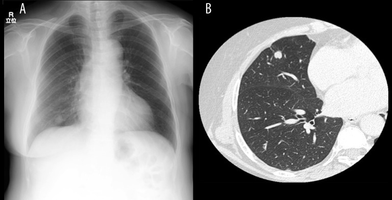
A 67-year-old female was referred to our hospital for an abnormal shadow on a chest X-ray (Figure 1A). She had an un-remarkable medical and family history and was a non-smoker. CT revealed an 18×13 mm nodule in the right middle lobe with no significant mediastinal lymphadenopathy (Figure 1B). One serum tumor marker, the ProGRP level, was significantly elevated (161 ng/ml), while all other tumor markers were within normal limits.
Figure 1.
(A) Chest X-ray showing an abnormal shadow in the right middle lobe. (B) Chest CT showing a nodule measuring 13×18 mm in size in the right middle lobe.
These findings were strongly indicative of malignancy, although the pathologic diagnosis was not confirmed on transbronchial lung biopsy (TBLB). Since there were no signs of any distant metastasis, the clinical stage was determined to be T1aN0M0, stage A based on the CT image findings. We performed a right middle lobectomy, because a pathological diagnosis of lung cancer was confirmed according to the results of a rapid frozen section biopsy of the lesion. There were no intraoperative complications, and the patient had an uneventful recovery. The final results of the pathologic examination showed an atypical carcinoid of pT1aN2M0, stage A. The level of ProGRP decreased (69 ng/ml) within 1 month after the surgery.
Discussion
The most effective treatment for pulmonary carcinoid tumors is surgical resection in cases of localized disease, and it is believed that chemotherapy and radiotherapy have no therapeutic contribution in patients with unresectable carcinoid lung tumors [4]. The long-term survival rates of patients with typical and atypical tumors are significantly different. For example, the 5-year survival rate for neuroendocrine tumors of the lung ranges from 87% to 97% for typical carcinoids and from 56% to 77% for atypical carcinoids [5,6]. Therefore, providing follow-up care after surgery is extremely important, especially in patients with atypical carcinoids.
Generally, measurements of serum tumor markers play a crucial role in detecting the recurrences and/or distant metastasis of malignancy after surgery. However, no serum tumor markers are highly specific or sensitive for detecting recurrences or distant metastasis of pulmonary carcinoid tumors. In the present case, the serum level of ProGRP was elevated at 161 pg/ml (normal range: <46 pg/ml) before treatment and subsequently decreased to 69 pg/ml at 1 month after the surgery.
ProGRP is a precursor form of gastrin-releasing peptide (GRP), a neuropeptide hormone that was originally isolated from porcine gastric tissue [7]. GRP is widely distributed throughout the gastrointestinal and pulmonary tracts. In general, ProGRP is a well-known biomarker of small cell lung cancer. However, according to Polak [8], typical carcinoids in which ProGRP is produced by the carcinoid cells account for 16% of lesions, while atypical carcinoids with this characteristic account for 63% of such tumors. Therefore, the serum ProGRP level may be elevated in such cases due to its correlation with the tissue ProGRP level. In addition, although Kamata et al. reported that the serum ProGRP level increases with the progression of renal dysfunction [9], in the present case, the patient’s renal function was found to be in the normal range. The ProGRP level also decreased due to the effects of treatment, and the level did not quickly return to the normal ranges because ProGRP has a long half-life of 19–28 days.
Consequently, measuring the ProGRP level may be useful for monitoring carcinoid tumors in cases in which this parameter is elevated above the normal range, although more thorough scientific documentation is needed to clarify the relationship between carcinoid tumors and ProGRP due to the limited number of cases [10].
Conclusions
In conclusion, ProGRP should be further evaluated as a biomarker in a larger series of patients to determine whether it demonstrates any significant correlation with cancer recurrence.
Footnotes
Conflict of interest statement
Naohiro Taira and the other co-authors have no conflicts of interest to declare.
References:
- 1.Lips CJM, Lentjes EGWM, Höppener JWM. The spectrum of carcinoid tumours and carcinoid syndromes. Ann Clin Biochem. 2003;40:612–27. doi: 10.1258/000456303770367207. [DOI] [PubMed] [Google Scholar]
- 2.Asamura H, Kameya T, Matsuno Y, et al. Neuroendocrine neoplasms of the lung: a prognostic spectrum. J Clin Oncol. 2006;24(1):70–76. doi: 10.1200/JCO.2005.04.1202. [DOI] [PubMed] [Google Scholar]
- 3.Katai M, Sakurai A, Inaba H, et al. Octreotide as a rapid and effective pain-killer for metastatic carcinoid tumor. Endocr J. 2005;52:277–80. doi: 10.1507/endocrj.52.277. [DOI] [PubMed] [Google Scholar]
- 4.Thomas CF, Jr, Tazelaar HD, Jett JR. Typical and atypical pulmonary carcinoids: outcome in patients presenting with regional lymph node involvement. Chest. 2001;119(4):1143–50. doi: 10.1378/chest.119.4.1143. [DOI] [PubMed] [Google Scholar]
- 5.Filosso PL, Rena O, Donati G. Bronchial carcinoid tumors: surgical management and long-term out come. J Thorac Cardiovasc Surg. 2002;123:303–9. doi: 10.1067/mtc.2002.119886. [DOI] [PubMed] [Google Scholar]
- 6.Travis WD, Rush W, Flieder DB, et al. Survival analysis of 200 pulmonary neuroendocrine tumors with clarification of criteria for atypical carcinoid and its separation from typical carcinoid. Am J Surg Pathol. 1998;22:934–44. doi: 10.1097/00000478-199808000-00003. [DOI] [PubMed] [Google Scholar]
- 7.McDonald TJ, Jornvall H, Nilsson G, et al. Characterization of a gastrin releasing peptide from porcine non-antral gastric tissue. Biochem Biophys Res Commun. 1979;90(1):227–33. doi: 10.1016/0006-291x(79)91614-0. [DOI] [PubMed] [Google Scholar]
- 8.Polak JM, Hamid Q, Springal DR, et al. Localizationof bombesin-like peptides in tumors. Ann NY Acad Sci. 1988;547:32–35. doi: 10.1111/j.1749-6632.1988.tb23900.x. [DOI] [PubMed] [Google Scholar]
- 9.Kamata K, Uchida M, Takeuchi Y, et al. Increased serum concentrations of pro-gastrin-releasing peptide in patients with renal dysfunction. Nephrol Dial Transplant. 1996;11:1267–70. [PubMed] [Google Scholar]
- 10.Uchida N, Fukino S, Kodama W. [A case of typical pulmonarycarcinoid tumor with elevation of preoperative ProGRP] The Journal of the Japanese Association for Chest Surgery. 2008;22(5):839–42. [Google Scholar]


