Abstract
We report on three unrelated patients with the 22q11.2 microdeletion syndrome (del22q11) who have phenotypic anomalies compatible with oculo-auriculo-vertebral spectrum (OAVS). Hemifacial microsomia, unilateral microtia, hearing loss, congenital heart/aortic arch arteries defects, and feeding difficulties were present in all three patients. Additional anomalies occasionally diagnosed included coloboma of the upper eyelid, microphthalmia, cerebral malformation, palatal anomalies, neonatal hypocalcemia, developmental delay, and laryngomalacia. Several clinical features characteristic of OAVS have been described in patients with del22q11 from the literature, including ear anomalies, hearing loss, cervical vertebral malformations, conotruncal cardiac defects, renal malformations, feeding and respiratory difficulties. Atretic ear with facial asymmetry has been previously described in one patient. Thus, clinical expression of hemifacial microsomia and microtia resembling OAVS should now be included within the wide phenotypic expression of del22q11. The occurrence of this manifestation in del22q11 is currently low. Nevertheless, patients with hemifacial microsomia and microtia associated with clinical features typically associated with del22q11 should now have for specific cytogenetic testing.
Keywords: deletion 22q11.2, Goldenhar syndrome, hemifacial microsomia, oculo-auriculo-vertebral spectrum, velo-cardio-facial syndrome
Introduction
Deletion 22q11.2 syndrome (del22q11 OMIM #192430) (DiGeorge/velo-cardio-facial syndrome) is characterized by facial dysmorphisms, palatal anomalies, congenital heart defects (CHDs), hypoplastic thymus and immune deficit, neonatal hypocalcemia, speech and learning difficulties [Ryan et al., 1997; McDonald-McGinn et al., 1999]. Most of the structures primarily affected in del22q11 are derivatives of the branchial arch system, and a developmental relation to neural crest cell migrational anomalies has been noted [Van Mierop and Kutsche, 1986; Kirby and Waldo, 1990].
The oculo-auriculo-vertebral spectrum (OAVS, OMIM 164210) is a non-random association of unilateral microtia, hemifacial microsomia with mandibular hypoplasia, ocular epibulbar dermoid, and cervical vertebral malformations [Rollnick et al., 1987; Gorlin et al., 2001]. Cerebral, cardiac, and renal malformations can also be detected [Rollnick et al., 1987]. The condition is also known as Goldenhar syndrome, referring to its first description [Goldenhar, 1952]. Morphogenetic anomalies of the first and second branchial arches, usually of a unilateral nature, and neural crest cell migrational abnormalities have been suspected to be implicated in the etiopathogenesis of the condition [Lammer and Opitz, 1986], but chromosomal aneuploidies have been detected occasionally [Gorlin et al., 2001].
Here we report on three unrelated patients with del22q11 who have phenotypic anomalies compatible with OAVS.
Clinical Reports
Patient 1
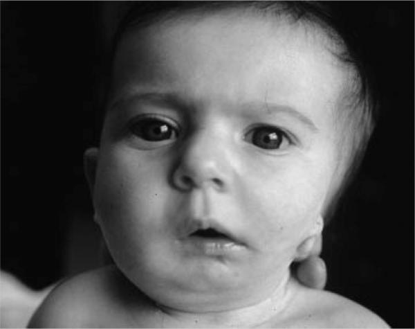
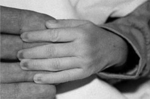
This 5-month-old Caucasian male is the third child of healthy unrelated parents. Family history was unremarkable. At birth the mother was 31 years old, the father 39. The baby was born by vaginal delivery after an uneventful pregnancy. Birth weight was 3,470 g (50th–75th centile), length 50 cm (50th–75th centile), head circumference 34 cm (50th centile). Apgar scores were 8 at 1 min, and 9 at 5 min. Echocardiography performed shortly after birth revealed a double outlet right ventricle with valvular and infundibular pulmonary stenosis, subaortic ventricular septal defect, and normally related great arteries. Hypocalcemia secondary to hypoparathyroidism was diagnosed during the third day of life, and treated by calcium gluconate supplementation. Serum calcium concentration normalized after 10 days of therapy. Cerebral and abdominal ultrasound studies were normal. Audiologic screening was normal on the right, hearing loss was demonstrated on the left. A CT scan of the left ear demonstrated absence of the external auditory meatus and dysgenesis of the inner ear system. Ophthalmologic examination and vertebral column X-ray disclosed no abnormalities. T-lymphocyte count was low, serum immunoglobulin concentrations were normal for age. Clinical examination at 5 months of life revealed left hemifacial microsomia, coloboma of the upper left eyelid, cleft palate, a small mouth, asymmetric mandibular hypoplasia, bilateral preauricular tags, microtia with aural atresia of the left ear (Fig. 1), and long fingers (Fig. 2). Standard chromosome analysis on peripheral blood lymphocytes revealed a normal 46,XY karyotype. Considering the association of conotruncal CHDs, cleft palate and facial anomalies, fluorescence in situ hybridization (FISH) with the N25 probe was performed, confirming the presence of a de novo 22q11.2 microdeletion.
FIG. 1.

Facial appearance of Patient 1.
FIG. 2.

Hands with long and tapering fingers in Patient 1.
Patient 2
This 35-day-old Caucasian male is the first child of healthy nonconsanguineous parents. At birth the mother was 25 years old, the father 29. Family history was unremarkable. The baby was born by vaginal delivery at the 38th week of a normal pregnancy. Birth weight was 3,090 g (50th centile), length 47.5 cm (25th centile), head circumference 35 cm (75th–90th centile). At 2 weeks of age the patient was admitted to the hospital, with a history of feeding and respiratory difficulties. Phenotypic examination demonstrated a prominent forehead with a receding hairline, mild facial asymmetry (right cheek smaller than the left), asymmetric crying facies with a right facial droop, microtia of the right ear with a preauricular tag and atresia, low-set left ear with an overfolded, thick superior helix, asymmetric gynecomastia, a sacral dimple, and long fingers. Gastrointestinal radiological investigations performed for failure to thrive revealed severe gastro-esophageal reflux requiring a Nissan fundoplication and G-tube placement. Doppler two-dimensional echocardiography revealed an aberrant right subclavian artery, patent foramen ovalis, and trivial tricuspid regurgitation. Barium swallow confirmed a posterior indentation of the proximal thoracic esophagus and trachea due to aberrant subclavian artery. No vascular ring was evident by chest CT scan. Endoscopy of the upper airway showed normal vocal cord movement, and laryngomalacia. Full-night polysomnography disclosed several apneic events with oxygen desaturations. Brain MRI revealed an open fronto-temporal operculum with persistent cavum septum pellucidum, mild widening of the foramen of Magendie and the cisterna magna. Right external and internal ear anomalies were also detected by MRI images, including absence of the right pinna, non-visualization of the external auditory canal, and a prominent vestibule with no lateral semicircular canal. No MRI anomalies of the left internal and external ear were noted. Audiologic screening revealed right-sided hearing loss, and left-sided normal hearing. Ophthalmologic examination, vertebral column X-ray, EEG, and renal ultrasonography were all normal. Laboratory investigations, including T-lymphocyte counts, thyroid and parathyroid hormones, prolactin and estriol dosages, were in the normal range. Chromosomal analysis from amniocytes and peripheral blood lymphocytes revealed a normal 46,XY karyotype. Microdeletion 22q11.2 was detected after birth using the N25 probe. Parental studies have not been obtained to date.
Patient 3
This 23-month-old male is the fourth child of healthy non-consanguineous parents. At birth the mother was 34 years old, the father 39. The baby was born by vaginal delivery at term of an uneventful pregnancy. Birth weight was 3,500 g (75th centile). Developmental miles stones were mildly delayed (sitting at 9 months, walking at 15 months, speech delay). Ophthalmological examination revealed bilateral optic nerve hypoplasia. Audiologic screening demonstrated hearing loss at the right ear. Echocardiography showed a small atrial septal defect ostium secundum type. Vertebral column X-ray revealed butterfly vertebra at the T8 and T10 levels. The baby had a history of dysphagia and feeding difficulties, resolved during the second year of life. Laryngomalcia required aryepiglottoplasty. Cerebral and renal ultrasonographies were normal. Serum calcium concentration and T- and B-cell subsets were normal. Clinical examination at 23 months of life showed left hemifacial microsomia, periorbital fullness, broad nasal root, bulbous nasal tip, hypoplastic nares, submucous cleft palate, small mouth, left microtia with aural atresia, small right ear with a simple protruding helix, tapered fingers, sacral dimple, and flat feet. Weight was 12,600 kg (25th–50th centile), length 86 cm (25th–50th centile), head circumference 49.6 cm (50th–75th centile). Standard chromosome analysis on peripheral blood lymphocytes showed a normal 46,XY karyotype. FISH with the N25 probe showed a de novo 22q11.2 microdeletion.
Discussion
Several clinical features characteristic of OAVS have been described in patients with the clinical diagnosis of DiGeorge/velo-cardio-facial syndrome and/or cytogenetic detection of del22q11, including ear anomalies with preauricular pits or tags [Black et al., 1975; Ryan et al., 1997; McDonald-McGinn et al., 1999], hearing loss [Burn et al., 1993; Wilson et al., 1993; Britt Ravnan et al., 1996; Ryan et al., 1997; Digilio et al., 1999], cervical vertebral malformations [Ming et al., 1997], conotruncal cardiac defects [Marino et al., 2005], renal malformations [Devriendt et al., 1996; Stewart et al., 1999; Wu et al., 2002], feeding and respiratory difficulties [Ryan et al., 1997; Rommel et al., 1999; Eicher et al., 2000; Digilio et al., 2001] (Table I). Microtia with aural atresia [Derbent et al., 2003] or microphthalmia [Beemer et al., 1986] with facial asymmetry has also been described sporadically. Recently, a 1.12-Mb deletion partially overlapping with the distal end of the common 3 Mb deletion involved in the DiGeorge/velo-cardio-facial syndrome has been reported in a child with clinical features of OAVS [Xu et al., 2008].
TABLE I.
Clinical Findings in the Present Patients With Oculo-Auriculo-Vertebral Spectrum and Deletion 22q11 Syndrome
| Clinical feature | Patient 1 | Patient 2 | Patient 3 | OAVS | Del22q11 |
|---|---|---|---|---|---|
| Hemifacial microsomia | + | + | + | + | − |
| Microphthalmia | + | − | − | + | + |
| Epibulbar dermoid | − | − | − | + | − |
| Unilateral microtia | + | + | + | + | − |
| Dysmorphic ear | + | + | + | + | + |
| Preauricular pit/tag | + | + | − | + | + |
| Hearing loss | + | + | + | + | + |
| Cerebral anomalies | − | + | − | + | + |
| Cervical malformations | − | − | − | + | + |
| Congenital heart defect | + | − | + | + | + |
| Aortic arch arteries anomalies | − | + | − | − | + |
| Renal malformations | − | − | − | + | + |
| Feeding difficulties | + | + | + | + | + |
| Laryngomalacia | − | + | + | + | + |
| Neonatal hypocalcemia | + | − | − | − | + |
| T-cell deficiency | + | − | − | + |
Del22q11, deletion 22q11 syndrome; OAVS, oculo-auriculo-vertebral spectrum.
Abnormal migration of neural crest cells is a developmental anomaly suggested to be implicated in the pathogenesis of both OAVS [Lammer et al., 1985] and DiGeorge/velo-cardio-facial syndrome [Lammer and Opitz, 1986; Van Mierop and Kutsche, 1986; Kirby and Waldo, 1990; Pizzuti et al., 1997], considering the phenotypically affected structures. With regard to del22q11, the differentiation of the branchial arches, including the precursor tissue of the ear, is controlled by neural crest cell development [Van Mierop and Kutsche, 1986; Kirby and Waldo, 1990]. Additionally, it must be pointed out that some of the genes located inside the 22q11.2 “critical region,” including TBX1 and UFD1L, are specifically expressed during embryogenesis in the primordial ear [Pizzuti et al., 1997; Yamagishi et al., 1999; Vitelli et al., 2003; Raft et al., 2004]. In fact, TBX1−/− mice have a missing outer and middle ear, with a very malformed inner ear, lacking a cochlea or vestibular system [Vitelli et al., 2003; Raft et al., 2004]. Furthermore, malformation of the vertebral column could also be due to TBX1 haploinsufficiency [Chapman et al., 1996; Ming et al., 1997].
An additional pathogenetic mechanism possibly linking OAVS and del22q11 is the occurrence of an early developmental anomaly in the embryonic vasculature. In fact, vascular anomalies have been described in del22q11 and many clinical aspects of this condition are thought to be explained by vascular insufficiency or disruption [Shprintzen, 2005]. In OAVS, a mechanism interfering with vascular supply and focal hemorrhage in the developing first and second branchial arch region seems to be implicated in causing hemifacial microsomia [Poswillo, 1975; Soltan and Holmes, 1986].
A heterogeneous spectrum of CHDs is reported in 5–58% of patients with OAVS [Shokeir, 1977; Morrison et al., 1992; Kumar et al., 1993; Digilio et al., 2008], and conotruncal defects, particularly tetralogy of Fallot, have been described. Cardiac involvement in the present patients is characterized by double-outlet right ventricle, atrial septal defect ostium secundum type, and an isolated aortic arch anomaly (Table I). Double-outlet right ventricle has occasionally been described in patients with del22q11 [Ryan et al., 1997; Matsuoka et al., 1998; McDonald-McGinn et al., 1999; Marino et al., 2001], usually defined as associated with a subaortic ventricular septal defect and normally related great arteries [Marino et al., 2005]. Anomalies of branching of the aortic arch and brachiocephalic arteries have been reported in association with del22q11 [Momma et al., 1999; McElhinney et al., 2001; Toscano et al., 2002], and prenatal anomalous regression and/or persistence of the primitive branchial arches can explain embryologically the pathogenesis of these defects [Momma et al., 1999].
The association with neonatal hypocalcemia, immune deficit, and cleft palate in patient 1 was particularly suggestive of del22q11. Interestingly, the epibulbar dermoid characteristic of OAVS was absent in all three patients. A possible explanation is the fact that this anomaly is not derived from defects of neural crest cell migration.
The term OAVS is used to define patients with a combination of anomalies including microtia, ocular abnormalities, mandibular hypoplasia, and skeletal defects [Gorlin et al., 2001]. Goldenhar syndrome is generally used for variants of OAVS with epibulbar dermoids [Goldenhar, 1952]. Nevertheless, the condition is extremely complex and heterogeneous, so that hemifacial microsomia and isolated microtia with mandibular hypoplasia may also be variants of the same dysmorphologic entity [Rollnick et al., 1987]. Additionally, family studies have identified isolated microtia or isolated mandibular hypoplasia in first-degree relatives of patients with OAVS [Rollnick and Kaye, 1983; Digilio et al., 2008]. For this reason, clinical features in patients in the present report are defined as OAVS or Goldenhar syndrome as manifestations of a spectrum, although ocular and cervical manifestations are lacking.
Facial appearance of the present patients combines characteristics of OAVS and del22q11. Facial anomalies of del22q11 are covered by hemifacial microsomia and mildly expressed, consisting in periorbital fullness in all cases, large nasal tip in two (with hypoplastic nares in one), and small mouth in two.
Fingers are long and tapering as usually observed in patients with del22q11. This is not a characteristic feature of OAVS, so that this sign could be useful to distinguish those patients with del22q11 from those without.
The occurrence of OAVS manifestations in patients with del22q11 is low. It must be considered that del22q11 and OAVS occur singularly very frequently (1/4,000 and 1/5,600, respectively), and coincidence cannot be excluded in the present patients. The possibility that both conditions occur together for chance in the same patient can be estimated as 1/22,400,000.
In conclusion, clinical expression of hemifacial microsomia and microtia resembling OAVS should be included within the wide phenotypic expression of del22q11. The occurrence of this manifestation in del22q11 is low. Nevertheless, patients with hemifacial microsomia and microtia associated with clinical features characteristic for del22q11 (conotruncal heart defects or anomalies of the aortic arch, neonatal hypocalcemia, immune deficit) should now be considered for cytogenetic testing FISH, MLPA, or array-based comparative genomic hybridization.
REFERENCES
- Beemer FA, de Nef JJEM, Delleman JW, Bleeker-Wagemakers EM, Shprintzen RJ. Additional eye findings in a girl with the velo-cardio-facial syndrome. Am J Med Genet. 1986;24:541–542. doi: 10.1002/ajmg.1320240319. [DOI] [PubMed] [Google Scholar]
- Black FO, Spanier SS, Kohut RI. Aural abnormalities in partial DiGeorge syndrome. Arch Otolaryngol. 1975;101:129–134. doi: 10.1001/archotol.1975.00780310051014. [DOI] [PubMed] [Google Scholar]
- Britt Ravnan J, Chen E, Golabi M, Lobo RV. Chromosome 22q11.2 microdeletions in velocardiofacial syndrome patients with widely variable manifestations. Am J Med Genet. 1996;66:250–256. doi: 10.1002/(SICI)1096-8628(19961218)66:3<250::AID-AJMG2>3.0.CO;2-T. [DOI] [PubMed] [Google Scholar]
- Burn J, Takao A, Wilson D, Cross I, Momma K, Wadey R, Scambler P, Goodship J. Conotruncal anomaly face syndrome is associated with a deletion within chromosome 22q11. J Med Genet. 1993;30:822–824. doi: 10.1136/jmg.30.10.822. [DOI] [PMC free article] [PubMed] [Google Scholar]
- Chapman DL, Garvey N, Hancock S, Alexiou M, Agulnik SI, Gibson-Brown JJ, Cebra-Thomas J, Bollag RJ, Silver LM, Papaioannou VE. Expression of the T-box family genes, Tbx1-Tbx5, during early mouse development. Dev Dyn. 1996;206:379–390. doi: 10.1002/(SICI)1097-0177(199608)206:4<379::AID-AJA4>3.0.CO;2-F. [DOI] [PubMed] [Google Scholar]
- Derbent M, Yilmaz Z, Baltaci V, Saygili A, Varan B, Tokel K. Chromosome 22q11.2 deletion and phenotypic features in 30 patients with conotruncal heart defects. Am J Med Genet Part A. 2003;116A:129–135. doi: 10.1002/ajmg.a.10832. [DOI] [PubMed] [Google Scholar]
- Devriendt K, Swillen A, Proesmans W, Gewillig M, Fryns JP. Renal and urological tract malformations caused by a 22q11 deletion. J Med Genet. 1996;33:349. doi: 10.1136/jmg.33.4.349. [DOI] [PMC free article] [PubMed] [Google Scholar]
- Digilio MC, Pacifico C, Tieri L, Marino B, Giannotti A, Dallapiccola B. Audiological findings in patients with microdeletion 22q11 (diGeorge/velocardiofacial syndrome) Br J Audiol. 1999;33:329–334. doi: 10.3109/03005369909090116. [DOI] [PubMed] [Google Scholar]
- Digilio MC, Marino B, Cappa M, Cambiaso P, Giannotti A, Dallapiccola B. Auxological evaluation in patients with DiGeorge/velocardiofacial syndrome (deletion 22q11.2 syndrome) Genet Med. 2001;3:30–33. doi: 10.1097/00125817-200101000-00007. [DOI] [PubMed] [Google Scholar]
- Digilio MC, Calzolari F, Capolino R, Toscano A, Sarkozy A, de Zorzi A, Dallapiccola B, Marino B. Congenital heart defects in patients with oculo-auriculo-vertebral spectrum (Goldenhar syndrome) Am J Med Genet Part A. 2008;146A:1815–1819. doi: 10.1002/ajmg.a.32407. [DOI] [PubMed] [Google Scholar]
- Eicher PS, McDonald-McGinn DM, Fox CA, Driscoll DA, Emanuel BS, Zackai EH. Dysphagia in children with a 22q11.2 deletion: Unusual pattern found on modified barium swallow. J Pediatr. 2000;137:158–164. doi: 10.1067/mpd.2000.105356. [DOI] [PubMed] [Google Scholar]
- Goldenhar M. Associations malformatives de l'oeil et de l'oreille, en particulier le syndrome dermoide epibulbaire-appendices auriculairesfistula auris congenita et ses relations avec la dysostose mandibulofaciale. J Genet Hum. 1952;1:243–282. [Google Scholar]
- Gorlin RJ, Cohen MM, Jr, Hennekam RCM. Syndromes of the head and neck. Oxford University Press; New York: 2001. [Google Scholar]
- Kirby ML, Waldo KL. Role of neural crest in congenital heart disease. Circulation. 1990;82:332–340. doi: 10.1161/01.cir.82.2.332. [DOI] [PubMed] [Google Scholar]
- Kumar A, Friedman JM, Taylor GP, Patterson MW. Pattern of cardiac malformation in oculoauriculovertebral spectrum. Am J Med Genet. 1993;46:423–426. doi: 10.1002/ajmg.1320460415. [DOI] [PubMed] [Google Scholar]
- Lammer EJ, Opitz JM. The DiGeorge anomaly as a developmental field defect. Am J Med Genet. 1986;2:113–127. doi: 10.1002/ajmg.1320250615. [DOI] [PubMed] [Google Scholar]
- Lammer EJ, Chen DT, Hoar RM, Agnish ND, Benke PJ, Braun JT, Curry CJ, Fernhoff PM, Grix AW, Lott IT, Richard JM, Sun SC. Retinoic acid embryopathy. N Engl J Med. 1985;313:837–841. doi: 10.1056/NEJM198510033131401. [DOI] [PubMed] [Google Scholar]
- Marino B, Digilio MC, Toscano A, Anaclerio S, Giannotti A, Feltri C, de Ioris MA, Angioni A, Dallapiccola B. Anatomic patterns of conotruncal defects associated with deletion 22q11. Genet Med. 2001;3:45–48. doi: 10.1097/00125817-200101000-00010. [DOI] [PubMed] [Google Scholar]
- Marino B, Mileto F, Digilio MC, Carotti A, Di Donato R. Congenital cardiovascular disease and velo-cardio-facial syndrome. In: Murphy KC, Scambler PJ, editors. Velo-cardio-facial syndrome: A model for understanding microdeletion disorders. University Press; Cambridge: 2005. pp. 47–82. [Google Scholar]
- Matsuoka R, Rimura M, Scambler PJ, Morrow BE, Imamura S-I, Minoshima S, Shimizu N, Yamagishi H, Joh-o K, Watanabe S, Oyama K, Saji T, Ando M, Takao A, Momma K. Molecular and clinical study of 183 patients with conotruncal anomaly face syndrome. Hum Genet. 1998;103:70–80. doi: 10.1007/s004390050786. [DOI] [PubMed] [Google Scholar]
- McDonald-McGinn DM, Kirschner R, Goldmuntz E, Sullivan K, Eicher P, Gerdes M, Moss E, Solot C, Wang P, Jacobs I, Handler S, Knightly C, Heher K, Wilson M, Ming JE, Grace K, Driscoll D, Pasquariello P, Randall P, Larossa D, Emanuel BS, Zackai EH. The Philadelphia story. The 22q11.2 deletion: Report on 250 patients. Genet Couns. 1999;10:11–24. [PubMed] [Google Scholar]
- McElhinney DB, Clark BJ, Weinberg PM, Kenton ML, McDonald-McGinn D, Driscoll DA, Zackai EH, Goldmuntz E. Association of chromosome 22q11 deletion with isolated anomalies of aortic arch laterality and branching. J Am Coll Cardiol. 2001;37:2114–2119. doi: 10.1016/s0735-1097(01)01286-4. [DOI] [PubMed] [Google Scholar]
- Ming JE, McDonald-McGinn DM, Megerian TE, Driscoll DA, Elias ER, Russell BM, Irons M, Emanuel BS, Markowitz RI, Zackai EH. Skeletal anomalies and deformities in patients with deletions 22q11. Am J Med Genet. 1997;72:210–215. doi: 10.1002/(sici)1096-8628(19971017)72:2<210::aid-ajmg16>3.0.co;2-q. [DOI] [PubMed] [Google Scholar]
- Momma K, Matsuoka R, Takao A. Aortic arch anomalies associated with chromosome 22q11 deletion (CATCH22) Pediatr Cardiol. 1999;20:97–102. doi: 10.1007/s002469900414. [DOI] [PubMed] [Google Scholar]
- Morrison PJ, Mulholland HC, Craig BG, Nevin NC. Cardiovascular abnormalities in the oculo-auriculo-vertebral spectrum (Goldenhar syndrome) Am J Med Genet. 1992;44:425–428. doi: 10.1002/ajmg.1320440407. [DOI] [PubMed] [Google Scholar]
- Pizzuti A, Novelli G, Ratti A, Amati F, Mari A, Calabrese G, Nicolis S, Silani V, Marino B, Scarlato G, Ottolenghi S, Dallapiccola B. UFD1L, a developmentally expressed ubiquitination gene, is deleted in CATCH22 syndrome. Hum Mol Genet. 1997;6:59–65. doi: 10.1093/hmg/6.2.259. [DOI] [PubMed] [Google Scholar]
- Poswillo D. Hemorraghe in development of the face. In: Bergsma D, editor. Morphogenesis and malformation of face and brain. Alan R. Liss, Inc.; New York: 1975. pp. 61–81. [Google Scholar]
- Raft S, Nowotschin S, Liao J, Morrow BE. Suppression of neural fate and control of inner ear morphogenesis by TBX1. Development. 2004;131:1801–1812. doi: 10.1242/dev.01067. [DOI] [PubMed] [Google Scholar]
- Rollnick BR, Kaye CI. Hemifacial microsomia and variants: Pedigree data. Am J Med Genet. 1983;15:233–253. doi: 10.1002/ajmg.1320150207. [DOI] [PubMed] [Google Scholar]
- Rollnick BR, Kaye CI, Nagatoshi K, Hauck W, Martin AO. Oculoauriculovertebral dysplasia and variants: Phenotypic characteristics of 294 patients. Am J Med Genet. 1987;26:361–375. doi: 10.1002/ajmg.1320260215. [DOI] [PubMed] [Google Scholar]
- Rommel N, Vantrappen G, Swillen A, Devriendt K, Feenstra L, Fryns JP. Retrospective analysis of feeding and speech disorders in 50 patients with VCFS. Genet Couns. 1999;10:71–78. [PubMed] [Google Scholar]
- Ryan AK, Goodship JA, Wilson DI, Philip N, Levy A, Seidel H, Schuffenhauer S, Oechsler H, Belohradsky B, Prieur M, Aurias A, Raymond FL, Clayton-Smith J, Hatchwell E, McKeown C, Beemer FA, Dallapiccola B, Novelli G, Hurst JA, Ignatius J, Green AJ, Winter RM, Brueton L, Brondum-Nielsen K, Stewart F, Van Essen T, Patton M, Paterson J, Scambler PJ. Spectrum of clinical features associated with interstitial chromosome 22q11 deletion: A European collaborative study. J Med Genet. 1997;34:798–804. doi: 10.1136/jmg.34.10.798. [DOI] [PMC free article] [PubMed] [Google Scholar]
- Shokeir MHK. The Goldenhar syndrome: A natural history. Birth Defects Orig Artic Ser. 1977;XIII:67–83. [PubMed] [Google Scholar]
- Shprintzen RJ. Velo-cardio-facial syndrome. In: Cassidy SB, Allanson JE, editors. Management of genetic syndromes. 2nd edition Wiley-Liss, Inc.; New York: 2005. pp. 615–631. [Google Scholar]
- Soltan HC, Holmes LB. Familial occurrence of malformations possibly attributable to vascular abnormalities. J Pediatr. 1986;108:112–114. doi: 10.1016/s0022-3476(86)80783-1. [DOI] [PubMed] [Google Scholar]
- Stewart TL, Irons MB, Cowan JM, Bianchi DW. Increased incidence of renal anomalies in patients with chromosome 22q11 microdeletion. Teratology. 1999;59:20–22. doi: 10.1002/(SICI)1096-9926(199901)59:1<20::AID-TERA6>3.0.CO;2-S. [DOI] [PubMed] [Google Scholar]
- Toscano A, Anaclerio S, Digilio MC, Giannotti A, Fariello G, Dallapiccola B, Marino B. Ventricular septal defect and deletion of chromosome 22q11: Anatomical types and aortic arch anomalies. Eur J Pediatr. 2002;161:116–117. doi: 10.1007/s00431-001-0877-5. [DOI] [PubMed] [Google Scholar]
- Van Mierop LHS, Kutsche LM. Cardiovascular anomalies in DiGeorge syndrome and importance of neural crest as a possible pathogenetic factor. Am J Cardiol. 1986;58:133–137. doi: 10.1016/0002-9149(86)90256-0. [DOI] [PubMed] [Google Scholar]
- Vitelli F, Viola A, Morishima M, Prampano T, Baldini A, Lindsay E. TBX1 is required for inner ear morphogenesis. Hum Mol Genet. 2003;12:62–73. doi: 10.1093/hmg/ddg216. [DOI] [PubMed] [Google Scholar]
- Wilson DI, Burn J, Scambler P, Goodship J. DiGeorge syndrome: Part of CATCH22. J Med Genet. 1993;30:852–856. doi: 10.1136/jmg.30.10.852. [DOI] [PMC free article] [PubMed] [Google Scholar]
- Wu H-Y, Rusnack SL, Bellah RD, Plachter N, McDonald-McGinn DM, Zackai EH, Canning DA. Genitourinary malformations in chromosome 22q11.2 deletion. J Urol. 2002;168:2564–2565. doi: 10.1016/S0022-5347(05)64215-2. [DOI] [PubMed] [Google Scholar]
- Xu J, Fan YS, Siu VM. A child with features of Goldenhar syndrome and a novel 1.12 Mb deletion in 22q11.2 by cytogenetics and oligonucleotide array CGH: Is this a candidate region for the syndrome? Am J Med Genet Part A. 2008;146A:1886–1889. doi: 10.1002/ajmg.a.32359. [DOI] [PubMed] [Google Scholar]
- Yamagishi H, Garg V, Matsuoka R, Thomas T, Srivastava D. A molecular pathway revealing a genetic basis for human cardiac and craniofacial defects. Science. 1999;283:1158–1161. doi: 10.1126/science.283.5405.1158. [DOI] [PubMed] [Google Scholar]


