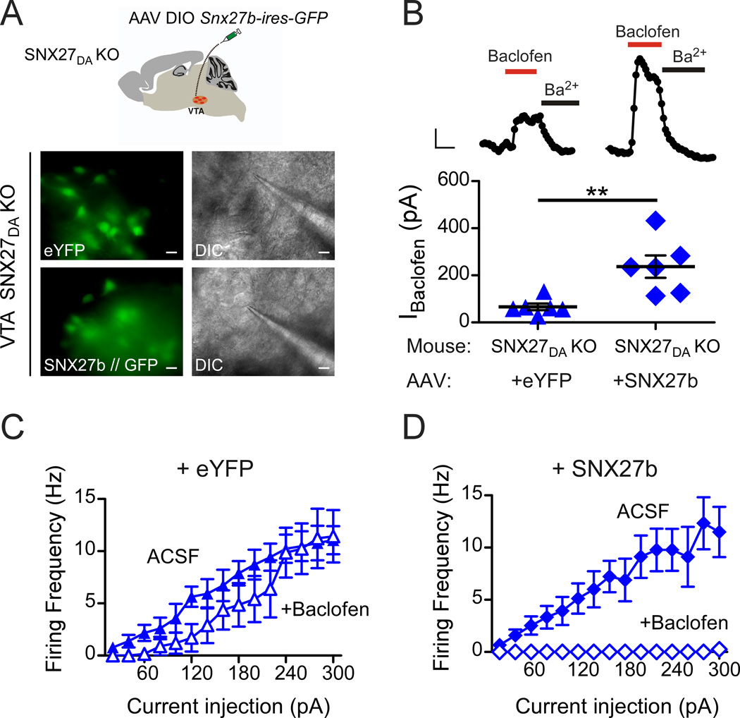
Figure 5. GABABR-GIRK currents and inhibition are restored with expression of SNX27b in VTA DA neurons of SNX27DA KO mice.
(A) Schematic shows stereotaxic injection of AAV DIO-Snx27b-ires-GFP into VTA of SNX27DA KO mice. Fluorescence and DIC images of eYFP/GFP+ cell selected for recording from ex vivo midbrain section of SNX27DA KO with either AAV DIO-eYFP or AAV DIO-Snx27b-ires-GFP (scale bar: 20 µm). (B) Top, whole-cell recordings from VTA DA neurons show baclofen-activated GIRK currents (IBaclofen; 300 µM) from WT and SNX27DA KO mice injected with AAV DIO-eYFP or AAV DIO-Snx27b-ires-GFP, and inhibition with Ba2+ (1 mM). Scale bar; 100 s and 50 pA. Bottom, baclofen-induced currents of Snx27b-ires-GFP positive cells are significantly larger compared to +eYFP (**p<0.01, Student’s t test). Scatter plot of IBaclofen for indicated DA neurons with average current indicated by solid black bar. (C,D) Input-output plots show firing frequency increase with larger current injections in the absence (solid circles, ACSF) and presence (filled circles) of baclofen for DA neurons infected with eYFP (C) (n=6) or SNX27-ires-GFP (D) (n=6) (p<0.01 absence versus presence baclofen, 160–300pA, 2-way ANOVA with Bonferroni post hoc test).

