Abstract
Prostate carcinoma is the second most common cause of cancer related mortality in males in the United States. The pattern of metastatic disease of prostate cancer is well recognized, frequently involving sclerotic bone lesions and abdomino-pelvic lymph nodes. Anti-1-amino-3-[18F]fluorocyclobutane-1-carboxylic acid (anti-3-[18F] FACBC) is a synthetic amino acid analog positron emission tomography (PET) radiotracer with reported utility in the detection of prostate carcinoma. We present two cases of unusual presentations of prostate carcinoma, one with malignant ascitis and omental implants and the other with lytic bone lesions detected with anti-3-[18F]FACBC.
Keywords: anti-3-[18F] FACBC, prostate cancer, PET
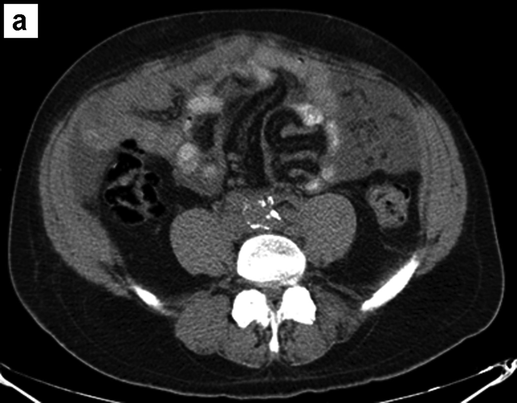
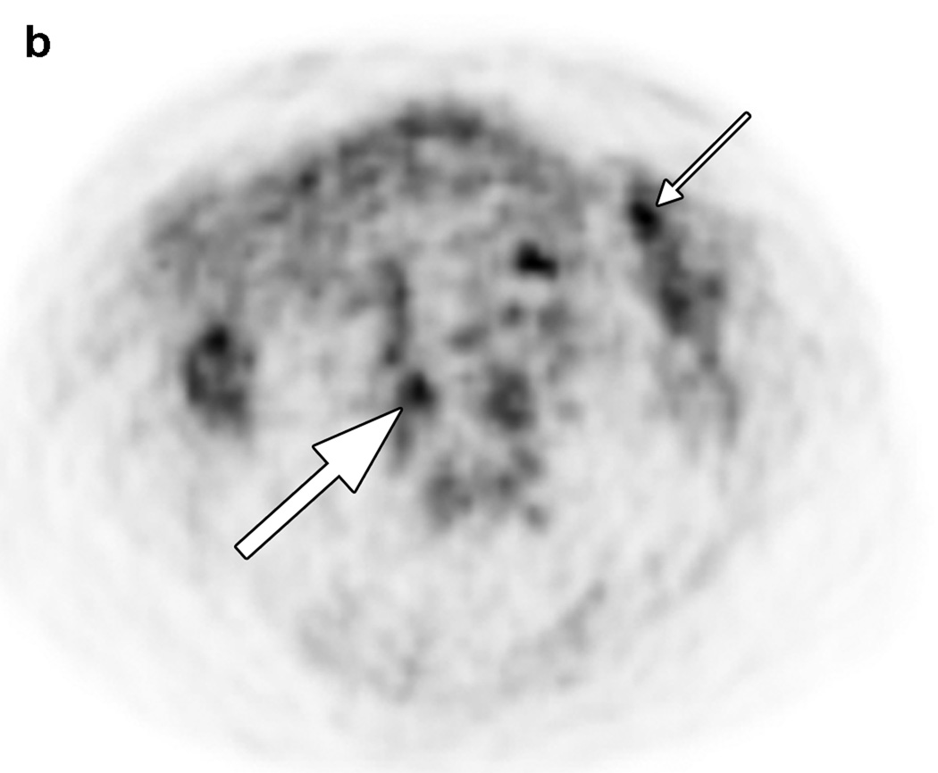
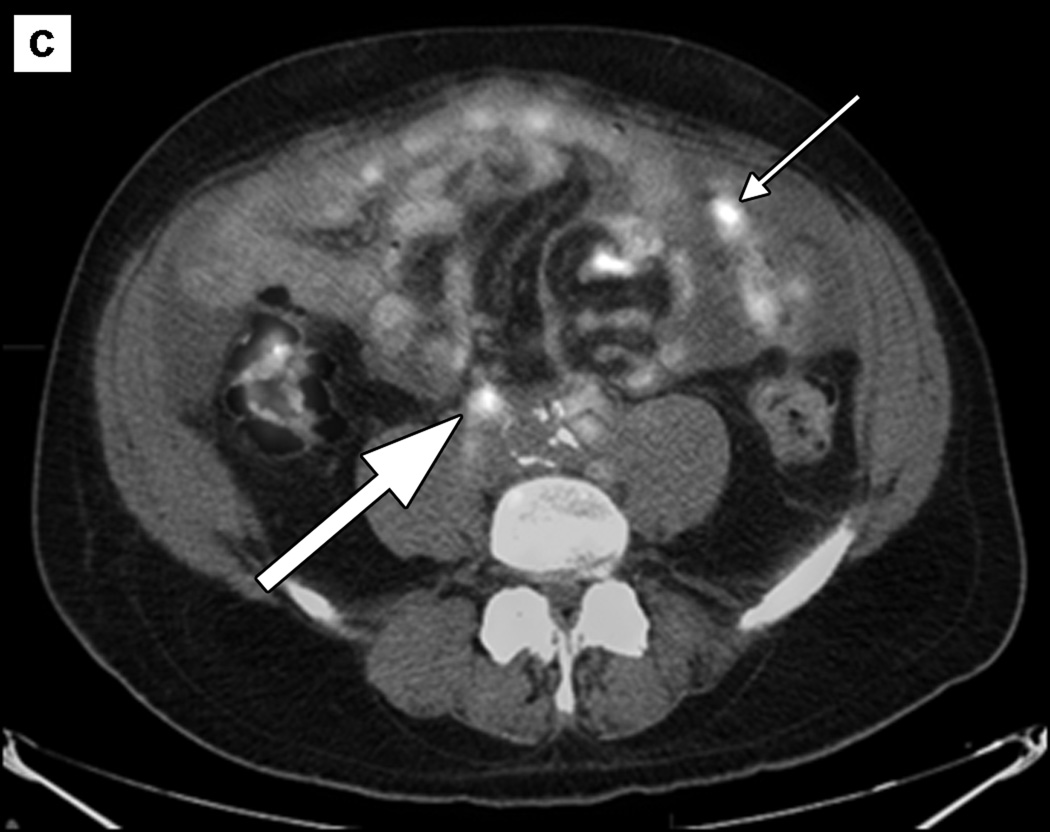
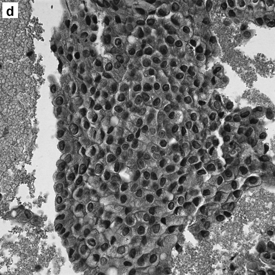
Figure 1.
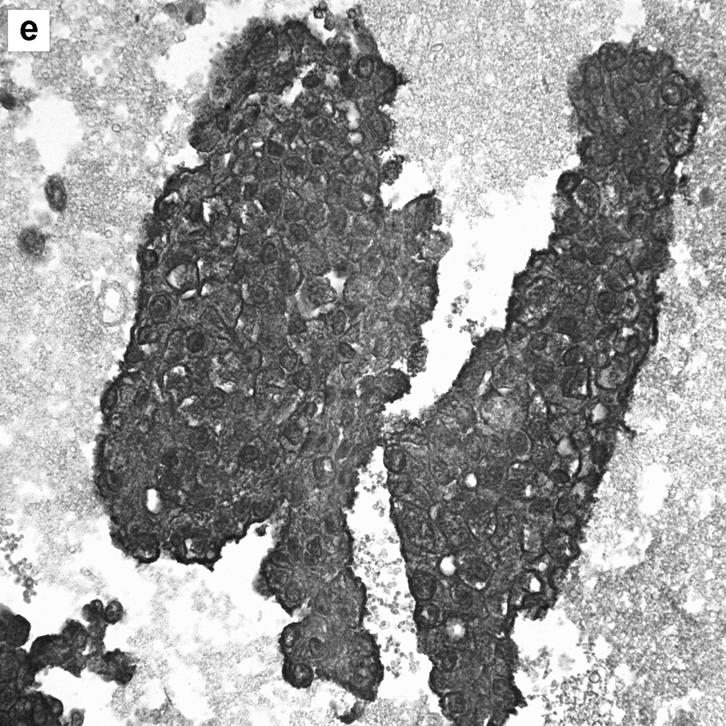
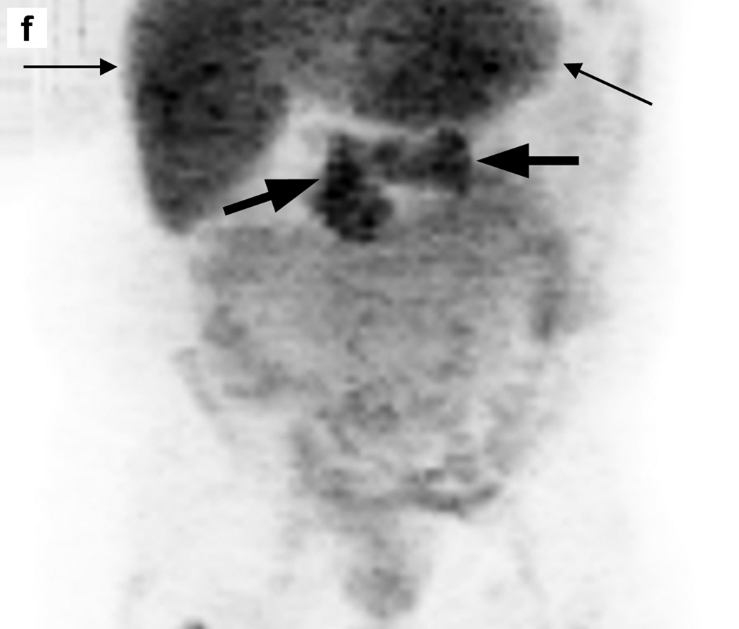
A 65 year-old male with a history of Gleason 8 (3+5) prostate adenocarcinoma treated with radiation therapy 5 years before, presented with a rising PSA (40ng/ml at presentation, 302ng/ml on day of anti-3-[18F]FACBC scan) and symptoms of partial bowel obstruction. Anti-3-[18F] FACBC PET-CT performed as part of an ongoing clinical trial with methods previously reported (1, 2, 7) revealed abnormal uptake in omental implants (SUVmax 7.7, small arrows), retroperitoneal lymph nodes (SUVmax 7.2 ,large arrows) and ascitis (SUVmax 3.1) in axial CT (a), anti-3-[18F]FACBC PET (b) and fused image (c). There was also uptake activity in a 5cm peri rectal mass (SUVmax 9.3) and the prostate (SUVmax 7.3) (not shown). Patient underwent negative colonoscopy and biopsy with findings of nonspecific mild architectural irregularity of the colonic mucosa. CT-guided FNA of a left paraaortic lymph node was positive for metastatic prostate adenocarcinoma as shown in the 400× histology image of the cell block H & E stain (d) and strongly positive PSA immunostain (e). Cytology of the ascitic fluid also revealed metastatic cells of prostatic origin. Maximum intensity projection (MIP) image (f) demonstrates typical biodistribution of anti-3-[18F] FACBC (8, 9) with intense hepatic and pancreatic uptake (arrows) and diffuse abdominal uptake secondary to malignant implants and ascitis. Advanced prostate carcinoma can present with a wide range of clinical features. The most common sites of metastasis include the axial skeleton, lymph nodes and lungs. Uncommon sites of metastatic disease include adrenal gland, kidney, brain, pancreas, genitalia, and breasts (3). Metastasis to the omentum and malignant ascitis without bone metastasis as seen in this case are rare presentations (4).
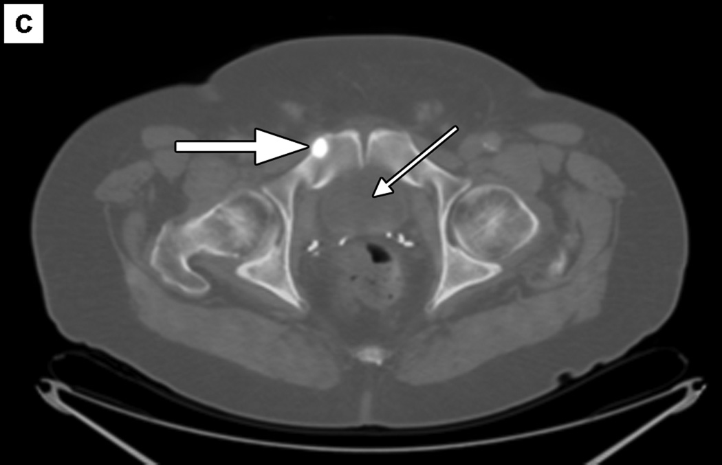
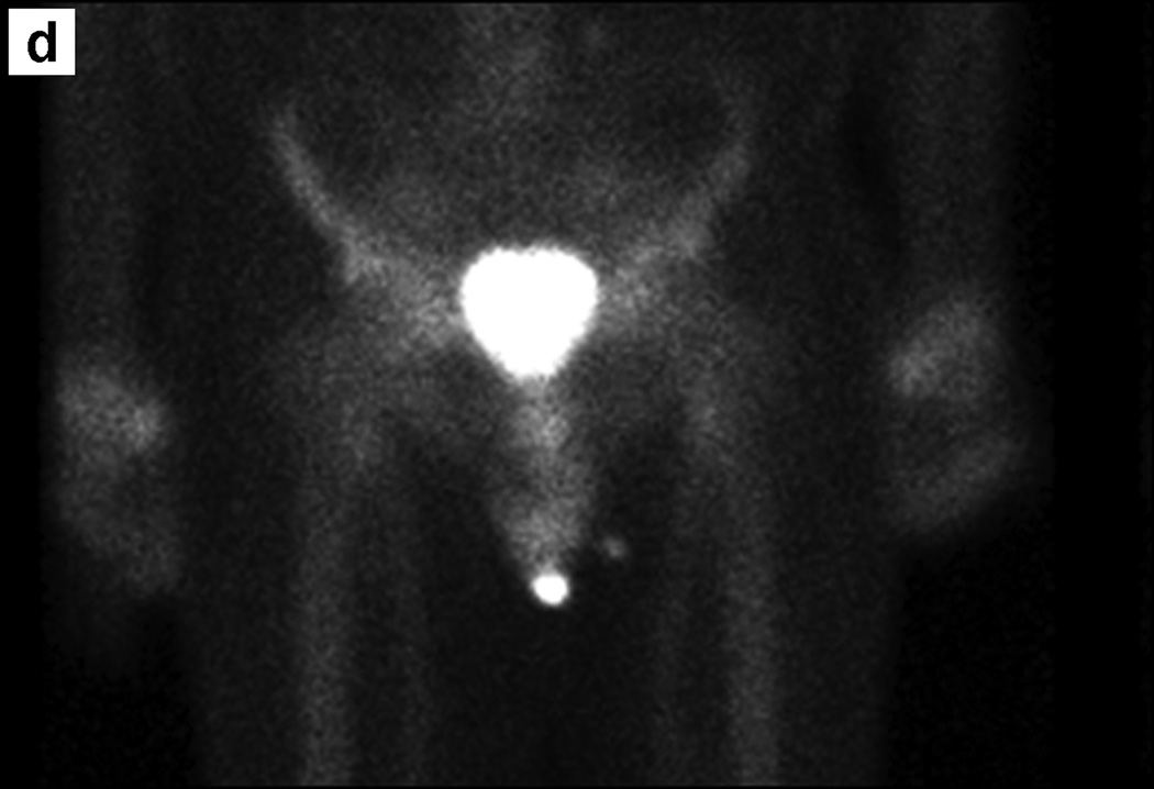
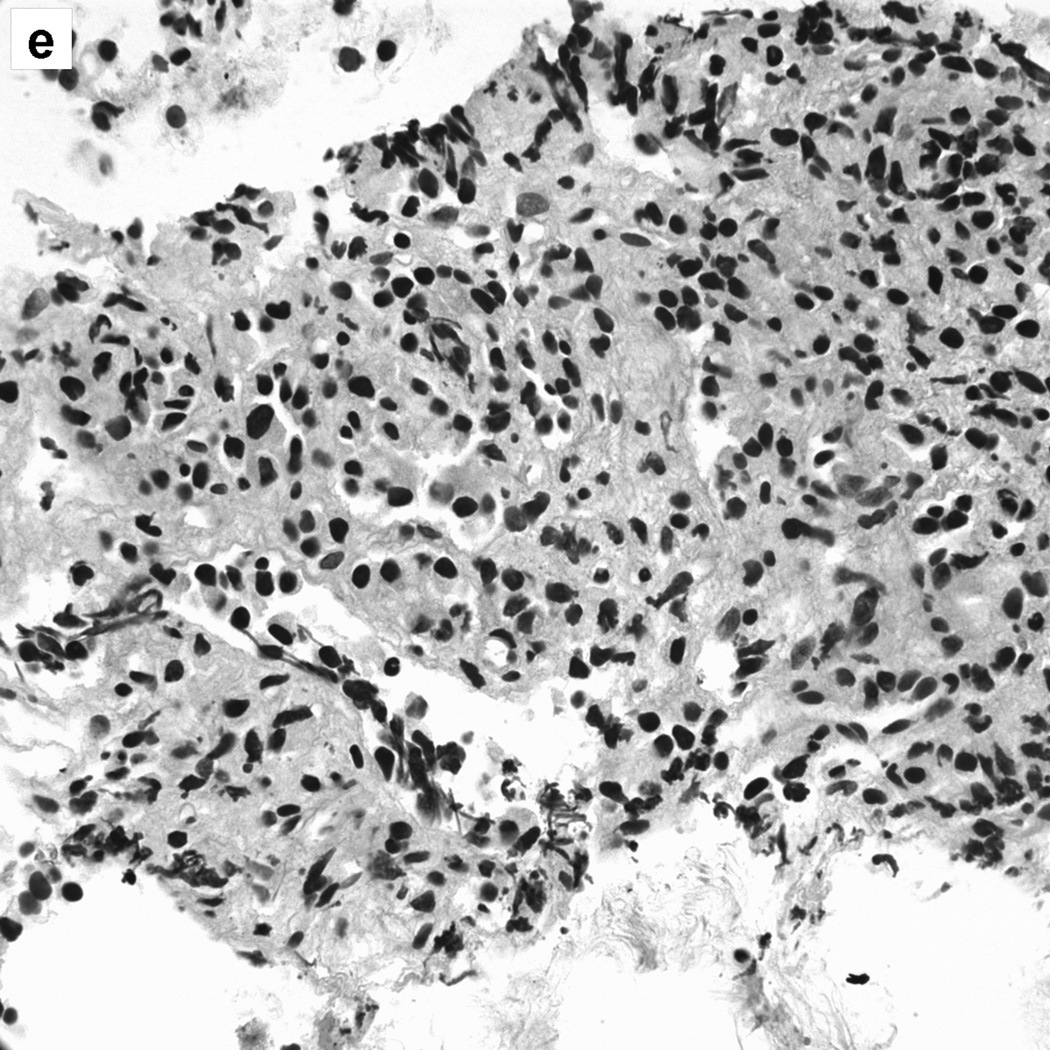
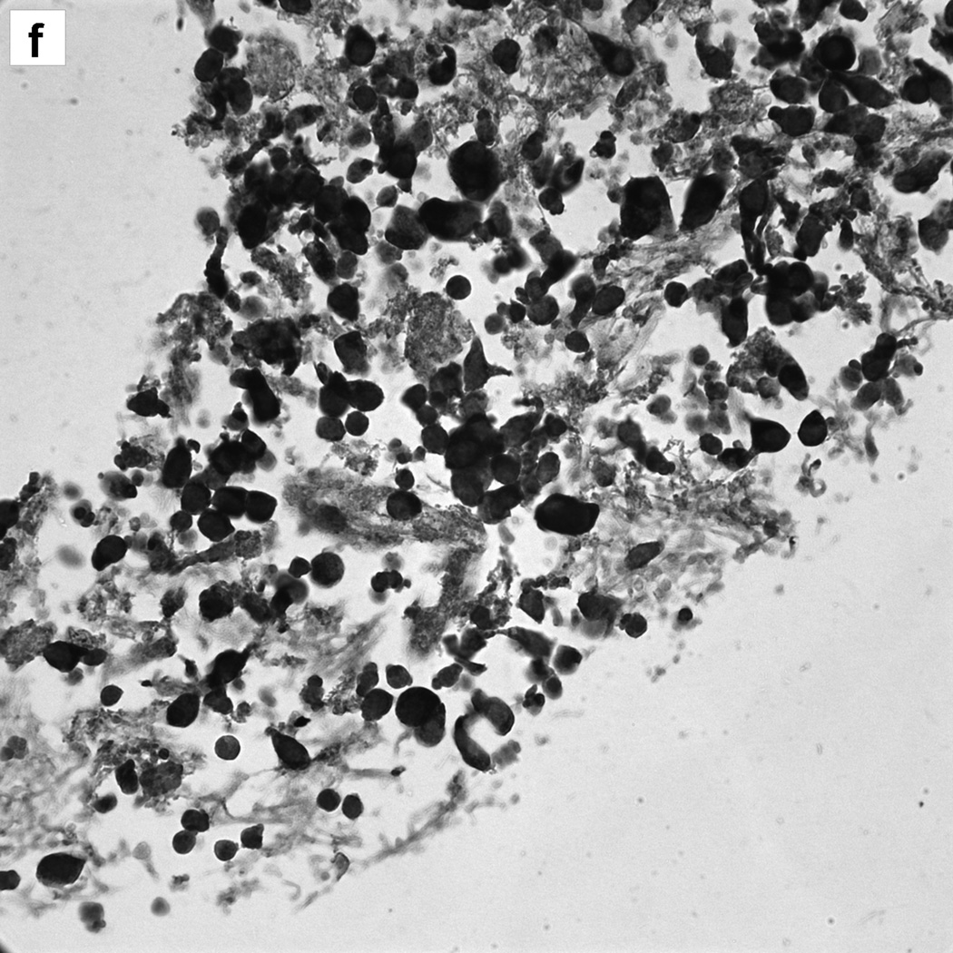
Figure 2.
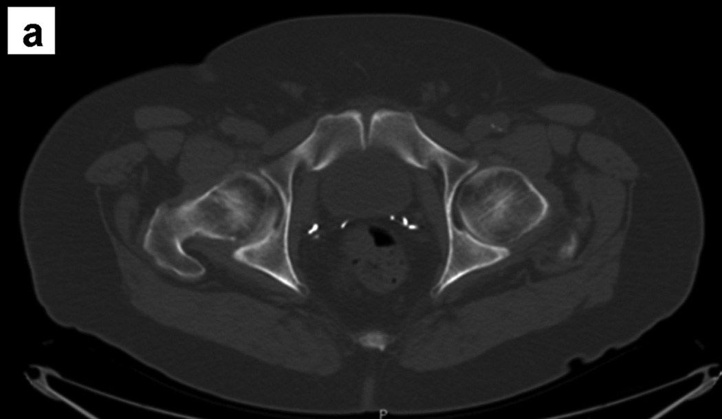
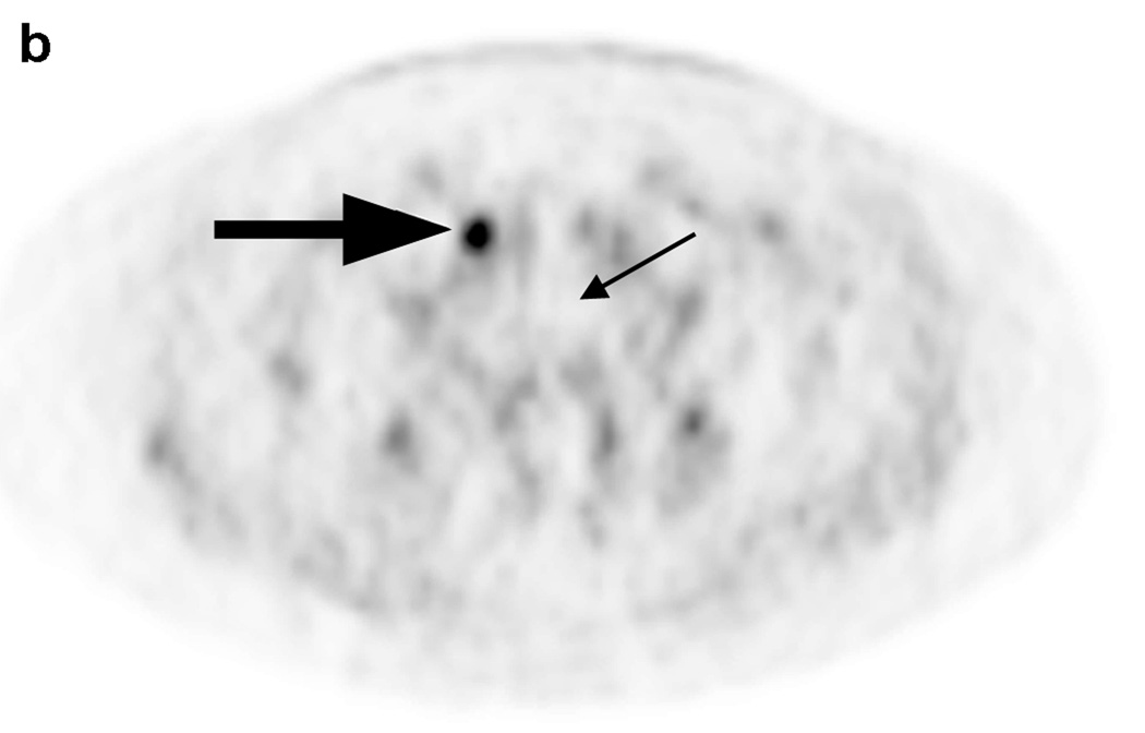
A 73 year old male presented with rising PSA of 2.97 after prostatectomy for Gleason 8 (3+5) prostate adenocarcinoma 5 years before. Scan demonstrated intense uptake in the antastomosic urethra (not shown), as well as intense focal uptake in a solitary lytic lesion in right superior pubic ramus (SUVmax 12.6,large arrows) in axial CT (a) anti-3-[18F]FACBC PET (b) and fused Image (c). Note relatively little to no bladder excretion (small arrows) which is an advantage of anti-3-[18F] FACBC for prostate cancer imaging. (1, 2, 7). Bone scan (d) showed no evidence of osteoblastic metastatic disease in the axial or appendicular skeleton although bladder may have obscured the lesion. Patient underwent CT – guided biopsy of the right pubic ramus lesion, with a histological confirmation of metastatic prostate disease as shown in the 400× histology image of the cell block H & E stain (e) and strongly positive PSA immunostain (f). Bone metastasis occurs in about 70% of patients with advanced prostate cancer (5). Prostate adenocarcinoma metastasis to bone are typically multiple and sclerotic with only about 10% presenting as solitary lesions (6). Presentation of a purely osteolytic solitary lesion is rare (6).
Acknowledgments
Research Support: Research sponsored by the NIH (1 R01 CA 129356-01) and the Georgia Cancer Coalition.
REFERENCES
- 1.Schuster DM, Votaw JR, Nieh PT, et al. Initial experience with the anti-1-amino-3-18F-fluorocyclobutane-1-carboxylic acid with PET/CT in prostate carcinoma. J Nucl Med. 2007;48(1):56–63. [PubMed] [Google Scholar]
- 2.Schuster DM, Savir-Baruch B, Nieh PT, et al. Report of a clinical trial of anti-1 amino 3 [18F]fluorocyclobutane-1-carboxylic acid (anti-[18F] FACBC) PET-CT in recurrent prostate carcinoma. J Nucl Med. 2010;51(Suppl.2):127P. [Google Scholar]
- 3.Saif MW. Malignant ascites associated with carcinoma of the prostate. The Journal of Applied Research. 2005;5(2):305. [PMC free article] [PubMed] [Google Scholar]
- 4.Kehinde EO, Abdeen SM, Al-Hunayan A, et al. Prostate cancer metastatic to the omentum. Scandinavian Journal of Urology and Nephrology. 2002;36(3):225. doi: 10.1080/003655902320131938. [DOI] [PubMed] [Google Scholar]
- 5.Roodman GD. Mechanisms of bone metastasis. The New England Journal of Medicine. 2004;350(16):1655. doi: 10.1056/NEJMra030831. [DOI] [PubMed] [Google Scholar]
- 6.Agheli A, Patsiornik Y, Chen Y, et al. Prostate carcinoma, presenting with a solitary osteolytic bone lesion to the right hip. Radiology Case Reports. 2009;4:288. doi: 10.2484/rcr.v4i4.288. [DOI] [PMC free article] [PubMed] [Google Scholar]
- 7.Jani AB, Fox TH, Whitaker D, et al. Case study of anti-1-amino-3-F-18 fluorocyclobutane-1-carboxylic acid (Anti-[F-18] FACBC) to guide Prostate Cancer Radiotherapy Target Design. Clin Nucl Med. 2009;34:279–284. doi: 10.1097/RLU.0b013e31819e51e3. [DOI] [PubMed] [Google Scholar]
- 8.Oka S, Hattori R, Kurosaki F, et al. A preliminary study of anti-1-amino-3-18F-fluorocyclobutyl-1-carboxylic acid for the detection of prostate cancer. J Nucl Med. 2007;48:46–55. [PubMed] [Google Scholar]
- 9.Nye JA, Schuster DM, Yu W, et al. Biodistribution and radiation dosimetry of the synthetic nonmetabolized amino acid analogue anti-F-18 FACBC in humans. J Nucl Med. 2007;48:1017–1020. doi: 10.2967/jnumed.107.040097. [DOI] [PubMed] [Google Scholar]


