Abstract
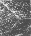
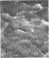
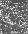
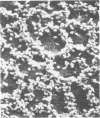
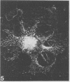
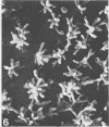
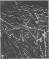
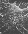
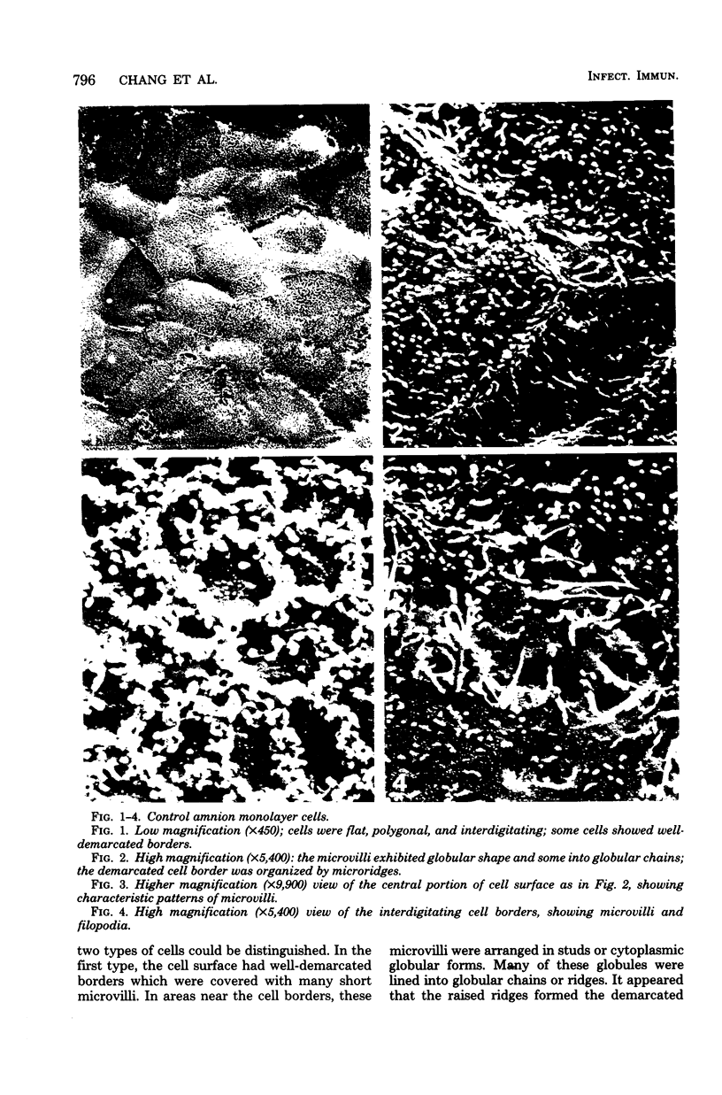
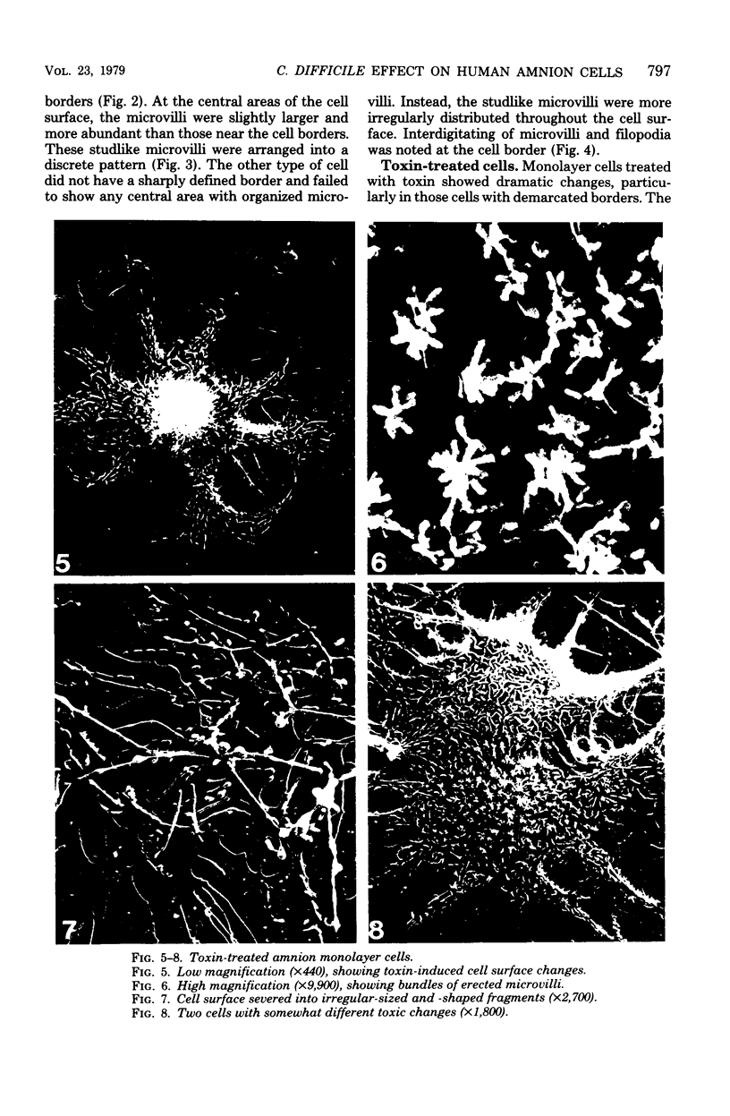
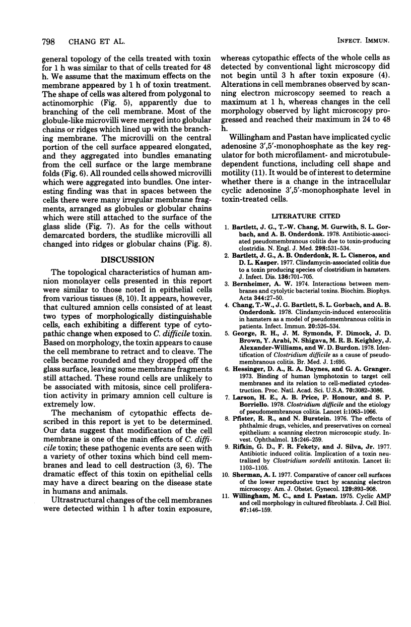
The ultrastructure of the surface of primary human amnion monolayer cells undergoing cytopathology induced by Clostridium difficile toxin was examined by scanning electron microscopy. Our observations indicated that the type and distribution of cell surface projections were altered dramatically by this toxin. The patterns of such surface changes were specific for the two different types of cells found in this cell culture. Cells with demarcated borders showed rearrangement of microvilli into globular chains or ridges which lined up with the branching membrane. Cells without demarcated borders exhibited studlike microvilli, all arranged into ridges or globular chains. These changes were noted after 1 h of toxin exposure and persisted without further progression, in spite of continued toxin exposure, up to 48 h. These data indicate that C. difficile produces a cytolytic toxin and that scanning electron microscopy may be useful in determining toxin-cell interactions.
Full text
PDF
Images in this article
Selected References
These references are in PubMed. This may not be the complete list of references from this article.
- Bartlett J. G., Chang T. W., Gurwith M., Gorbach S. L., Onderdonk A. B. Antibiotic-associated pseudomembranous colitis due to toxin-producing clostridia. N Engl J Med. 1978 Mar 9;298(10):531–534. doi: 10.1056/NEJM197803092981003. [DOI] [PubMed] [Google Scholar]
- Bartlett J. G., Onderdonk A. B., Cisneros R. L., Kasper D. L. Clindamycin-associated colitis due to a toxin-producing species of Clostridium in hamsters. J Infect Dis. 1977 Nov;136(5):701–705. doi: 10.1093/infdis/136.5.701. [DOI] [PubMed] [Google Scholar]
- Chang T. W., Bartlett J. G., Gorbach S. L., Onderdonk A. B. Clindamycin-induced enterocolitis in hamsters as a model of pseudomembranous colitis in patients. Infect Immun. 1978 May;20(2):526–529. doi: 10.1128/iai.20.2.526-529.1978. [DOI] [PMC free article] [PubMed] [Google Scholar]
- George R. H., Symonds J. M., Dimock F., Brown J. D., Arabi Y., Shinagawa N., Keighley M. R., Alexander-Williams J., Burdon D. W. Identification of Clostridium difficile as a cause of pseudomembranous colitis. Br Med J. 1978 Mar 18;1(6114):695–695. doi: 10.1136/bmj.1.6114.695. [DOI] [PMC free article] [PubMed] [Google Scholar]
- Hessinger D. A., Daynes R. A., Granger G. A. Binding of human lymphotoxin to target-cell membranes and its relation to cell-mediated cytodestruction. Proc Natl Acad Sci U S A. 1973 Nov;70(11):3082–3086. doi: 10.1073/pnas.70.11.3082. [DOI] [PMC free article] [PubMed] [Google Scholar]
- Larson H. E., Price A. B., Honour P., Borriello S. P. Clostridium difficile and the aetiology of pseudomembranous colitis. Lancet. 1978 May 20;1(8073):1063–1066. doi: 10.1016/s0140-6736(78)90912-1. [DOI] [PubMed] [Google Scholar]
- Pfister R. R., Burstein N. The effects of ophthalmic drugs, vehicles, and preservatives on corneal epithelium: a scanning electron microscope study. Invest Ophthalmol. 1976 Apr;15(4):246–259. [PubMed] [Google Scholar]
- Rifkin G. D., Fekety F. R., Silva J., Jr Antibiotic-induced colitis implication of a toxin neutralised by Clostridium sordellii antitoxin. Lancet. 1977 Nov 26;2(8048):1103–1106. doi: 10.1016/s0140-6736(77)90547-5. [DOI] [PubMed] [Google Scholar]
- Sherman A. I. Comparison of cancer cell surfaces of the lower reproductive tract by scanning electron microscopy. Am J Obstet Gynecol. 1977 Dec 15;129(8):893–908. doi: 10.1016/0002-9378(77)90522-1. [DOI] [PubMed] [Google Scholar]
- Willingham M. C., Pastan I. Cyclic amp and cell morphology in cultured fibroblasts. Effects on cell shape, microfilament and microtubule distribution, and orientation to substratum. J Cell Biol. 1975 Oct;67(1):146–159. doi: 10.1083/jcb.67.1.146. [DOI] [PMC free article] [PubMed] [Google Scholar]


