Abstract
The present investigation was an attempt to elucidate oxidative stress induced by bisphenol A on erythrocytes and its amelioration by green tea extract. For this, venous blood samples from healthy human adults were collected in EDTA vials and used for preparation of erythrocytes suspension. When erythrocyte suspensions were treated with different concentrations of BPA/H2O2, a dose-dependent increase in hemolysis occurred. Similarly, when erythrocytes suspensions were treated with either different concentrations of H2O2 (0.05–0.25 mM) along with BPA (50 μg/mL) or 0.05 mM H2O2 along with different concentrations of BPA (50–250 μg/mL), dose-dependent significant increase in hemolysis occurred. The effect of BPA and H2O2 was found to be additive. For the confirmation, binding capacity of bisphenol A with erythrocyte proteins (hemoglobin, catalase, and glutathione peroxidase) was inspected using molecular docking tool, which showed presence of various hydrogen bonds of BPA with the proteins. The present data clearly indicates that BPA causes oxidative stress in a similar way as H2O2 . Concurrent addition of different concentrations (10–50 μg/mL) of green tea extract to reaction mixture containing high dose of bisphenol A (250 μg/mL) caused concentration-dependent amelioration in bisphenol A-induced hemolysis. The effect was significant (P < 0.05). It is concluded that BPA-induced oxidative stress could be significantly mitigated by green tea extract.
1. Introduction
Bisphenol A (BPA 2,2-bis(4-hydroxyphenyl) propane), a xenoestrogen, is an important monomer of polycarbonate plastics and a constituent of epoxy and polystyrene resins [1]. It is widely used to line metal cans, water pipes [2], baby bottles, drinking cups [3], dental sealants [4, 5], and many other household appliances. Studies have shown that for incomplete polymerization and for degradation of the polymer, bisphenol A can leach out from food and beverage containers [6], as well as from dental sealants and composites under normal conditions of use. Bisphenol A has been found not only in environmental samples, including air, water, sewage sludge, soil, and dust [7–9], but also in specimens of human body fluids, such as plasma, umbilical cord blood, placental tissue, amniotic fluid, follicular fluid, and breast milk. Due to the widespread use and building toxicological database, a need arises to investigate mechanism of bisphenol A-induced toxicity.
Studies revealed that bisphenol A causes adverse effects on brain, reproductive system, and liver [10, 11]. Mathur and D′Cruz [12] have reported that BPA causes an imbalance in the prooxidant and antioxidant status of the testis and increased amount of reactive oxygen species. A study carried out by Verma and Sangai [13] showed that treatment with bisphenol A causes cytotoxicity in human erythrocytes which may be due to the oxidative stress. But none of the studies have stated the exact mechanism of bisphenol A-induced oxidative damage.
We propose that bisphenol A causes generation of hydroxyl radical causing peroxidation of lipids. Erythrocytes have been used as a cellular model to investigate oxidative damage in biomembranes due to the presence of both high concentration of polyunsaturated fatty acids and the oxygen transport associated with redox active hemoglobin molecules, which are potent promoters of reactive oxygen species formation [14]. Along with that, H2O2, a potent endogenous oxidant of erythrocytes producing deleterious oxidative changes in the membrane by well-studied pathway, was chosen as standard compound to investigate/compare mechanism of bisphenol A activity.
Herbal medicines derived from plants are being increasingly utilized to treat wide variety of clinical diseases. More attention has been paid to the protective effects of natural antioxidants against drug-induced toxicities especially whenever free radical generation is involved [15]. Green tea demonstrated stronger free radical scavenging activity comparatively at lower doses in vitro [16]. Green tea leaves possess many beneficial properties to human health owning to the presence of many bioactive components, particularly the catechins that have intrinsic property of scavenging the reactive oxygen species produced in our body by various systems [17]. Green tea extract is known to have a broad spectrum of biological activities such as antioxidant, antiviral [18], anticancer [19], antibacterial [20], antifungal [21], and neuroprotective effects [22].
In the present study, we elucidate experimentally as well as by docking that bisphenol A induces oxidative stress similarly as H2O2 and green tea ameliorates changes caused by bisphenol A. Virtual screening of bisphenol A was targeted against hemoglobin, catalase, and glutathione peroxidase of erythrocytes via molecular docking studies and the docked conformations were evaluated in terms of binding efficiency. Xenobiotics-protein interaction studies have significance in obtaining a better insight in the mechanisms of toxicities. Thus, the molecular docking predictions were used to find the probable interacting sites of bisphenol A with hemoglobin, catalase, and glutathione peroxidase of erythrocytes.
2. Materials and Methods
2.1. Extract Preparation
The green tea extract was prepared according to the method of Bhargava and Singh [23]. Briefly, 5 g of green tea powder (Brook-bond Company, Darjeeling green tea, India) was mixed with 100 mL 50% ethanol in water for 12 h with intermittent shaking every 2 h. The mixture was filtered with Whatman number 42 filter paper. The pooled filtrate was concentrated by evaporating below 50°C to obtain residue which was stored under refrigerated conditions. Residue was resuspended in normal saline before use.
2.2. Experimental Sample
Volunteers (healthy adult well-nourished human beings of 23–25 years of age) were recruited to participate in the study after approval of the Institutional Ethics Committee of the Zoology Department of Gujarat University, Ahmedabad. The venous whole blood samples were collected in syringes containing EDTA and centrifuged at 1,000 ×g for 10 min at 4°C to remove plasma and buffy coat. The RBCs were washed twice with cold saline (0.9% NaCl). The pellets were diluted with normal saline to obtain cell density of 2 × 10⁴ RBC/mL [24].
2.3. In Vitro Incubation of Erythrocytes with BPA, H2O2, and Green Tea Extract
The effects of BPA (Hi-media Laboratories Pvt. Ltd., Mumbai, India) and H2O2 (Merck Specialties Pvt. Ltd., Mumbai, India) on erythrocytes were investigated by incubating 2 mL of erythrocyte suspensions with (i) different concentrations of BPA (0–250 μg/mL) and (ii) different concentrations of H2O2 (0–0.25 mM). Further, to investigate any interaction between BPA and H2O2, 2 mL of erythrocyte suspensions was incubated with (iii) 50 μg/mL of BPA along with different concentrations of H2O2 (0–0.25 mM) and (iv) 0.05 mM of H2O2 along with different concentrations of BPA (0–250 μg/mL). To study ameliorative effect of green tea extract against BPA, 2 mL of erythrocyte suspensions was incubated with different concentrations of green tea extract (0–50 μg/mL) along with 250 μg/mL concentration of BPA.
Total volume of each reaction mixture was made up to 4 mL with additional saline. All reaction mixtures were incubated at 37°C for 4 h with intermittent shaking.
2.4. Measurement of Hemolysis
The suspensions were centrifuged at 1,000 ×g for 10 min. The color density of the supernatant was read spectrophotometrically at 540 nm. Percent hemolysis was calculated by using the following formula:
| (1) |
To obtain 100% hemolysis, 2 mL of erythrocytes suspension was incubated with 2 mL of distilled water.
2.5. Statistical Analysis
Each experiment was repeated ten times. The results were expressed as the means ± SEM. The data were statistically analyzed using one-way analysis of variance (ANOVA) followed by Tukey's post hoc test. The level of significance was accepted with P < 0.05. Correlation coefficient was measured to estimate the strength of linear association between two variables.
2.6. Preparation of Protein Target Structure and Ligands
The X-ray crystal structure of hemoglobin (PDB id-2HHB), catalase (PDB id-1QQW), and glutathione peroxidase (PDB id-2f8A) was retrieved from the Protein Data Bank (PDB ID-2YK0) [25]. The Data was subjected to energy minimization using GROMOS96 utility (without reaction field) implemented in Swiss-Pdb Viewer 4.0.1. [26].
Ligand structure of bisphenol A (2D structure) was retrieved in structure data format (SDF) from NCBI PubChem [27] and 3D structure was derived using ChemSketch v. 10 [28]. The structure of bisphenol A was energy minimized using molecular mechanics geometry optimization module implemented in HyperChem [29]. AMBER force field with distant dependent dielectric constant, scale factor for electrostatic and van der Waals forces set to 0.5 and without any cutoffs to bond types, and its lengths were chosen to determine global minimum energy conformation. Subsequently, all the structures were minimized and exported to hard disk.
2.7. Active Site Prediction
This prediction was not carried out for protein structures cocrystallized with ligand as the ligand binding site was implicated as active site for docking with the ligand dataset. Structures with unbound ligands were computationally analyzed for active site using Q-SiteFinder [30]. It detects pockets on the protein surface through calculation of van der Waals interaction energies using a methyl probe and probes with favorable interaction energies were clustered and ranked.
2.8. Virtual Screening
Bisphenol A under study was virtually screened (docked) into the binding site of the target proteins as hemoglobin (PDB id-2HHB), catalase (PDB id-1QQW), and glutathione peroxidase (PDB id-2f8A) using ArgusLab 4.0.1 from Planaria Software LLC [31]. To enable fast sampling, the binding site was constructed which consists of all residues that have at least one atom within 3.5 Å from any atom in the cocrystallized ligand. This procedure of constructing binding site was applied for protein structures with cocrystallized ligand. A different approach was performed for proteins with unbound ligand whose active site was predicted using Q-SiteFinder. Best 3 scored pockets were computationally analyzed for each protein target using Jmol Java plugin implemented in Q-SiteFinder and the amino acids embedded in the predicted cavity volume were utilized as active site residues.
These two approaches generally gave a good representation of the important residues in the binding pocket for a protein target. A grid box of size X = 22 × Y = 15 × Z = 16 with atom scaling of 0.40 Å was generated and high precision ArgusDock engine with a score as scoring function was selected. After grid generation, the ligands were flexibly docked with the protein and 1000 poses were generated, among which best 10 poses of low energy which were clustered in rank 1 were examined. ArgusDock engine makes use of ligand torsionality as a hierarchical tree in which the root's node (group of bonded atoms that do not have rotatable bonds) is placed in a search point inside a grid comprised of residues of the active site. A set of diverse and energetically favorable translations are generated and poses that survive in torsional search through an approximate exhaustive search are retained and finally clustered.
3. Results
3.1. Effect on Erythrocytes
When erythrocyte suspensions were incubated with normal saline, ambient supernatant was clear with intact erythrocytes pellet in the bottom of the tube. Almost no hemolysis was observed in this mixture.
Addition of various concentrations of H2O2 (0.05–0.25 mM) to erythrocyte suspensions caused significant (P < 0.05) increase in hemolysis (Figure 1). Maximum hemolysis (77.88%) was obtained on addition of 0.25 mM of H2O2 (Table 1). The results showed that H2O2 induced hemolysis in a concentration-dependent manner (r = 0.95).
Figure 1.
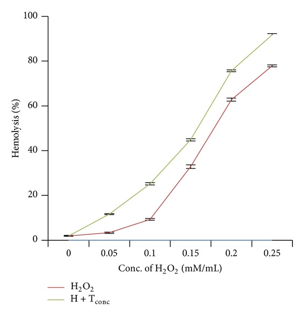
Effect of hydrogen peroxide and its combination with bisphenol A on red blood corpuscles. Results are expressed as mean ± SEM; n = 10. ∗Significant at the level P < 0.05 as compared to control.
Table 1.
H2O2 induced hemolysis.
| Concentration of H2O2 (mM) | Hemolysis (%) |
|---|---|
| 0 (control) | 1.97 ± 0.14 |
| 0.05 | 3.36 ± 0.28 |
| 0.10 | 9.33 ± 0.37∗ |
| 0.15 | 32.85 ± 0.76∗ |
| 0.20 | 62.74 ± 0.68∗ |
| 0.25 | 77.88 ± 0.44∗ |
Results are expressed as mean ± SEM; n = 10.
∗Significant at the level P < 0.05 as compared to control.
Addition of different concentrations (50–250 μg/mL) of bisphenol A to erythrocyte suspensions resulted in significant (P < 0.05) increase in hemolysis (Figure 2). With increasing concentrations of bisphenol A, amount of intact erythrocytes settled down at the bottom of the tubes got reduced drastically resulting in reddish supernatant. Maximum hemolysis (98.75%) was achieved on addition of 250 μg/mL of bisphenol A (Table 2). The result showed that hemolysis induced by bisphenol A is dose-dependent (r = 0.91).
Figure 2.
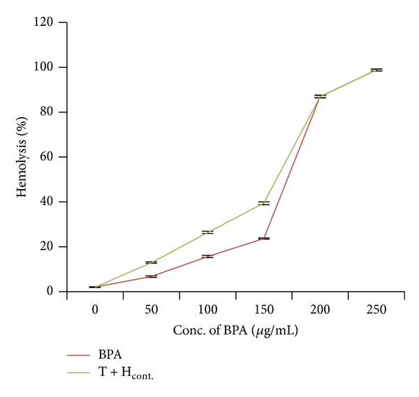
Effect of bisphenol A alone and its combination with hydrogen peroxide on red blood corpuscles. Results are expressed as mean ± SEM; n = 10. ∗Significant at the level P < 0.05 as compared to control.
Table 2.
Bisphenol A induced hemolysis.
| Concentration of bisphenol A (μg/mL) | Hemolysis (%) |
|---|---|
| 0 (control) | 1.97 ± 0.14 |
| 50 | 6.71 ± 0.33∗ |
| 100 | 15.60 ± 0.49∗ |
| 150 | 23.59 ± 0.30∗ |
| 200 | 86.97 ± 0.55∗ |
| 250 | 98.75 ± 0.47∗ |
Results are expressed as mean ± SEM; n = 10.
∗Significant at the level P < 0.05 as compared to control.
In another set of experiments, we studied effect of cotreatments of H2O2 and bisphenol A on hemolysis. When erythrocyte suspensions were incubated with different concentrations of H2O2 (0.05–0.2 mM) along with the 50 μg/mL of bisphenol A, it caused significant (P < 0.05) increase in hemolysis (Figure 1). Table 3 showed that when erythrocytes were treated with 0.05, 0.10, and 0.15 mM/mL of H2O2, hemolysis achieved was 3.36%, 9.33%, and 32.85%, which reached 12.93%, 25.18%, and 44.82% upon addition of 50 μg/mL of BPA, respectively. The effect was also dose-dependent (r = 0.98).
Table 3.
H2O2 induced hemolysis along with cotreatment of bisphenol A.
| Treatment | Hemolysis (%) | |
|---|---|---|
| H2O2 concentration (mM) | Bisphenol A concentration (µg/mL) | |
| 0 (control) | 0 (control) | 1.97 ± 0.14 |
| 0 | 50 | 6.71 ± 0.33∗ |
| 0.05 | 50 | 12.93 ± 0.37∗ |
| 0.10 | 50 | 25.18 ± 0.50∗ |
| 0.15 | 50 | 44.81 ± 0.50∗ |
| 0.20 | 50 | 75.69 ± 0.31∗ |
| 0.25 | 50 | 91.73 ± 0.47∗ |
Results are expressed as mean ± SEM; n = 10.
∗Significant at the level P < 0.05 as compared to control.
In the same manner, when erythrocyte suspensions were cotreated with 0.05 mM of H2O2 along with different concentrations (50–250 μg/mL) of bisphenol A, it caused significant (P < 0.05) increase in hemolysis (Figure 2). Table 4 showed that when erythrocytes were treated with 50, 100, and 150 μg/mL of bisphenol A, hemolysis achieved was 6.71%, 15.60%, and 23.59%, which reached 12.93%, 26.37%, and 39.37% upon addition of H2O2 (0.05 mM), respectively. The effect was dose-dependent (r = 0.97). Tables 3 and 4 showed that this acceleration of hemolysis could be due to the additive effect of both the compounds.
Table 4.
Bisphenol A induced hemolysis along with cotreatment of H2O2.
| Treatment | Hemolysis (%) | |
|---|---|---|
| Bisphenol A concentration (μg/mL) | H2O2 concentration (mM) | |
| 0 (control) | 0 (control) | 1.97 ± 0.14 |
| 0 | 0.05 | 3.36 ± 0.28 |
| 50 | 0.05 | 12.93 ± 0.37∗ |
| 100 | 0.05 | 26.37 ± 0.45∗ |
| 150 | 0.05 | 39.37 ± 0.58∗ |
| 200 | 0.05 | 86.89 ± 0.58∗ |
| 250 | 0.05 | 98.73 ± 0.42∗ |
Results are expressed as mean ± SEM; n = 10.
∗Significant at the level P < 0.05 as compared to control.
Addition of only green tea extract into erythrocyte suspensions resulted in hemolysis (1.67%) almost equal to control tubes treated with normal saline. When erythrocytes were incubated with different concentrations of green tea extract (10–50 μg/mL) along with 250 μg/mL of bisphenol A, it caused significant (P < 0.05) and concentration-dependent reduction in hemolysis (r = 0.99) (Figure 3). This protective effect was highest at 50 μg/mL concentration of green tea extract.
Figure 3.
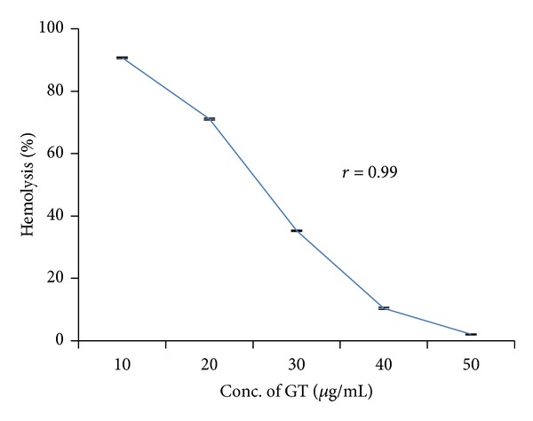
Retardation in bisphenol A-induced hemolysis by green tea extract. Results are expressed as mean ± SEM; n = 10. ∗Significant at the level P < 0.05 as compared to control.
3.2. Computational Docking and Modeling Studies
We implemented a structure-based computational approach to investigate the potential interaction pattern of bisphenol A against hemoglobin, catalase, and glutathione peroxidase. The ligand dataset was virtually screened with the protein targets using ArgusLab software and the binding energy values were analyzed for each docked conformation. To identify the precise binding sites on hemoglobin, catalase, and glutathione peroxidase, molecular docking simulations were done with the binding mode of bisphenol A. Bisphenol A was docked into the X-ray crystal structures of hemoglobin (PDB id-2HHB), catalase (PDB id-1QQW), and glutathione peroxidase (PDB id-2f8A).
The results showed that bisphenol A has higher binding affinity with the cavity present in hemoglobin and possessed greater energy (−79.5491 kcal/mol) value than the others, namely, catalase (−36.9179 kcal/mol) and glutathione peroxidase (−71.8106 kcal/mol) (Table 5). Conformations having low energy and exhibiting favorable hydrogen bonding with the amino acids side chain and its amide nitrogen were considered. The docking stimulations revealed the most likely binding site within the cavity of enzyme. Table 5 shows the amino acid residues interacting with the binding site of hemoglobin were Lys 99, Thr 137, Ser 133, and Asp126 (H-bond and steric interaction—Figure 4), binding sites of catalase were Lys 237, Tyr 215, Arg 203, Phe 198, Ser 201, and Phe 446 (H-bond and stearic interactions—Figure 5), and binding sites of glutathione peroxidase were Arg 20, Phe 110, Glu 111, His 121, and Glu 114 (H-bond and stearic interactions—Figure 6). The essential driving force of this binding site was hydrogen bond.
Table 5.
Docking results of human erythrocyte (PDB id-2HHB), catalase (PDB id-1QQW), and glutathione peroxidase (PDB id-2F8A) with bisphenol A.
| Name | Target molecules | Docking score | Interaction | H-bond | Amino acid interactions | |
|---|---|---|---|---|---|---|
| H-bond interactions | Steric interaction | |||||
| Bisphenol A | Hemoglobin | −79.5491 | −99.0041 | −4.80926 | Thr 137, Ser 133, Asp 126 | Lys 99, Thr 137 |
| Bisphenol A | Catalase | −36.9179 | −54.8291 | −3.275 | Lys 237, Tyr 215, Ser 201, | Tyr 215, Arg 203, Phe 198, Phe 446 |
| Bisphenol A | Glutathione peroxidase | −71.8106 | −86.9671 | −3.44834 | Arg 20, Glu 111, Glu 114 | Phe 110, Glu 114, His 121, |
Figure 4.
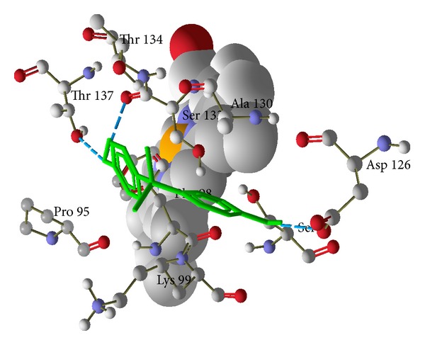
Hydrogen bond and steric interactions of human deoxyhaemoglobin (PDBid-2HHB) with bisphenol A. Green color is bisphenol A and whole structure is hemoglobin.
Figure 5.
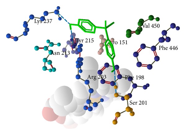
Hydrogen bond and steric interaction of human catalase (PDB id-1QQW) with bisphenol A. Green color is bisphenol A and whole structure is of catalase.
Figure 6.
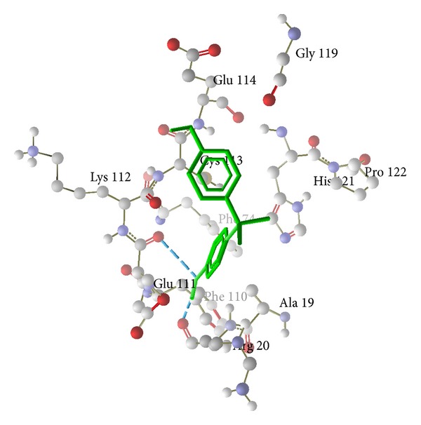
Hydrogen bond and steric interaction of human glutathione peroxidase (PDB id-2F8A) with bisphenol A. Green color is bisphenol A and whole structure is glutathione peroxidase.
4. Discussion
Results of the present study revealed that treatment of H2O2 to the erythrocyte suspensions caused dose-dependent increase in hemolysis. Aslan et al. [32] have also reported increased osmotic fragility of erythrocyte suspensions treated with H2O2. Interaction of H2O2 with iron of heme results in the generation of more potent hydroxyl radicals [33, 34], causing increased lipid peroxidation by attacking membrane polyunsaturated fatty acids. This is a known mechanism of action of H2O2 causing oxidative stress.
Being lipophilic in nature [35], BPA may get stabilized in the hydrophobic plasma membrane of the cell and may initiate peroxide formation. It may cross the lipid membrane and bind to iron of hemoglobin resulting in dissociation of hemoglobin subunits and release of iron which itself acts as prooxidant and produces high amount of hydroxyl radicals. Generated hydroxyl radicals may disturb the lipid membrane and initiate lipid peroxidation making the cell osmotically more fragile and ultimately causing hemolysis.
Earlier, it has been shown that bisphenol A induced oxidative stress in different tissues in rat [36, 37] and induced cellular apoptosis in hepatocytes [38]. Moon et al. [39] indicated that bisphenol A can induce hepatic damage and mitochondrial dysfunction by increasing oxidative stress in the liver. Sajiki [40] confirmed that bisphenol A binds to ferric heme of red blood corpuscle and Obata and Kubota [41] reported that bisphenol A increased hydroxyl radical formation in the rat striatum in a study using in vivo microdialysis. Another study done by Verma and Sangai [13] reported significant and concentration-dependent rise in lipid peroxidation in liver and kidney homogenates treated with bisphenol A in in vitro experiments, which supports our study.
The combinational studies (BPA and H2O2, Tables 3 and 4) have accelerated hemolysis which could be due to additive effect of both the compounds. The above hypothesis was clearly proven as the hemolysis induced by bisphenol A and H2O2 was highly correlated (Figures 7 and 8).
Figure 7.
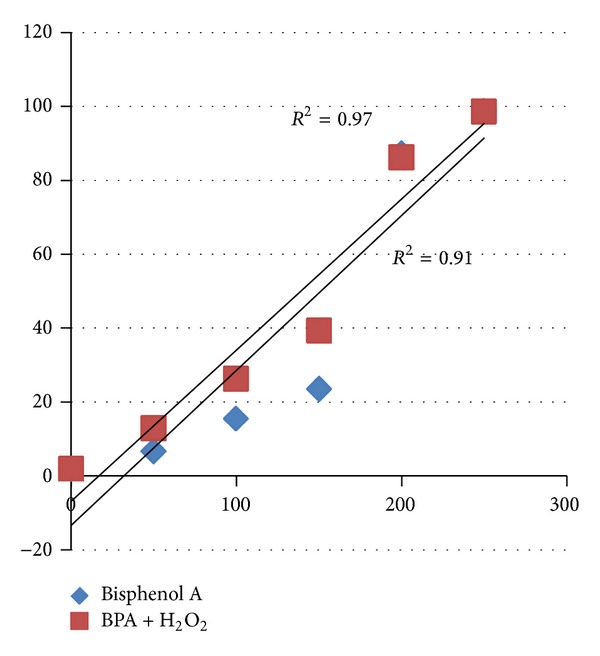
Correlation between bisphenol A and different concentration of bisphenol A along with the H2O2 (0.05 mM).
Figure 8.
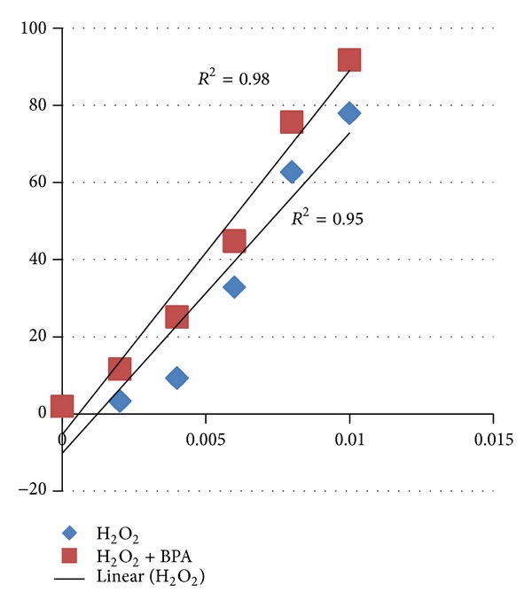
Correlation between H2O2 and different concentration of H2O2 along with the bisphenol A (50 μg/mL).
As shown in Figure 3, green tea extract significantly (P < 0.05) reduced hemolysis induced by bisphenol A. Another study of Verma and Sangai [13] has reported ameliorative effect of black tea extract (0–200 μg/mL) on bisphenol A-induced cytotoxicity. Maximal retardation in hemolysis was observed with 200 μg/mL concentration in case of black tea extract, which was 50 μg/mL concentration in green tea extract. It clearly indicates that green tea extract is comparatively more potent than the black tea extract. Polyphenols found in green tea show 20 times more powerful antioxidant activity than vitamin C [42]. Several studies [43–45] have reported environmental contaminants-induced cytotoxic effects on erythrocytes and their amelioration by natural antioxidants.
Green tea extract contains catechins, well known for their antioxidative property, stabilized plasma membrane of erythrocytes, and reducing hemolysis [46]. Catechins can also chelate metal ions such as iron (III) to form inactive complexes and prevent the generation of potentially damaging free radicals [46, 47]. Green tea extract is considered as potent scavengers of reactive oxygen species, such as superoxide, hydrogen peroxide, hydroxyl radicals, and nitric oxide produced by various chemicals [48]. In recent study, supplementation of green tea extract attenuates cyclosporine A-induced oxidative stress in rats [49]. Ostrowska et al. [50] revealed that green tea may protect liver and brain cells against oxidative stress induced by ethanol intoxication.
Mathur et al. [51] explained that bisphenol A elicits depletion of antioxidant defense system and induces oxidative stress. Another study done by Hassan et al. [52] reported that bisphenol A induces hepatotoxicity through oxidative stress and ultimately decreases the antioxidant enzymes. Catalase and glutathione peroxidase are important enzymes of antioxidant defense systems, which protect tissue against oxidative stress induced by reactive oxygen species. Both these enzymes catalyze the hydrolysis of H2O2 into water and oxygen molecules to prevent the tissue injury.
We propose that bisphenol A increases hemolysis by increasing the formation of hydroxyl radicals which may directly or indirectly (through the inhibition of glutathione peroxidase and catalase) induce lipid peroxidation. For confirmation of this hypothesis, we have performed molecular docking of bisphenol A with hemoglobin, catalase, and glutathione peroxidase. Molecular docking approach typically uses an energy-based scoring function to identify the energetically most favorable ligand conformation with the bound target. Binding energies of the protein-ligand interactions are important to describe how fit the ligand binds to the target macromolecule. The general hypothesis is that lower energy scores represent better protein-ligand bindings compared to higher energy values. The ligand-binding mode with the lowest energy docking simulations of bisphenol A against proteins (hemoglobin (PDB id-2HHB), catalase (PDB id-1QQW), and glutathione peroxidase (PDB id-2f8A)) were evaluated based on the binding compatibility (docked energy (kcal/mol)) with the receptor (Figures 4, 5, and 6). On the basis of the result, we conclude that the binding affinity of bisphenol A is higher with the hemoglobin (−79.5491 kcal/mol) compared to the catalase (−36.9179 kcal/mol) and glutathione peroxidase (−71.8106 kcal/mol) as shown in the result. The docking results revealed that no van der Waals, hydrophobic, or electrostatic interactions existed but only hydrogen bond played a major role in the binding of bisphenol A against hemoglobin, catalase, and glutathione peroxidase. We sought to determine and explore the mechanism of bisphenol A by binding more efficiently with heme than the catalase and glutathione peroxidase, produce hydroxyl radicals like H2O2, and induce lipid peroxidation.
In conclusion, we report that bisphenol A exerts cytotoxicity on human erythrocytes through the production of hydroxyl radical in the same manner as hydrogen peroxide. Green tea extracts have beneficial effects on bisphenol A-induced cytotoxicity due to their potent antioxidant properties and ability to reduce oxidative stress.
Conflict of Interests
The authors declare that they have no conflict of interests regarding the publication of this paper.
References
- 1.Ranjit N, Siefert K, Padmanabhan V. Bisphenol—a and disparities in birth outcomes: a review and directions for future research. Journal of Perinatology. 2010;30(1):2–9. doi: 10.1038/jp.2009.90. [DOI] [PMC free article] [PubMed] [Google Scholar]
- 2.Bae B, Jeong JH, Lee SJ. The quantification and characterization of endocrine disruptor bisphenol-A leaching from epoxy resin. Water Science and Technology. 2002;46(11-12):381–387. [PubMed] [Google Scholar]
- 3.Vandenberg LN, Hauser R, Marcus M, Olea N, Welshons WV. Human exposure to bisphenol A (BPA) Reproductive Toxicology. 2007;24(2):139–177. doi: 10.1016/j.reprotox.2007.07.010. [DOI] [PubMed] [Google Scholar]
- 4.Sasaki N, Okuda K, Kato T, et al. Salivary bisphenol—a levels detected by ELISA after restoration with composite resin. Journal of Materials Science: Materials in Medicine. 2005;16(4):297–300. doi: 10.1007/s10856-005-0627-8. [DOI] [PubMed] [Google Scholar]
- 5.Joskow R, Barr DB, Barr JR, Calafat AM, Needham LL, Rubin C. Exposure to bisphenol A from bis-glycidyl dimethacrylate-based dental sealants. The Journal of the American Dental Association. 2006;137(3):353–362. doi: 10.14219/jada.archive.2006.0185. [DOI] [PubMed] [Google Scholar]
- 6.Carwile JL, Luu HT, Bassett LS, et al. Polycarbonate bottle use and urinary bisphenol A concentrations. Environmental Health Perspectives. 2009;117(9):1368–1372. doi: 10.1289/ehp.0900604. [DOI] [PMC free article] [PubMed] [Google Scholar]
- 7.Goodman JE, Witorsch RJ, McConnell EE, et al. Weight-of-evidence evaluation of reproductive and developmental effects of low doses of bisphenol A. Critical Reviews in Toxicology. 2009;39(1):1–75. doi: 10.3109/10408440903279946. [DOI] [PubMed] [Google Scholar]
- 8.Vandenberg LN, Chahoud I, Heindel JJ, Padmanabhan V, Paumgartten FJR, Schoenfelder G. Urinary, circulating, and tissue biomonitoring studies indicate widespread exposure to bisphenol A. Environmental Health Perspectives. 2010;118(8):1055–1070. doi: 10.1289/ehp.0901716. [DOI] [PMC free article] [PubMed] [Google Scholar]
- 9.Loganathan SN, Kannan K. Occurrence of bisphenol a in indoor dust from two locations in the Eastern United States and implications for human exposures. Archives of Environmental Contamination and Toxicology. 2011;61(1):68–73. doi: 10.1007/s00244-010-9634-y. [DOI] [PubMed] [Google Scholar]
- 10.Lang IA, Galloway TS, Scarlett A, et al. Association of urinary bisphenol a concentration with medical disorders and laboratory abnormalities in adults. Journal of the American Medical Association. 2008;300(11):1303–1310. doi: 10.1001/jama.300.11.1303. [DOI] [PubMed] [Google Scholar]
- 11.Nakagawa Y, Tayama S. Metabolism and cytotoxicity of bisphenol A and other bisphenols in isolated rat hepatocytes. Archives of Toxicology. 2000;74(2):99–105. doi: 10.1007/s002040050659. [DOI] [PubMed] [Google Scholar]
- 12.Mathur PP, D’Cruz SC. The effect of environmental contaminants on testicular function. Asian Journal of Andrology. 2011;13(4):585–591. doi: 10.1038/aja.2011.40. [DOI] [PMC free article] [PubMed] [Google Scholar]
- 13.Verma RJ, Sangai NP. The ameliorative effect of black tea extract and quercetin on bisphenol A—induced cytotoxicity. Acta Poloniae Pharmaceutica: Drug Research. 2009;66(1):41–44. [PubMed] [Google Scholar]
- 14.Sadrzadeh SMH, Graf E, Panter SS, Hallaway PE, Eaton JW. Hemoglobin: a biologic Fenton reagent. The Journal of Biological Chemistry. 1984;259(23):14354–14356. [PubMed] [Google Scholar]
- 15.Frei B, Higdon JV. Antioxidant activity of tea polyphenols in vivo: evidence from animal studies. The Journal of Nutrition. 2003;133(10):3275S–3284S. doi: 10.1093/jn/133.10.3275S. [DOI] [PubMed] [Google Scholar]
- 16.Valentão P, Fernandes E, Carvalho F, Andrade PB, Seabra RM, Bastos ML. Hydroxyl radical and hypochlorous acid scavenging activity of small centaury (Centaurium erythraea) infusion. A comparative study with green tea (Camellia sinensis) Phytomedicine. 2003;10(6-7):517–522. doi: 10.1078/094471103322331485. [DOI] [PubMed] [Google Scholar]
- 17.Guleria RS, Jain A, Tiwari V, Misra MK. Protective effect of green tea extract against the erythrocytic oxidative stress injury during Mycobacterium tuberculosis infection in mice. Molecular and Cellular Biochemistry. 2002;236(1-2):173–181. doi: 10.1023/a:1016119718321. [DOI] [PubMed] [Google Scholar]
- 18.Fassina G, Buffa A, Benelli R, Varnier OE, Noonan DM, Albini A. Polyphenolic antioxidant (-)-epigallocatechin-3-gallate from green tea as a candidate anti-HIV agent. AIDS. 2002;16(6):939–941. doi: 10.1097/00002030-200204120-00020. [DOI] [PubMed] [Google Scholar]
- 19.Fujiki H. Green tea: health benefits as cancer preventive for humans. Chemical Record. 2005;5(3):119–132. doi: 10.1002/tcr.20039. [DOI] [PubMed] [Google Scholar]
- 20.Sakanaka S, Juneja LR, Taniguchi M. Antimicrobial effects of green tea polyphenols on thermophilic spore-forming bacteria. Journal of Bioscience and Bioengineering. 2000;90(1):81–85. doi: 10.1016/s1389-1723(00)80038-9. [DOI] [PubMed] [Google Scholar]
- 21.Li X-C, ElSohly HN, Nimrod AC, Clark AM. Antifungal activity of (-)-epigallocatechin gallate from Coccoloba dugandiana . Planta Medica. 1999;65(8, article 780) doi: 10.1055/s-2006-960871. [DOI] [PubMed] [Google Scholar]
- 22.Silvia A, Avramovich-Tirosh MY, Reznichenko L, et al. Multifunctional activities of green tea catechins in neuroprotection: modulation of cell survival genes, iron-dependent oxidative stress and PKC signaling pathway. NeuroSignals. 2005;14(1-2):46–60. doi: 10.1159/000085385. [DOI] [PubMed] [Google Scholar]
- 23.Bhargava KP, Singh N. Anti-stress activity of Ocimum sanctum Linn. The Indian Journal of Medical Research. 1981;73:443–451. [PubMed] [Google Scholar]
- 24.Verma RJ, Raval PJ. Cytotoxicity of aflatoxin on red blood corpuscles. Bulletin of Environmental Contamination and Toxicology. 1991;47(3):428–432. doi: 10.1007/BF01702206. [DOI] [PubMed] [Google Scholar]
- 25.Bernstein FC, Koetzle TF, Williams GJB, et al. The protein data bank: a computer based archival file for macromolecular structures. European Journal of Biochemistry. 1977;80(2):319–324. doi: 10.1111/j.1432-1033.1977.tb11885.x. [DOI] [PubMed] [Google Scholar]
- 26.Guex N, Peitsch MC. Swiss-Pdb viewer: a fast and easy to-use PDB viewer for macintosh and PC. Protein Data Bank Quaterly Newsletter. 1996;77:7–9. [Google Scholar]
- 27.Bolton EE, Wang Y, Thiessen PA, Bryant SH. Chapter 12 PubChem: integrated platform of small molecules and biological activities. Annual Reports in Computational Chemistry. 2008;4:217–241. [Google Scholar]
- 28.ACD/ChemSketch, version 10. Advanced Chemistry Development, Inc., Toronto, Canada, http://www.acdlabs.com, accessed on June 10, 2011. 10. Ahmedabad, India: Hyperchem 8. 0. 7-Licensed version at The Gujarat Cancer Research Institute; 2008. [Google Scholar]
- 29.2011-10 Hyperchem. 8.0.7. Licensed version at The Gujarat Cancer Research Institute. Asarwa, Ahmedabad, India.
- 30.Laurie ATR, Jackson RM. Q-SiteFinder: an energy-based method for the prediction of protein-ligand binding sites. Bioinformatics. 2005;21(9):1908–1916. doi: 10.1093/bioinformatics/bti315. [DOI] [PubMed] [Google Scholar]
- 31.Thompson MA. Molecular docking using ArgusLab, an efficient shape-based search algorithm and the A Score scoring functions. ACS Meeting; 2004; Philadelphia, Pa, USA. p. p. 172. CINF 42. [Google Scholar]
- 32.Aslan M, Thornley-Brown D, Freeman BA. Reactive species in sickle cell disease. Annals of the New York Academy of Sciences. 2000;899:375–391. doi: 10.1111/j.1749-6632.2000.tb06201.x. [DOI] [PubMed] [Google Scholar]
- 33.Van den Berg JJM, Op den Kamp JAF, Lubin BH, Roelofsen B, Kuypers FA. Kinetics and site specificity of hydroperoxide—induced oxidative damage in red blood cells. Free Radical Biology and Medicine. 1992;12(6):487–498. doi: 10.1016/0891-5849(92)90102-m. [DOI] [PubMed] [Google Scholar]
- 34.Halliwell B, Gutteridge JMC. Oxygen free radicals and iron in relation to biology and medicine: some problems and concepts. Archives of Biochemistry and Biophysics. 1986;246(2):501–514. doi: 10.1016/0003-9861(86)90305-x. [DOI] [PubMed] [Google Scholar]
- 35.Doerge DR, Fisher JW. Background paper on metabolism and toxicokinetics of bisphenol A. Proceedings of the WHO/HSE/FOS/11.1 Expert Meeting on Bisphenol A (BPA '10); 2010. [Google Scholar]
- 36.Bindhumol V, Chitra KC, Mathur PP. Bisphenol A induces reactive oxygen species generation in the liver of male rats. Toxicology. 2003;188(2-3):117–124. doi: 10.1016/s0300-483x(03)00056-8. [DOI] [PubMed] [Google Scholar]
- 37.Rashid H, Ahmad F, Rahman S, et al. Iron deficiency augments bisphenol A-induced oxidative stress in rats. Toxicology. 2009;256(1-2):7–12. doi: 10.1016/j.tox.2008.10.022. [DOI] [PubMed] [Google Scholar]
- 38.Asahi J, Kamo H, Baba R, et al. Bisphenol A induces endoplasmic reticulum stress-associated apoptosis in mouse non-parenchymal hepatocytes. Life Sciences. 2010;87(13-14):431–438. doi: 10.1016/j.lfs.2010.08.007. [DOI] [PubMed] [Google Scholar]
- 39.Moon MK, Kim MJ, Jung IK, et al. Bisphenol a impairs mitochondrial function in the liver at doses below the no observed adverse effect level. Journal of Korean Medical Science. 2012;27(6):644–652. doi: 10.3346/jkms.2012.27.6.644. [DOI] [PMC free article] [PubMed] [Google Scholar]
- 40.Sajiki J. Simple and accurate determination of bisphenol A in red blood cells prepared with basic glycine buffer using liquid chromatography-electrochemical detection. Journal of Chromatography B. 2003;783(2):367–375. doi: 10.1016/s1570-0232(02)00639-6. [DOI] [PubMed] [Google Scholar]
- 41.Obata T, Kubota S. Formation of hydroxy radicals by environmental estrogen-like chemicals in rat striatum. Neuroscience Letters. 2000;296(1):41–44. doi: 10.1016/s0304-3940(00)01619-0. [DOI] [PubMed] [Google Scholar]
- 42.Chopde V, Phatak A, Upaganlawer A, Tankar A. Green tea (Camellia Sinensis): chemistry, traditional, medicinal uses and its pharmacological activities—a review. Pharmacognoncy Review. 2008;2(3):157–162. [Google Scholar]
- 43.Asnani V, Verma RJ. Aqueous ginger extract ameliorates paraben induced cytotoxicity. Acta Poloniae Pharmaceutica—Drug Research. 2006;63(2):117–119. [PubMed] [Google Scholar]
- 44.Verma RJ, Trivedi MH, Chinoy NJ. Amelioration by black tea extract of sodium fluoride—induced hemolysis of human RBC: in vitro experiments. Fluoride. 2006;39(4):261–265. [Google Scholar]
- 45.Verma RJ, Chakraborty D. Protection front oxidative damage using Emblica officinalis Gaertn. extracts in case of ochratoxin induced toxicity in normal human RBC. Natural Product Radiance. 2007;6(4):310–314. [Google Scholar]
- 46.Guo Q, Zhao B, Li M, Shen S, Wenjuan X. Studies on protective mechanisms of four components of green tea polyphenols against lipid peroxidation in synaptosomes. Biochimica et Biophysica Acta. 1996;1304(3):210–222. doi: 10.1016/s0005-2760(96)00122-1. [DOI] [PubMed] [Google Scholar]
- 47.Yang CS, Maliakal P, Meng X. Inhibition of carcinogenesis by tea. Annual Review of Pharmacology and Toxicology. 2002;42:25–54. doi: 10.1146/annurev.pharmtox.42.082101.154309. [DOI] [PubMed] [Google Scholar]
- 48.Schroeder P, Klotz L, Sies H. Amphiphilic properties of (-)-epicatechin and their significance for protection of cells against peroxynitrite. Biochemical and Biophysical Research Communications. 2003;307(1):69–73. doi: 10.1016/s0006-291x(03)01132-x. [DOI] [PubMed] [Google Scholar]
- 49.Mohamadin AM, El-Beshbishy HA, El-Mahdy MA. Green tea extract attenuates cyclosporine A-induced oxidative stress in rats. Pharmacological Research. 2005;51(1):51–57. doi: 10.1016/j.phrs.2004.04.007. [DOI] [PubMed] [Google Scholar]
- 50.Ostrowska J, Łuczaj W, Kasacka I, Rózański A, Skrzydlewska E. Green tea protects against ethanol-induced lipid peroxidation in rat organs. Alcohol. 2004;32(1):25–32. doi: 10.1016/j.alcohol.2003.11.001. [DOI] [PubMed] [Google Scholar]
- 51.Mathur PP, Chitra KC, Latchoumycandane C. Induction of oxidative stress by bisphenol A in the epididymal sperm of rats. Toxicology. 2003;185(1-2):119–127. doi: 10.1016/s0300-483x(02)00597-8. [DOI] [PubMed] [Google Scholar]
- 52.Hassan ZK, Elobeid MA, Virk P, et al. Bisphenol A induces hepatotoxicity through oxidative stress in rat model. Oxidative Medicine and Cellular Longevity. 2012;2012:6 pages. doi: 10.1155/2012/194829.194829 [DOI] [PMC free article] [PubMed] [Google Scholar]


