Abstract
In recent years, it has become increasingly important to determine the age of living people for a variety of reasons, including identifying criminal and legal responsibility and for many other social events such as birth certificate, marriage, beginning a job, joining the army and retirement
Objectives:
The aim of this study was to assess the developmental stages of mandibular third molar for estimation of dental age (DA) in different age groups and to evaluate the possible correlation between DA and chronological age (CA) in South Indian population.
Materials and Methods:
Digital orthopantomography of 330 subjects (165 males, 165 females) who fit the study and the criteria were obtained. Assessment of mandibular third molar development was performed using Demirjian et al., modified method and DA was assessed using tooth specific stages.
Results and Discussion:
The present study showed a significant correlation between DA and CA in both males and females. Third molar development commenced around 9 years and root completion takes place around 18.9 years in males and in females 9 years and 18.6 years respectively. Demirjian modified method underestimated the mean age of males by 0.8 years and females by 0.5 years and also showed that females mature earlier than males in selected population.
Conclusion:
Digital radiographic assessment of mandibular third molar development can be used to generate mean DA using Demirjian modified method and also the estimated age range for an individual of unknown CA. Since the Demirjian method is based on French-Canadian population, to enhance the accuracy of forensic age estimates based on third molar development, the use of population-specific standards is recommended.
Keywords: Age estimation, chronological age, dental age, forensic odontology, mandibular third molar, south Indian population
Introduction
The application of forensic odontology is expanding as the science develops. Teeth and bones are most commonly used for identification of an unknown individual and for age determination.[1,2,3] Dental age (DA) is useful for evaluating a child's growth status and for assessing the ages of subjects in anthropological, forensic and medico-legal situations. DA is a practical method of gauging a child's degree of maturity. Dental maturation is a complex sequence of events from initial mineralization of a tooth, crown formation, root growth, eruption of tooth in to the mouth and root apex maturation.[4] Among these, developing teeth are generally considered to be the most useful and reliable indicators of maturation and thus the biological and chronological age (CA) because they are less affected than other body tissues by endocrinopathies and environmental insults, exogenic factors such as malnutrition or disease.[5,6,7,8] Tooth formation used for assessing dental maturation because it is a continuous and progressive process that can be followed radiographically and most teeth can be evaluated at each examination.[9] Dividing tooth formation in to discrete maturity events such as crown and root stages provides the opportunity to assess maturity from childhood to early adulthood. The most widely used method is the assessment of crown and root formation stage.[10,11]
Radiology plays an indispensable role in human age determination. Radiographic analysis of third molar development expands the years of age estimation to 9-23 years as crown and root development can be studied independent of eruption. The third molar is of particular interest because (a) it is the last and most variable tooth to form and (b) it is the only tooth to complete formation after puberty, which has made it attractive in forensic and legal circles as an estimator of adulthood. The basis for dental identification is the theory that human dentition is never same in any two individuals. The morphology and arrangement of teeth vary from person-to-person.[12] This study aims to assess the developmental stages of mandibular third molar in young adults and adolescents at different ages for the estimation of DA and to evaluate the possible correlation with CA and estimated DA.
Materials and Methods
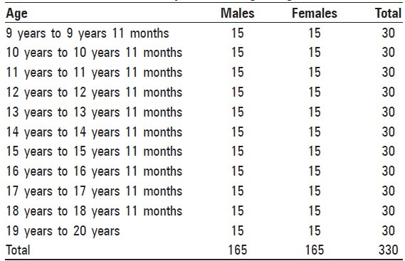
Study consisted of 330 randomly selected subjects (165 males and 165 females) with age ranging from 9 years to 20 years divided in to 11 groups according to age [Table 1]. Informed consent was taken from all the individuals and the study was approved by the Ethical Committee of Narayana Dental College and Hospital, Nellore Andhra Pradesh. Patients with: (a) serious medical illness. (b) History of extraction of permanent teeth. (c) History of trauma to face. (d) Impacted or ankylosed teeth or transposition of teeth. (e) Congenital absence of third molars was excluded from the study. Clinical examination of all 330 subjects was performed and name, sex, date of birth of each individual and date of X-ray was recorded. 330 digital orthopantomographs (OPG) were taken with a Planmeca digital machine.
Table 1.
Distribution of sample according to age and sex
DA determination by Demirjian's modified method
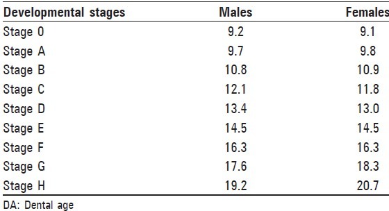
CA of an individual was calculated by subtracting the birth date from the date, on which the radiographs were exposed for that particular individual. Decimal age was taken for simplicity of statistical calculation and ages were estimated on a yearly basis, e.g. 9 years 9 months as 9.09 years and it was considered in 9 years age group. To avoid observer bias, each digital OPG of an individual was coded with a numerical identity number (1-330) to ensure that the examiner was blind to sex, name and age of subjects. Evaluators were given written instructions for staging, including drawings and written descriptions of the stages of tooth development of Demirjian's modified method that supplements the graphic representations with archetypical radiographs for each stage. The third molar was scored"0"to"H,"depending on the stage of calcification [Table 2].[13,14,15] In this study, “48” tooth was assessed for staging of molar with no history of extraction of tooth on that side. DA of each subject was assessed by comparing developmental stages of mandibular third molar with the rating system [Table 3].[1,13,16] For statistical computations, stages were assigned a numeric value where stage 0 = 1, stage A = 2, stage B = 3, stage C = 4, stage D = 5, stage E = 6, stage F = 7, stage G = 8 and stage H = 9. To test intra-examiner reliability, each examiner unknowingly re-evaluated 20 of their images after 1 month.
Table 2.
Schematic drawings and brief description of stages of crown and root formation used to score third molar development (modified Demirjian et. al. method)
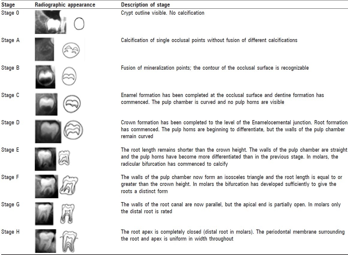
Table 3.
Assessment of DA from third molar developmental stage
Results
The significance of the difference between the means of different ages was determined using a paired sample t-test. Pearson's correlation between means of different ages was also calculated. To test the reliability of the DA assessment, the examiner re-evaluated 20 randomly selected radiographs of the same subjects. The correlation between first and second assessments was determined using Kappa statistics. The differences were considered significant when the P value is less than 0.05.
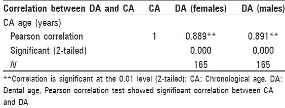
In males, mandibular third molar development commenced around 9.4 years and root completion took place around 18.9 years [Table 4] and in females third molar development commenced around 9 years and root completion took place around 18.6 years [Table 5], which shows that there is no significant sex difference in third molar development. Standard deviation between estimated DA and CA in males is increased in groups 7, 10 and in females 4, 5, 6 groups. Demirjian's modified method was found to underestimate age with mean accuracy of 0.8 years for males and 0.5 years for females. Reliability of the Demirjian et al., method was verified by testing intra-and inter-observer agreement. The Felli's Kappa value for intra-observer agreement was 92.9% and confidential limit was 0.914-0.941 [Table 6]. Inter-observer agreement was 92.4% [Table 7]. Pearson correlation test showed a significant correlation between CA and DA [Table 8].
Table 4.
Comparison between DA using the Demirjian method and CA (in years) in males
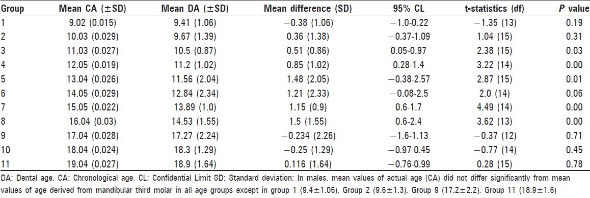
Table 5.
Comparison between DA using the Demirjian method and CA (in years) in females
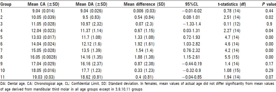
Table 6.
Intra-observer correlation coefficient

Table 7.
Inter-observer correlation (Cohen Kappa)

Table 8.
Pearson correlation for females and males
Discussion
Age estimation has become increasingly important to determine the age of living individuals. Identification of age is very important for a variety of reasons, including identifying criminal and legal responsibility, determining the emotional support needed for the victim of a sexual assault and for many other social events such as birth certificate, marriage, beginning a job, joining the army and retirement.[6,12,17,18] Radiographic analysis of third molar development expands the years of age estimation to 9-23 years as crown and root development can be studied independent of eruption. After the early teens, most teeth have calcified and erupted except for the third molars. This makes the third molar development the most important choice for age assessment among young children and adolescents with greater accuracy from the late teens to the early twenties.[12,16,19]
A large number of dental stages make it very difficult to discriminate the tooth development (Moorrees et al. 1963). Hence, in this study, Demirjian's modified method is used as it is widely accepted as the maturity scoring system and it is universal in application. Advantage of this method was that the predicted DA was relatively accurate since it was not based on the eruption process of teeth. Digital imaging provided high quality images and greatest flexibility with reduced exposure.
Only a few studies compare dental maturity of individual teeth in populations using average age entering tooth stages and most find similarities between regional and ethnic groups (Liversidge 2008, 2011). Levesque et al.(1981) determined the age of alveolar and gingival eruption and mineralization state of the third molars based on the evidence from 4640 OPG from 2278 male to 2362 female Franco-Canadians of ages ranging from 7 years to 25 years.[16] In this study, developmental stages of mandibular third molar in different age groups were assessed. In males, third molar development commenced around 9 years and root completion took place around 18.9 years and in females 9 years and 18.6 years respectively, which showed females mature slightly earlier than males, in accordance with Hassen et al. (2007). The mean ages for the lower third molar developmental stages in this study seem to be within the age ranges reported by previous authors (Moorrees et al. 1963, Koski 1963) Figures 1–4.
Figure 1.
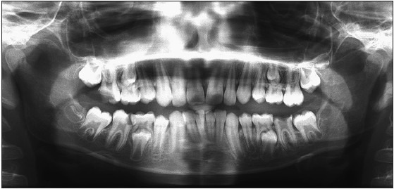
Digital OPG showing right Mandibular Third molar in Stage B
Figure 4.
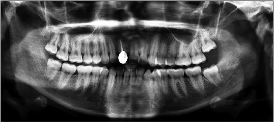
Digital OPG showing right mandibular third molar in stage G
Figure 2.
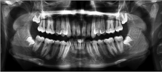
Digital OPG showing right Mandibular third molar in stage D
Figure 3.
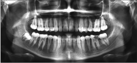
Digital OPG showing right Mandibular third molar in Stage E
In the present study, we found a significant correlation between DA and CA in both males and females i. e., r = 0.89 for males and r = 0.88 for females [Table 8] similar to Tritiana (1989), Orhan (2007), Engstrom et al.(1983), Lamons Gray (1958), Green (1961), Krogman (1967), Cheraskin (1972), Malagola (1989), Jaeger (1990), Carvaho et al.(1990), Zaborra and Terranova (1989).[20] In this study, we found that Demirjian's modified method showed the highest Kappa value (very good agreement) for intra-observer readings for the mandibular third molar. Intra-observer correlation coefficient is 92.9% and was statistically significant.
The inclination of developing third molar relative to X-ray film may result in the crown appearing tilted on the radiograph making crown stage difficult to assess. Roots of third molar are less divergent than other molars and are often found making root stage assessment more difficult especially if stages include an estimation of root length. Greatest limit of the method seems to be the operator experience in determining the dental stages of development.
In previous studies, Demirjian's method has been tested in different populations. In all populations, the authors have obtained an overestimation of DA ranging from 0.02 to 3 years. Koshy and Tandon recorded an overestimation of 2.82 years for females and 3.04 years for males in Indian population.[21] Liversidge et al. explained that overestimation in DA using Demirjian's method in different populations may be due to positive secular trend in growth and development during last 25 years. However, in this present study, there is underestimation of DA similar to Acharya.[22] X-ray standards to individuals of a socio-economic status lower than that of the reference population usually may lead to underestimation of that person's age.
According to International study Group on Forensic Age Diagnostics, a forensic age diagnosis for the purpose of criminal investigations consist of clinical examination, evaluation of signs of sexual maturity, X-ray examination of hand and evaluation of dental status of an individual.[23,24,25,26,27] Most important aspect of DA estimation is to remember that he or she should not be restricted to only one age estimation technique, but to apply different techniques available and perform repetitive measurements and calculations in order to establish more reproducibility. Comprehensive age estimation will utilize all available methods for a particular case and third molar development data compliments the skeletal maturation to give a complete assessment of age of unknown individuals.
Demirjian method is based on standards of French-Canadian population. When this method is applied to South Indian population, mean difference between true age and assessed DA is minimal [0.8 years in males and 0.5 years in females] and also showed significant positive correlation. The present study supports the use of Demirjian modified method for assessment of DA in selected population.
Conclusion
Teeth represent useful material for age estimation. Digital radiographic assessment of development of mandibular third molar can be used a better choice for predicting biological age of an individual because of its simplicity, reliability and reduced radiation exposure to the individual and should be the preferential method in forensic applications. Third molar root development can be reliably used to generate mean age and the estimated age range for an individual of unknown CA. Third molar can be commonly available biologically valid tool for age estimation in late teens to early adult group. Hence, it should be used with caution because of considerable variability among individuals.
Footnotes
Source of Support: Nil
Conflict of Interest: None declared
References
- 1.Raut DL, Mody RN. Radiographic evaluation of cervical vertebrae, carpal metacarpal bones and mandibular third molar during adolescence and in young adults. J Indian Acad Oral Med Radiol. 2006;18:24–9. [Google Scholar]
- 2.Rai B, Anand S. Age estimation in children from dental radiograph: Are gression equation. Internet J Biol Anthropol. 2008;1:2. [Google Scholar]
- 3.Chaillet N, Willems G, Demirjian A. Dental maturity in Belgian children using Demirjian's method and polynomial functions: New standard curves for forensic and clinical use. J Forensic Odontostomatol. 2004;22:18–27. [PubMed] [Google Scholar]
- 4.Liversidge HM. The assessment and interpretation of Demirjian, Goldstein and Tanner's dental maturity. Ann Hum Biol. 2012;39:412–31. doi: 10.3109/03014460.2012.716080. [DOI] [PubMed] [Google Scholar]
- 5.Demirjian A, Goldstein H. New systems for dental maturity based on seven and four teeth. Ann Hum Biol. 1976;3:411–21. doi: 10.1080/03014467600001671. [DOI] [PubMed] [Google Scholar]
- 6.Eid RM, Simi R, Friggi MN, Fisberg M. Assessment of dental maturity of Brazilian children aged 6 to 14 years using Demirjian's method. Int J Paediatr Dent. 2002;12:423–8. doi: 10.1046/j.1365-263x.2002.00403.x. [DOI] [PubMed] [Google Scholar]
- 7.Sciulli PW. Relative dental maturity and associated skeletal maturity in prehistoric native Americans of the Ohio valley area. Am J Phys Anthropol. 2007;132:545–57. doi: 10.1002/ajpa.20547. [DOI] [PubMed] [Google Scholar]
- 8.Agarwal V, Srivastava BK, Nagarappa R, Tangade P, Ravishankar TL. Age estimation from teeth of Indian adults following Gustafson's method. J Indian Assoc Public Health Dent. 2009;13:95–8. [Google Scholar]
- 9.Rai B, Kaur J, Cameriere R, Singh J. Radiographic method evaluation of teeth development in north Indian children and young people. Indian J Forensic Odontol. 2009;2:55–7. [Google Scholar]
- 10.Liversidge HM. Dentalmaturation of 18 th and 19 th century British children using Demirjian's method. Int J Paediatr Dent. 1999;9:111–5. doi: 10.1046/j.1365-263x.1999.00113.x. [DOI] [PubMed] [Google Scholar]
- 11.Liversidge HM, Lyons F, Hector MP. The accuracy of three methods of age estimation usin gradiographic measurements of developing teeth. Forensic Sci Int. 2003;131:22–9. doi: 10.1016/s0379-0738(02)00373-0. [DOI] [PubMed] [Google Scholar]
- 12.Thevissen PW, Pittayapat P, Fieuws S, Willems G. Estimating age of majority on third molars developmental stages in young adults from Thailand using a modified scoring technique. J Forensic Sci. 2009;54:428–32. doi: 10.1111/j.1556-4029.2008.00961.x. [DOI] [PubMed] [Google Scholar]
- 13.McGettigan A, Timmins K, Herbison P, Liversidge H, Kieser J. Wisdom tooth formation as a method of estimating age in a New Zealand population. Dent Anthropol. 2011;24:33–41. [Google Scholar]
- 14.Willems G. A review of the most commonly used dental age estimation techniques. J Forensic Odontostomatol. 2001;19:9–17. [PubMed] [Google Scholar]
- 15.Garn SM, Lewis AB, Bonne B. Third molar formation and its development course. Angle Orthod. 1962;32:270–9. [Google Scholar]
- 16.Levesque GY, Demirijian A, Tanguay R. Sexual dimorphism in the development, emergence, and agenesis of the mandibular third molar. J Dent Res. 1981;60:1735–41. doi: 10.1177/00220345810600100201. [DOI] [PubMed] [Google Scholar]
- 17.Michie CA. Age assessment: Time for progress? Arch Dis Child. 2005;90:612–3. doi: 10.1136/adc.2003.041921. [DOI] [PMC free article] [PubMed] [Google Scholar]
- 18.Azrak B, Victor A, Willershausen B, Pistorius A, Hörr C, Gleissner C. Usefulness of combining clinical and radiological dental findings for a more accurate noninvasive age estimation. J Forensic Sci. 2007;52:146–50. doi: 10.1111/j.1556-4029.2006.00300.x. [DOI] [PubMed] [Google Scholar]
- 19.Kasper KA, Austin D, Kvanli AH, Rios TR, Senn DR. Reliability of third molar development for age estimation in a Texas Hispanic population: A comparison study. J Forensic Sci. 2009;54:651–7. doi: 10.1111/j.1556-4029.2009.01031.x. [DOI] [PubMed] [Google Scholar]
- 20.Koupis NS, Papadopoulos MA. The use of dental age as maturity indicator. A comprehensive review. Hell Orthod Rev. 1998;1:159–73. [Google Scholar]
- 21.Koshy S, Tandon S. Dental age assessment: The applicability of Demirjian's method in south Indian children. Forensic Sci Int. 1998;94:73–85. doi: 10.1016/s0379-0738(98)00034-6. [DOI] [PubMed] [Google Scholar]
- 22.Acharya AB. Age estimation in Indians using Demirjian's 8-teeth method. J Forensic Sci. 2011;56:124–7. doi: 10.1111/j.1556-4029.2010.01566.x. [DOI] [PubMed] [Google Scholar]
- 23.Olze A, Bilang D, Schmidt S, Wernecke KD, Geserick G, Schmeling A. Validation of common classification systems for assessing the mineralization of third molars. Int J Legal Med. 2005;119:22–6. doi: 10.1007/s00414-004-0489-5. [DOI] [PubMed] [Google Scholar]
- 24.Schmeling A, Reisinger W, Loreck D, Vendura K, Markus W, Geserick G. Effects of ethnicity on skeletal maturation: Consequences for forensic age estimations. Int J Legal Med. 2000;113:253–8. doi: 10.1007/s004149900102. [DOI] [PubMed] [Google Scholar]
- 25.Olze A, Schmeling A, Taniguchi M, Maeda H, van Niekerk P, Wernecke KD, et al. Forensic age estimation in living subjects: The ethnic factor in wisdom tooth mineralization. Int J Legal Med. 2004;118:170–3. doi: 10.1007/s00414-004-0434-7. [DOI] [PubMed] [Google Scholar]
- 26.Schmeling A, Grundmann C, Fuhrmann A, Kaatsch HJ, Knell B, Ramsthaler F, et al. Criteria for age estimation in living individuals. Int J Legal Med. 2008;122:457–60. doi: 10.1007/s00414-008-0254-2. [DOI] [PubMed] [Google Scholar]
- 27.Schmeling A, Olze A, Pynn BR, Kraul V, Schulz R, Heinecke A, et al. Dental age estimation based on third molar eruption in First Nation people of Canada. J Forensic Odontostomatol. 2010;28:32–8. [PubMed] [Google Scholar]


