Although most of us are more comfortable walking forward, in part because we can see where we are going, people also have the capacity to walk, and even run, in a backward direction. This skill comes in handy when we get stuck in tight dead-end spaces, and is even a trendy exercise routine for some (1). Perhaps most famously, Michael Jackson immortalized the move in his iconic Moonwalker dance. Not surprisingly, the capacity for backward locomotion is not limited to humans and now, thanks to elegant experiments carried out in the fruit fly by Bidaye et al. and reported on page XXX in this issue (2), we have some understanding of how animals choose between forward and backward locomotion.
Experimental systems as diverse as mice, locusts, stick insects, and flies have taught us a lot about the kinematics and underlying neural control of forward walking (3– 7). Each leg joint is controlled by motor neurons triggering contractions of opposing flexor and extensor muscles. The challenge for the nervous system, and perhaps more so for a six-legged fly compared to the two-legged Mr. Jackson, is to coordinate each of these joint bends, both within a leg (e.g., hip and knee) and between legs (e.g., left and right). Interneurons acting locally within the central nervous system (CNS), where the motor neuron cell bodies reside, somehow coordinate all of these limb movements, assisted by sensory neurons in the legs that report load and joint angle back to the CNS (7, 8).
Importantly, walking backward is not a simple reversal of walking forward: For example, when we walk forward or backward, our knees bend the same way with each step, but the muscles in our hips that move our thighs work oppositely, depending on the direction (9). Such reversals in the movement of a proximal leg joint relative to more distal leg joints are also observed in backward-walking stick insects (10, 11). In other words, when our brains tell our motor systems to change direction, they selectively modulate parts of the locomotor circuit. How do nervous systems manage to accomplish this?
To begin to answer this question, Bidaye et al. turned to the fruit fly to exploit its powerful genetic toolkit. They began by watching what happened to the locomotor behavior of flies in which different combinations of neurons were artificially activated. To execute this screen, the authors used the yeast transcription factor Gal4 and its cognate UAS binding site to drive the expression of the thermally activated cation channel TrpA1 in subsets of neurons. From about 3500 Gal4/UAS-TrpA1 transformed fly lines, each expressing TrpA1 in a stereotyped set of neurons, they found one line that caused flies to walk backward instead of forward, a phenotype they dubbed “moonwalker.” Conversely, silencing these neurons greatly inhibited backward walking in situations, such as hitting a dead end, where wild-type flies normally choose to walk backward.
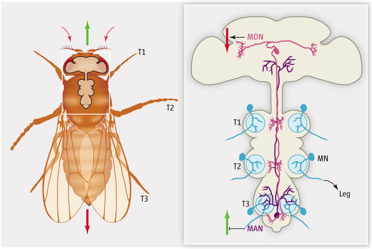
By expressing green fluorescent protein (GFP) under the control of Gal4/UAS instead of TrpA1, they found that the moonwalker line was active in seven morphologically distinct neurons. Subsequent experiments designed to identify which of the seven were important revealed that bilaterally activating only a specific pair of neurons was sufficient to make flies “do the moonwalk.” While one of these neurons was located in the brain and sent its axon posteriorly into the ventral nerve cord (VNC), where motor neuron cell bodies reside, the second neuron had the opposite orientation: Its cell body resided in a posterior region of the VNC and sent its axon anteriorly, into the brain (see the figure). On the basis of these two distinct orientations, the authors named these neurons MDN and MAN, for “moonwalker descending neuron” and “moonwalker ascending neuron,” respectively (figure).
Figure. A neural circuit for moonwalking.
Flies walk forward (green arrow) or backward (red arrow) in response to sensory cues (small red arrows). MDN and MAN are neurons that control walking direction, presumably by indirectly coordinating the activities of motor neurons (MN) via the leg neuropil (blue circles).
Bidaye et al. then used more precise genetic tools to tease out the individual contributions of MDN and MAN in the control of walking direction. Activation of MDN, alone, was sufficient to induce a significant amount of backward walking, while activating MAN, alone, was not. However, activating either MDN or MAN was sufficient to interfere with forward walking; flies in which either neuron was activated still walked forward, but for shorter distances. Thus, it seems that whereas MDN activity triggers a switch from forward to backward walking, MAN activity contributes to the moonwalker phenotype mainly by inhibiting forward walking (figure). MDN, with its cell body in the brain, may receive sensory cues from, for example, the eyes or antennae, that inform the fly it is approaching a dead end. As such, MDN may be a “command neuron” analogous to command interneurons in Caenorhabditis elegans that promote backward crawling in response to touching the worm's head (12– 14).
Although the findings of Bidaye et al. provide the first glimpse into how flies, and perhaps other legged animals, control walking direction, many questions remain. For one, none of the upstream or downstream neurons that make functional connections with MDN or MAN are known. Of particular interest is whether and how MDN and MAN selectively modify only parts of the locomotor circuit to induce flies to change direction. In C. elegans, forward and backward crawling require distinct motor neurons that receive information from different command interneurons (13, 14). To figure out if something similar is happening in limbed locomotion, we need a better understanding of the circuitry that controls forward walking. Answers will no doubt come from a wide variety of approaches, including ones similar to those used by Bidaye et al., to provide cellular resolution to complex motor outputs such as walking.
Acknowledgments
R.S.M. acknowledges support from NIH R01NS070644.
Footnotes
A pair of neurons in the CNS of fl ies controls and coordinates their ability to walk backward.
References and Notes
- 1.Neporent L. No Gain in Backward Exercise, Experts Say. The New York Times. 1998 Oct 13; http://www.nytimes.com/1998/10/13/health/no-gain-in-backward-exercise-experts-say.html.
- 2.Bidaye SS, Machacek C, Wu Y, Dickson BJ. Science. 2014;344:xxx. doi: 10.1126/science.1249964. [DOI] [PubMed] [Google Scholar]
- 3.Kiehn O. Curr Opin Neurobiol. 2011;21:100. doi: 10.1016/j.conb.2010.09.004. [DOI] [PubMed] [Google Scholar]
- 4.Goulding M. Nat Rev Neurosci. 2009;10:507. doi: 10.1038/nrn2608. [DOI] [PMC free article] [PubMed] [Google Scholar]
- 5.Büschges A, Akay T, Gabriel JP, Schmidt J. Brain Res Brain Res Rev. 2008;57:162. doi: 10.1016/j.brainresrev.2007.06.028. [DOI] [PubMed] [Google Scholar]
- 6.Strauss R. Curr Opin Neurobiol. 2002;12:633. doi: 10.1016/s0959-4388(02)00385-9. [DOI] [PubMed] [Google Scholar]
- 7.Mendes CS, Bartos I, Akay T, Márka S, Mann RS. Elife. 2013;2:e00231. doi: 10.7554/eLife.00231. [DOI] [PMC free article] [PubMed] [Google Scholar]
- 8.Zill S, Schmitz J, Büschges A. Arthropod Struct Dev. 2004;33:273. doi: 10.1016/j.asd.2004.05.005. [DOI] [PubMed] [Google Scholar]
- 9.Lee M, Kim J, Son J, Kim Y. Gait Posture. 2013;38:674. doi: 10.1016/j.gaitpost.2013.02.014. [DOI] [PubMed] [Google Scholar]
- 10.Rosenbaum P, Wosnitza A, Büschges A, Gruhn M. J Neurophysiol. 2010;104:1681. doi: 10.1152/jn.00362.2010. [DOI] [PubMed] [Google Scholar]
- 11.Akay T, Ludwar BCh, Göritz ML, Schmitz J, Büschges A. J Neurosci. 2007;27:3285. doi: 10.1523/JNEUROSCI.5202-06.2007. [DOI] [PMC free article] [PubMed] [Google Scholar]
- 12.Piggott BJ, Liu J, Feng Z, Wescott SA, Xu XZ. Cell. 2011;147:922. doi: 10.1016/j.cell.2011.08.053. [DOI] [PMC free article] [PubMed] [Google Scholar]
- 13.Wicks SR, Rankin CH. J Neurosci. 1995;15:2434. doi: 10.1523/JNEUROSCI.15-03-02434.1995. [DOI] [PMC free article] [PubMed] [Google Scholar]
- 14.Chalfie M, et al. J Neurosci. 1985;5:956. doi: 10.1523/JNEUROSCI.05-04-00956.1985. [DOI] [PMC free article] [PubMed] [Google Scholar]


