Abstract
BACKGROUND
Cigarette smoking increases the risk of ventricular fibrillation and sudden cardiac death (SCD). QT dispersion (QTD) is an important predictor of cardiac arrhythmia. The aim of this study was to assess the acute effect of smoking a single standard cigarette containing 1.7 mg nicotine on QT interval and QTD in healthy smokers and nonsmokers.
METHODS
The study sample population consisted of 40 healthy male hospital staff, including 20 smokers and 20 nonsmokers. They were asked to refrain from smoking at least 6 h before attending the study. A 12-lead surface electrocardiogram (ECG), recorded at paper speed of 50 mm/s, was obtained from all participants before and 10 min after smoking of a single complete cigarette. QT interval, corrected QT interval, QTD, and corrected QT dispersion (QTcD) were measured before and after smoking.
RESULTS
Smokers and nonsmokers did not have any significant differences in heart rate (HR) (before smoking = 67.35 ± 5.14 vs. 67.70 ± 5.07, after smoking = 76.70 ± 6.50 vs. 76.85 ± 6.50, respectively), QTD (before smoking = 37.75 ± 7.16 vs. 39.15 ± 6.55, after smoking = 44.75 ± 11.97 vs. 45.50 ± 9.58, respectively), and QTcD (before smoking = 39.85 ± 7.40 vs. 41.55 ± 6.57, after smoking = 50.70 ± 14.31 vs. 51.50 ± 11.71, respectively). However, after smoking a single cigarette, HR, mean QTD, and QTcD significantly increased (all had P value <0.001) in comparison to the measures before smoking.
CONCLUSION
Smoking of a single complete cigarette in both smokers and nonsmokers results in significant QTD increase, which can cause arrhythmia and SCD.
Keywords: Cardiac, Death, Electrocardiography, Smoking, Sudden
Introduction
Effects of smoking on various organ-systems such as the cardiovascular system are evaluated in a vast range of researches. Chronic cigarette smoking, as a risk factor of atherosclerosis and endothelial dysfunction, can cause acute coronary syndrome and sudden death.1,2 Acute effects of smoking on cardiovascular function are complex. It can transiently raise systemic blood pressure, peripheral vascular resistance, heart rate (HR), it also can change parameters of HR variability and also cause echocardiographic evidences of diastolic dysfunction.3-5 It is already known that smoking not only increases mortality due to coronary artery disease but also increases mortality secondary to sudden cardiac death (SCD).6 On the other hand, long QT interval is reported as a predictor of arrhythmia risk. Prolonged QT interval dispersion (QTD) can predict ventricular arrhythmia due to excessive loss of synchronization of ventricular repolarization.7,8 Our study was conducted to determine the acute effect of smoking on QTD in the smoker and nonsmoker healthy males.
Materials and Methods
A prospective cohort study with before and after the design was performed in Taleghani Hospital, Tehran, Iran in April and May 2012. The sample size was calculated as 20 subjects in each group considering α = 5%, d = 1, statistical power of 0.925.
Hence, 40 persons included 20 professional smokers (with smoking habit of at least 5 pack/year before the study onset), and 20 nonsmokers (with smoking habit of 0.5 pack/year or less prior to the study onset), selected from 63 young male hospital staff (age <40 years) volunteers after considering exclusion criteria randomly. All selected cases did not have a history of hypertension, cardiac, or pulmonary disease with normal resting electrocardiogram (ECG) and normal echocardiography. Cases with diabetes mellitus, renal failure, and those with signs or symptoms of coronary artery disease were excluded. None of the subjects was on chronic medication. Two cardiologists confirmed health of all participants according to routine cardiovascular examination.
To control the effect of other confounding factors such as recent smoking, tea, caffeinated beverage, and body position, all participants were asked to refrain from smoking at least 6 h before attending the study and were also asked not to consume tea and caffeinated beverages for 3 h before the study. Test was done in the morning, all subjects rested in the supine position to stabilize HR. After 10 min, a baseline standard 12-lead surface ECG was obtained during normal respiration. Later, participants were asked to smoke a single complete cigarette containing 1.7 mg nicotine in a sitting position and again lay down in the supine position. Ten minutes after smoking, second ECG records were obtained.
This research was accomplished with the budget of Shaheed Beheshti University of Medical Sciences, and all stages were in accordance with ethical issues of the university Ethics Committee (Ethical Approval Code: 308/290-2011). The participants were informed about the study protocol and then requested to sign the consent form.
ECG tracing
ECGs were traced with the speed of 50 mm/s and amplitude of 20 mA. QT interval was measured manually in each lead from the onset of QRS to the end of T-wave. Termination of T-wave was defined as its return to the TP isoelectric baseline. At the presence of U-wave, nadir of the curve between T- and U-waves was defined as the end of T-wave. In biphasic T-wave, the final return to the baseline was selected. Leads, in which due to low amplitude T-wave, QT interval could not be measured reliably, were omitted from the analysis. QTD was defined as a difference between maximum and minimum interval of measured QT intervals in the 12 lead ECGs. Corrected QT (QTc) and Corrected QT interval dispersion (QTcD) were calculated according to Bazett’s formula by dividing QT by the square root of the RR interval.9 QTc = QT/√RR.
ECG parameters were measured by two blind cardiologists who were oriented to the QT measurement method. If there was a difference in their measurement, the mean value accepted.
Statistics
All data are expressed as mean ± SD. Data analysis was performed by using SPSS statistical software (version 17.0, SPSS Inc., Chicago, IL, USA). Normal distribution was checked by Kolmogorov-Smirnov test. Data before and after smoking were compared by paired Student’s t-test. As QT dispersion and QTc dispersion had not a Gaussian distribution, Wilcoxon signed-rank test was used. The magnitude of change in QTD and QTcD with smoking was compared by Mann-Whitney U test. Pearson correlation coefficients were calculated to determine the strength of linear relationships between changes of QTD, QTcD, and HR changes before and after smoking. Results were considered as significant at an error probability level of P < 0.05.
Results
All 40 participants completed the study, and there were not any missing value. The mean age of professionals was 31.6 ± 4.8 years and of nonsmokers was 31.0 ± 5.6 years. Mean baseline and post-interventional measures (HR, QTD, QTcD) in both groups are summarized in table 1. There was no statistically significant difference in the baseline measures and measures obtained after smoking in both groups.
Table 1.
Electrocardiogram (ECG) findings obtained before and after smoking in professional smokers and nonsmokers separately
| Before smoking |
P | After smoking |
P | |||
|---|---|---|---|---|---|---|
| Nonsmokers | Professional smokers | Nonsmokers | Professional smokers | |||
| HR (bpm) | 67.70 ± 5.07 | 67.35 ± 5.14 | 0.82 | 76.85 ± 6.50 | 76.70 ± 6.50 | 0.97 |
| QTD (ms) | 39.15 ± 6.55 | 37.75 ± 7.16 | 0.69 | 45.50 ± 9.58 | 44.75 ± 11.97 | 0.84 |
| QTcD (ms) | 41.55 ± 6.57 | 39.85 ± 7.40 | 0.55 | 51.50 ± 11.71 | 50.70 ± 14.31 | 0.81 |
HR: Heart rate; QTD: QT interval dispersion; QTcD: Corrected QT interval dispersion; Values are mean ± SD
As there was no significant difference between the indices of the two groups, we pooled data from both professional and nonprofessional smokers to evaluate the effect of smoking on our variables.
Among 40 subjects, the mean HR, mean QTD, and mean QTcD were 67.53 ± 5.04, 38.45 ± 6.81, and 40.70 ± 6.92 respectively. After smoking a single cigarette, the same parameters increased significantly (P < 0.001) to 76.78 ± 6.41, 45.13 ± 10.71, and 51.10 ± 12.91, respectively (Table 2).
Table 2.
Electrocardiogram (ECG) findings obtained before and after smoking
| Before smoking | After smoking | Difference | P | |
|---|---|---|---|---|
| Mean HR (bpm) | 67.53 ± 5.04 | 76.78 ± 6.41 | 9.25 ± 4.27 | 0.001 |
| QTD (ms) | 38.45 ± 6.81 | 45.13 ± 10.71 | 6.67 ± 7.99 | 0.001 |
| QTcD (ms) | 40.70 ± 6.92 | 51.10 ± 12.91 | 10.40 ± 10.32 | 0.001 |
HR: Heart rate; QTD: QT interval dispersion; QTcD: Corrected QT interval dispersion; Values are mean ± SD
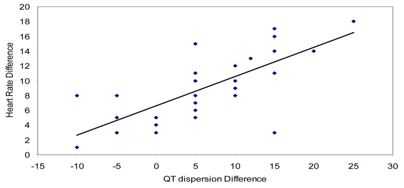
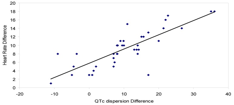
The difference between HR before and after smoking had positive linear correlation with QTD difference (r = 0.741) and changes in QTcD (r = 0.812) (Figures 1 and 2).
Figure 1.
Scatter plots of correlation between difference of heart rates before and after smoking and QT dispersion difference
Figure 2.
Scatter plots of correlation between difference of heart rates before and after smoking and QTc dispersion difference QTc: Corrected QT
Discussion
As smoking can increase coronary artery disease, it may cause some harmful changes in electrical function of myocardial cells. It may predispose to ventricular fibrillation and SCD by altering ventricular recovery time dispersion indices. QTD is an important parameter that reflects heterogeneity of ventricular repolarization and predicts ventricular arrhythmia and sudden death.10 Apart from the cumulative effect of smoking on the cardiovascular system, it should be considered that even a single cigarette induces the potential of SCD and arrhythmia by prolongation of QTD. Products of nicotine, tar, and nitric oxide derived free radicals interfere with normal chemical interactions of the body after smoking.11,12 For example, it is known that nicotine is a nonspecific blocker of potassium channels and has several pathophysiologic effects, including tachycardia, increased blood pressure, and catecholamine release, particularly within the short period after smoking. It can also prolong the action potential duration and depolarize membrane, so may cause QTD too.1,13 It has been reported that QT dispersion > 80 ms increases risk of cardiac death.8
In our research in accordance with previous researches, acute smoking significantly increased QTD and QTcD,14,15 but in contrast to Singh findings, in our study this increase was significant in both smokers and nonsmokers.14 And in addition to QTD and QTcD values, which were measured in Khosropanah and Barkat study, we have also assessed the relationship between QT interval increase and acute smoking, which was significant.15 Our results differ from Karakaya et al. findings of no significant difference between QT interval and QTD measures before and after smoking.16
Our study results showed that, the difference between HR before and after smoking had a strong correlation with QTD and QTcD. Consequently with more increase in HR after smoking, there would be more increase in QTD and QTcD and vice versa.
According to our results, QTD increase occurs in both groups of smokers and nonsmokers. Hence, it seems that there is no cardiac adaptation or tolerance to effects of smoking even in professional smokers, and smoking of even a single cigarette, regardless of past history of smoking increases QT dispersion, which is a predictor of cardiac arrhythmia and SCD.
Limitation of Study
Our study has some limitations. First, we included a small number of participants. So, we pooled two groups of professional and nonprofessional smokers for analysis. However, despite this small size, we were able to detect a significant change of QT dispersion associated with smoking. Second, we studied on young healthy participants. Therefore, our results should be interpreted with caution in other groups. Third, this study simply observed the acute change of QT with smoking, there is always chance that this change was due to some unobserved confounding variable or systematic bias.
Acknowledgments
This work was financially supported by Shahid Beheshti University of Medical Sciences, Tehran, Iran.
Footnotes
Conflicts of Interest
Authors have no conflict of interests.
REFERENCES
- 1.Ambrose JA, Barua RS. The pathophysiology of cigarette smoking and cardiovascular disease: an update. J Am Coll Cardiol. 2004;43(10):1731–7. doi: 10.1016/j.jacc.2003.12.047. [DOI] [PubMed] [Google Scholar]
- 2.Yagi H, Komukai K, Hashimoto K, Kawai M, Ogawa T, Anzawa R, et al. Difference in risk factors between acute coronary syndrome and stable angina pectoris in the Japanese: smoking as a crucial risk factor of acute coronary syndrome. J Cardiol. 2010;55(3):345–53. doi: 10.1016/j.jjcc.2009.12.010. [DOI] [PubMed] [Google Scholar]
- 3.Xue C, Sun YH, Ding RJ, Fu YY, Hu DY. Impact of acute smoking on artery function in healthy chronic smokers. Zhonghua Xin Xue Guan Bing Za Zhi. 2011;39(5):410–3. [PubMed] [Google Scholar]
- 4.Karakaya O, Barutcu I, Kaya D, Esen AM, Saglam M, Melek M, et al. Acute effect of cigarette smoking on heart rate variability. Angiology. 2007;58(5):620–4. doi: 10.1177/0003319706294555. [DOI] [PubMed] [Google Scholar]
- 5.Ghaidari ME, Akbarzadeh MA, Yazdani Sh, Asadpour Piranfar M, Aslani A. Effect of Acute Smoking on Diastolic Function. International Cardivascular Research Journal. 2010;4(2):81–5. [Google Scholar]
- 6.D'Alessandro A, Boeckelmann I, Hammwhoner M, Goette A. Nicotine, cigarette smoking and cardiac arrhythmia: an overview. Eur J Cardiovasc Prev Rehabil. 2011 doi: 10.1177/1741826711411738. [DOI] [PubMed] [Google Scholar]
- 7.Pye M, Quinn AC, Cobbe SM. QT interval dispersion: a non-invasive marker of susceptibility to arrhythmia in patients with sustained ventricular arrhythmias? Br Heart J. 1994;71(6):511–4. doi: 10.1136/hrt.71.6.511. [DOI] [PMC free article] [PubMed] [Google Scholar]
- 8.Elming H, Holm E, Jun L, Torp-Pedersen C, Kober L, Kircshoff M, et al. The prognostic value of the QT interval and QT interval dispersion in all-cause and cardiac mortality and morbidity in a population of Danish citizens. Eur Heart J. 1998;19(9):1391–400. doi: 10.1053/euhj.1998.1094. [DOI] [PubMed] [Google Scholar]
- 9.Bazett HC. An analysis of the time-relations of electrocardiograms. Annals of Noninvasive Electrocardiology. 1997;2(2):177–94. [Google Scholar]
- 10.Okin PM, Devereux RB, Howard BV, Fabsitz RR, Lee ET, Welty TK. Assessment of QT interval and QT dispersion for prediction of all-cause and cardiovascular mortality in American Indians: The Strong Heart Study. Circulation. 2000;101(1):61–6. doi: 10.1161/01.cir.101.1.61. [DOI] [PubMed] [Google Scholar]
- 11.Srivastava SK, Barua RS, Saha DC, Eales-Reynolds LJ, DeVoe MC, Ambrose JA. Endogenous free radical generating sources are involved in smoking-mediated dysfunction of nitric oxide biosynthesis in human coronary artery endothelial cells: An in vitro demonstration. J Am Coll Cardiol. 2003;41(6s1):306. [Google Scholar]
- 12.Grassi D, Desideri G, Ferri L, Aggio A, Tiberti S, Ferri C. Oxidative stress and endothelial dysfunction: say NO to cigarette smoking. Curr Pharm Des. 2010;16(23):2539–50. doi: 10.2174/138161210792062867. [DOI] [PubMed] [Google Scholar]
- 13.Wang H, Shi H, Wang Z. Nicotine depresses the functions of multiple cardiac potassium channels. Life Sci. 1999;65(12):L143–L149. doi: 10.1016/s0024-3205(99)00370-7. [DOI] [PubMed] [Google Scholar]
- 14.Singh K. Effect of smoking on QT interval, QT dispersion and rate pressure product. Indian Heart J. 2004;56(2):140–2. [PubMed] [Google Scholar]
- 15.Khosropanah Sh, Barkat M. The Acute Effect of Cigarette Smoking on QT Dispersion. Iran J Med Sci. 2002;27(2):56–59. [Google Scholar]
- 16.Karakaya O, Saglam M, Metin Esen A, Barutcu I, Ozdemir N, Yaymaci B, et al. Acute effect of cigarette smoking on ventricular repolarization paramaters. Kosuyolu Heart Journal. 2005;9(1):1–7. [Google Scholar]


