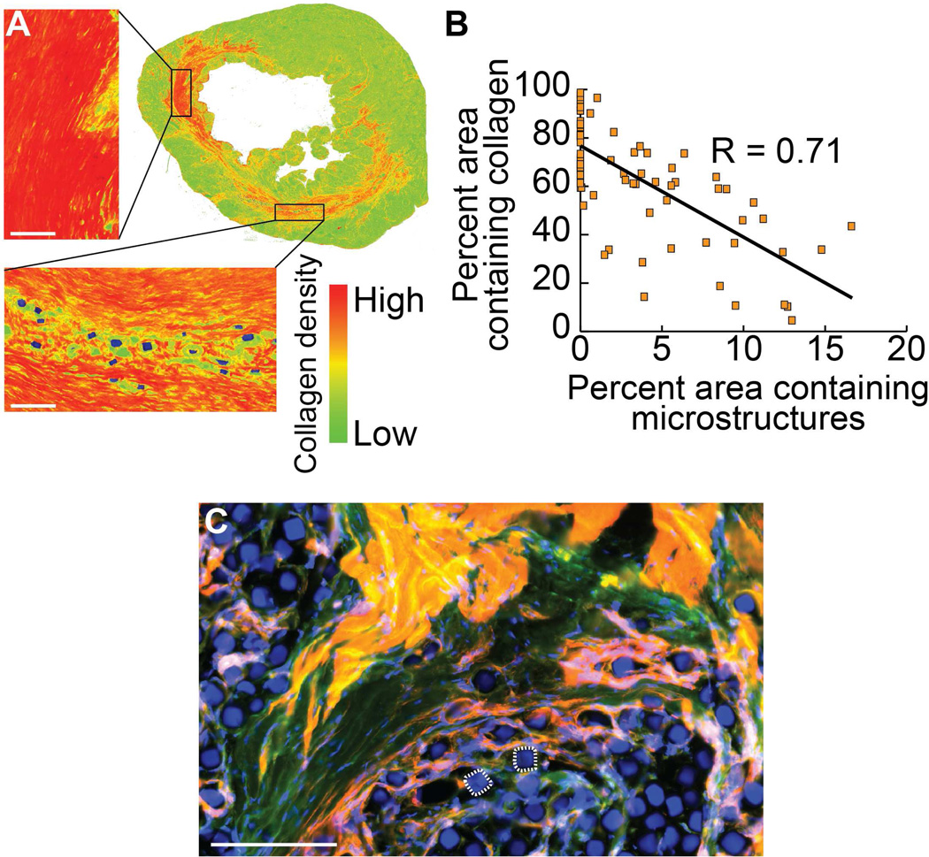
Fig. 4. Collagen production reduced proximal to microstructures in vivo.
Local changes in the makeup of the scar tissue are seen in the presence of microstructures. A representative aniline blue stained left ventricle of an infarcted rat treated with a single injection of microcubes is false-colored to depict collagen density based on colorimetric stain intensity (A), showing an inverse relationship between local collagen content and coverage area by microstructures after six weeks in the scarred region surrounding the injection site (Pearson Correlation Coefficient: R = 0.71) (B). Microstructures make independent and intimate contact with surrounding fibrotic and healthy cardiac tissue at the cellular level, analogous to the interactivity seen in the in vitro culture model. Two microcubes are outlined for reference (C). Scale bar = 100 µm (red = rhodamine phalloidin, green = myosin heavy chain antibody, blue = nuclei and microstructures).

