Abstract
Electrokinetic preconcentration coupled with mobility shift assays can give rise to very high detection sensitivities. We describe a microfluidic device that utilizes this principle to detect cellular kinase activities by simultaneously concentrating and separating substrate peptides with different phosphorylation states. This platform is capable of reliably measuring kinase activities of single adherent cells cultured in nanoliter volume microwells. We also describe a novel method utilizing spacer peptides that significantly increase separation resolution while maintaining high concentration factors in this device. Thus, multiplexed kinase measurements can be implemented with single cell sensitivity. Multiple kinase activity profiling from single cell lysate could potentially allow us to study heterogeneous activation of signaling pathways that can lead to multiple cell fates.
Kinases are an important family of proteins that regulate the majority of cell signaling pathways. They transmit information by catalyzing the phosphorylation of a specific substrate, thus modulating its activity. Interactions of multiple kinases in the signal transduction network lead to different outcomes in response to stimuli, which affects cell fate. Due to their importance in cell decision processing, there is tremendous interest in measuring cellular kinase activity levels.
Recent studies have found that many anticancer drugs kill most but not all the cells in a tumor, often resulting in relapse of cancer.1,2 It has been proposed that nongenetic cell-to-cell variability in protein activity, among other things, lead to this different response to drugs.1 As most conventional techniques provide only a population-averaged measurement of the signals within the regulatory pathway, they do not reflect an accurate picture of a heterogeneous population of cells being in different states of intracellular processing.3−5 Analysis of the overall changes in phosphorylation of population of cells may also miss cellular subpopulations that are in different signaling states due to the asynchronous nature of the response.6
To address the issues related to cellular heterogeneity in signal transduction, one would need measurements of various kinase activities at the single cell level. Microfluidic systems provide great potential and promise for analyzing single cell molecular content with an unparalleled speed, accuracy and throughput. Confinement in microchambers has been shown to increase the effective concentrations of target biomolecules and enable ultrasensitive detection of intracellular proteins from single cells.7,8 However, these methods require cells to be detached into suspension prior to analysis, an event which could activate many signaling pathways and perturb the biochemical process to be studied. Another major drawback of these assays is that adherent phenotypes such as morphology, and individual cell migration behaviors cannot be correlated with their biological activities. Furthermore, these methods rely on either special fluoregenic substrates8 that cannot be used for multiplexed detection or phosphospecific antibody methods7 that do not necessarily reflect the actual enzyme activity.
A more accurate approach that would provide crucial information about the kinetics and state of the signal transduction network is the direct kinase activity assay, which measures the ability of kinases to catalyze phosphorylation of a target protein or peptide. Currently, the most advanced methods for single cell kinase activity measurement involve imaging live cells that are genetically encoded for a substrate molecule that can report the activity changes within the cytoplasm.9,10 These live-cell imaging methods could yield spatiotemporal information about kinase activation; however, they are limited in the number and types of enzymes that can be measured simultaneously in single cells. In addition, expressing a reporter molecule involves laborious genetic engineering of a cell line to encode a fluorescent protein, and could alter the normal function of the cell. An alternative strategy that has been developed involves microinjecting fluorescent kinase substrates into single cells, lysing them and performing capillary electrophoresis (CE) to separate and quantify the phosphorylated and unphosphorylated substrates.11−13 It is possible to perform simultaneous measurements of several enzymes within the same cell due to the separation capability of CE. In both kinase activity assays described above, substrate specificity is an issue because there is significant substrate cross-reactivity among intracellular kinases. In addition, intracellular kinase substrate reporters could be subjected to other cellular processes such as proteolysis and dephosphorylation during intracellular kinase reaction,11 thus obfuscating the actual activity of the target kinase. Very recently, it is demonstrated that a microfluidic probe can lyse single adherent cells and capture the contents to perform single-cell kinase activity assay.14 This device is readily applicable to existing cells on a standard tissue culture plate; however, it remains a challenge to scale up for high throughput, multiplex single cell kinase activity measurements.
Apart from cell handling, novel phenomena in microfluidics have been exploited to enable ultrasensitive detection modalities. We have previously described a concentration-enhanced mobility shift platform in a microfluidic device that can amplify signals in homogeneous electrophoretic assays.15 Here we report that this platform can be applied to simultaneously measure multiple kinase activities from single adherent cells. The increased sensitivity of the concentration-enhanced mobility shift platform allows for detection of kinase activities from single cell lysate even upon dilution into a much larger reaction chamber. The strength of this platform include: (1) the ability to directly measure kinase activities from adherent cells without detaching them (2) improved reaction specificity by addition of off-target kinase inhibitors and various protease and phosphatase inhibitors to the lysate (3) do not require microinjection or genetic modification, and (4) ability to simultaneously measure several kinase activities within the same cell. We expect the platform that we described to be an important tool that would complement the existing single cell kinase assay methods.
Experimental Section
Details of the microchip operation, peptide synthesis and buffer recipes are included in the Supporting Information.
Recombinant Kinase Activity Assays
Varying concentrations of 0.5 μL recombinant kinase (Invitrogen, Carlsbad, CA) was mixed with 4.5 μL of the premixed assay buffer (Buffer A supplemented with 1 μM substrate, please refer to the Supporting Information for buffer recipes) and incubated at room temperature for 60 min. After 1 h of incubation, 1 μL of the reaction mixture was diluted in 99 μL of Buffer B and 30 μL of this final sample was loaded into each sample reservoir of the microfluidic device. The results of the assay were taken at the end of 15 min of electrokinetic concentration. Substrate cross-reactivity measurements were performed as described in the Supporting Information.
Bulk Cell Lysate Kinase Activity Assays
Bulk cell lysate was prepared as described in the Supporting Information. Assay buffer (Buffer C with 4 μM PKC inhibitor, 4 μM calmidazolium, 1 μM fluorescent substrate peptide, 1 mM DTT; for Akt and MK2, 0.4 μM PKI-tide, 5 μM GF109203X; for PKA, 5 μM GF109203X) was prepared in bulk and 18 μL volumes were aliquoted into separate microcentrifuge tubes. To begin each reaction, 10% (v/v) lysis buffer (Buffer D) or lysate (diluted in lysis buffer to stated concentrations) was added and the contents were mixed gently. Reaction was carried out at 37 °C for 60 min. After that, reaction was stopped by diluting 1 μL of this mixture in 99 μL of Buffer B. Thirty microliters of this final sample was used for the concentration-enhanced mobility shift assay.
Single Cell Culture and Kinase Assay
Single adherent HepG2 cells were isolated and grown in custom-made cell culture chambers defined by silicone gasket wells on coverglass (internal volume = 40 nL). Single cells confined in the sealed nanoliter size microwells were ultrasonically lysed in a water bath so that the released intracellular kinases can catalyze phosphorylation of peptide substrates that were preadded to the microwells just before lysis. The kinase reaction product from each of the 40 nL chambers were individually retrieved, diluted, and used for concentration-enhanced mobility shift assay. Please refer to Figure 3a and the Supporting Information for the detailed protocol.
Figure 3.
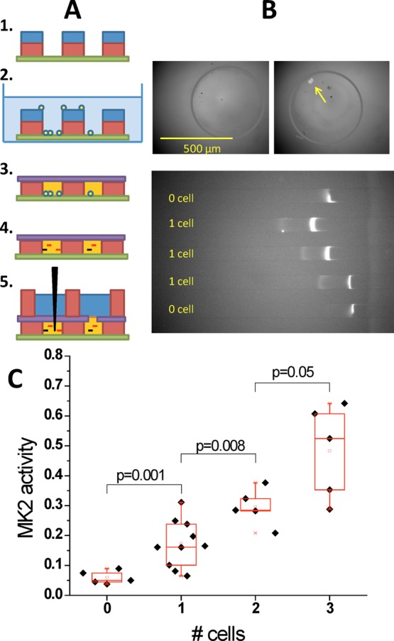
Demonstration of single cell concentration-enhanced kinase activity assay. (a) Procedures for single cell culture, lysis and kinase reaction. Individual cells are isolated and cultured in individual open microwells (∼40 nL) for microscopic observation. For kinase assay, buffer in the wells is replaced with kinase assay buffer, sealed with a Kapton tape, and the cells are ultrasonically lysed. After incubation, to allow kinase reaction, the reaction product is diluted into larger volumes and loaded into the microflluidic device for readout. (b) Detection of 0 vs 1 cell Akt activities from different microwells. Measured activity signals (marked by arrows) can be clearly differentiated between 0 and 1 cell cases. (c) MK2 activity vs cell number after normalizing by the average cell volume. Different numbers of cells have significantly different normalized kinase activities by Student’s two-tailed t-test, demonstrating that this assay has both single cell sensitivity and resolution.
Results and Discussion
Concentration-Enhanced Mobility Shift Assay
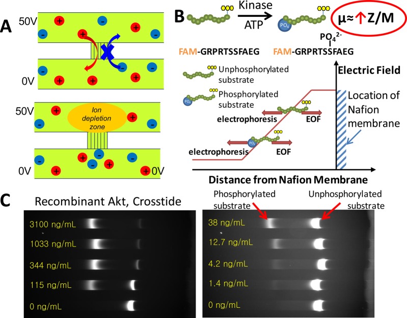
Figure 1 describes the key operation of the poly(dimethylsiloxane) (PDMS) microfluidic electrokinetic concentration chip.15 The chip consists of two microchannels bridged by a Nafion ion selective membrane that is only permeable to positive ions. The top microchannel and bottom microchannel are prefilled with sample solution and buffer solution, respectively. Under the voltage configuration shown in Figure 1a, positive ions from the sample channel at the vicinity of the Nafion membrane migrate to the buffer channel but the negative ions from the buffer channel are prevented from migrating to the sample channel. As a result, an ion depletion zone is created in the sample channel near the ion selective membrane. The conductivity gradient at the boundary of the ion depletion zone gives rise to a stable electric field gradient that can effectively focus negatively charged biomolecules at separate locations where electrophoretic velocity balances bulk flow velocity.15 In this work, fluorescently labeled peptides that contained the recognition sequences for specific kinases are used as substrates. Target kinases in the sample catalyze the phosphorylation of these peptides, leading to an increase in electrophoretic mobility. Phosphorylated peptides, which have a higher electrophoretic mobility due to the negative-charged phosphoryl group, are concentrated at the low electric field region. On the other hand, unphosphorylated peptides have a lower mobility; therefore, they concentrate nearer to the cation selective membrane where the electric field is higher (Figure 1b). In this manner, we can obtain good separation between phosphorylated and unphosphorylated substrates, while at the same time achieve continuous signal enhancement for greater sensitivity.
Figure 1.
(a) Ion selective membrane creates a local ion depletion zone with high electric field upon applying a voltage. (b) Unphosphorylated and phosphorylated peptide substrates are different in terms of their electrophoretic mobilities (additional charge), therefore they continuously concentrate at different locations in the electrokinetic concentration device where the peptide electric force balance the electroosmotic flow, (c) With increasing recombinant Akt concentration, the fluorescence intensity of the phosphorylated substrate peak increases while the fluorescence intensity of the unphosphorylated substrate peak decreases.
The microfluidics platform demonstrated in this paper has several advantages in this application compared to other techniques that combine preconcentration and separation. Previous reports have established that this platform is capable of achieving very high preconcentration factors (million fold),16 and has been successfully used in many biological assays including enhancing protein binding kinetics,17 improving sensitivity of enzyme assays,18−20 and reducing the limit of detection of mobility shift assays.15 Unlike other techniques such as transient isotachophoresis21 and temperature gradient focusing,22 this method does not require multiple buffer setup or temperature sensitive buffer. A major advantage of this platform is the ability to continuously concentrate and separate the peptide substrates from the entire sample channel over the experiment duration, leading to increasing detection sensitivity with time.
Development of Recombinant Kinase Activity Assay
We first performed an experiment with recombinant Akt and its fluorescent peptide substrate Crosstide (5-FAM-GRPRTSSFAEG). Recombinant enzyme assays were conducted as described in the Experimental Section. As shown in Figure 1c, two distinct bands corresponding to the phosphorylated (left) and unphosphorylated substrate (right) were formed in each sample channel during 15 min of preconcentration. The fluorescence intensities of these bands increased linearly with time. The fraction of phosphorylated substrate for each sample can be extracted by dividing the total fluorescence intensity of the phosphorylated substrate band by the total fluorescence intensity of both bands. The average kinase reaction rate can be directly calculated from these multiplexed measurements without any need for calibrations. There could be variations in the exact locations of the substrate bands as they depend on a delicate balance of hydrostatic pressure, electric field, position and condition of the nafion membrane as well as electroosmotic flow due to microchannel surface. However, because the assay is ratiometric, variation in band position and external influences such as light intensity do not affect calculation of the phosphorylation fraction. Figure 2a showed a dose response curve for the recombinant Akt assay. The limit of detection (LOD) for recombinant Akt was calculated to be 1 ng/mL (16.9 pM).
Figure 2.
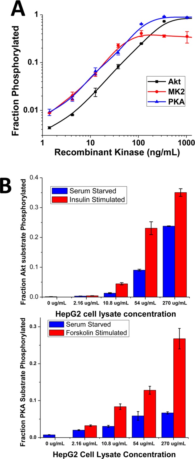
(a) Dose response curve for recombinant Akt, MK2 and PKA activity assay. The limits of detection are 1, 1.2 and 0.7 ng/mL, respectively. (b) Dose response curves showing upregulation of Akt activity by insulin and upregulation of PKA activity by forskolin (25 μM, 30 min). Plotted values indicate the mean ± s.e.m. for duplicate measurements.
To demonstrate the general applicability of this assay, separate experiments were performed with recombinant MK2 and PKA with their respective fluorescent peptide substrates, MK2tide and Kemptide (MK2: 5-FAM-AHLQRQLSIA, PKA: 5-FAM-LRRASLG). Except for new substrates, the recombinant MK2 and PKA assay was done with the same conditions as the recombinant Akt experiment. From the dose response curve in Figure 2a, the LODs for recombinant MK2 and PKA were 1.2 ng/mL (24.5 pM) and 0.7 ng/mL (16 pM), respectively, within 15 min of electrokinetic concentration.
To qualitatively assess the substrate specificity of our kinase of interest, the pairwise cross-reactivity between each substrate and kinase was measured as described in the Supporting Information. From Figure S1 (Supporting Information), the degree of substrate phosphorylation is greatest when a kinase reacts with its target substrate (Kemptide for PKA, Crosstide for Akt, MK2tide for MK2). However, this assay also detected a small amount (<10%) of off-target phosphorylation.
Due to the reduced specificity in using short peptides instead of full-length protein as kinase activity probes,23 it is important to implement strategies to prevent interfering cross-reactive kinases from phosphorylating the target substrate. Off-target kinase reactivity could present an accuracy problem when measuring specific kinase activities in complex samples such as crude cell lysate. Furthermore, the presence of other intracellular enzymes such as proteases and phosphatase could affect the stability and phosphorylation state of the substrates. This is one major limitation of many existing single cell kinase activity assays, where substrates that are microinjected into or expressed within the cell are subjected to the activity of other intracellular enzymes such as off-target kinases, proteases and phosphatases.
Immunoprecipitation–kinase assays offers a highly specific way to measure activity from only the target kinase, but it is challenging to apply to single cell lysate assay due to the limited amount of sample available. For single cell lysate assay, a more easily implemented strategy to improve specificity is to include a customized inhibitor cocktail of off-target kinases, proteases and phosphatases to the reaction buffer in order to minimize the effects of interfering enzymes. This strategy has been shown to effectively suppress the activity of the interfering enzymes without affecting the activity of the kinase of interest,24−26 and is routinely applied in commercial cell lysate kinase activity assays (Omnia (Life Technologies) and Upstate (Millipore) Kinase Assay Kits). In our subsequent experiments using bulk cell lysate and single cells, a cocktail of previously verified protease, phosphatase and off-target kinase inhibitors was added to improve the assay specificity. The protease and phosphatase inhibitor cocktail was used at the recommended concentration for bulk cell lysis, and has been shown to provide adequate protection in capillary electrophoresis based cell lysate kinase activity assay.27 The kinase inhibitor cocktail used in this paper was adapted from previous work24 that showed that this inhibitor combination significantly improves specificity (reduce nonspecific phosphorylation by interfering kinases by 75%) and does not inhibit the kinase that is being assayed for. An immunodepletion experiment from the same report24 also showed that any substrate phosphorylation observed is predominantly due to the specific activity of the target kinase. Potential users interested in implementing this strategy to improve specificity would need to perform the necessary biochemical assays to determine the suitable inhibitor concentrations to be included in their assay to minimize off-target contributions from endogeneous kinases.
Kinase Activity Assay from Bulk Cell Lysate
To show the application of this assay with physiological samples, the kinase activity experiments were repeated with diluted HepG2 cell lysate. The procedures for bulk cell lysate activity assay are described in the Experimental Section. Insulin stimulation is known to increase the activity of Akt in vivo. A comparison between the activity of serum starved HepG2 cell lysate and insulin stimulated HepG2 cell lysate is show in Figure S2 (Supporting Information). No phosphorylated substrate band was observed for the negative control samples (containing lysis buffer only). For the same final cell lysate concentration (down to the lowest tested concentration of 2.16 μg/mL), the insulin stimulated sample consistently showed a higher fraction of phosphorylated substrate compared to the serum starved sample, confirming the utility of our assay to measure changes in cellular kinase activities in response to stimuli (Figure 2b). The cells used in this experiment have about 1 ng of protein per cell (Lysis of ∼400 000 HepG2 cells resulted in recovery of 360 μg of total protein). Detection from a final volume of 30 μL run buffer, after a total of 100-fold dilution of a 2.16 μg/mL of cell lysate, represents kinase assay from ∼0.6 cell. This demonstrates the sensitivity of our assay for single cell level studies of cell signaling pathways.
In another experiment, we measured the effect of forskolin stimulation, which is known to increase the intracellular level of cAMP and upregulate PKA activity. Figure 2b shows that the forskolin stimulated sample consistently showed a higher fraction of phosphorylated PKA substrate compared to the serum starved sample. This shows that our platform can be generally applied to measure activities of different kinases with high sensitivity from physiological samples.
Kinase Activity Assay from Single Cell Lysate
Most mammalian cells are adherent cells, and need some types of matrices for growth. Although cell handling and various operations such as lysis have been demonstrated on suspension cells or detached adherent cells,28−31 they are considerably more challenging for adherent cells. Our assay is designed to be compatible with current methods of cell culture, keeping to biocompatible materials such as polystyrene, glass, silicone and avoiding complicated fluidic handling systems such as growing cells in enclosed microfluidic channels.
We developed a technique to grow single cells in open, nanoliter size microwells, as described in the Experimental Section (Figure 3a). Single cells are ultrasonically lysed in 40 nL chambers (Figure S4, Supporting Information), so that the released intracellular kinases can catalyze phosphorylation of peptide substrates that we preadded to the wells just before lysis. Because the enzymes and substrates are confined in nanoliter sized chambers preventing further dilution, they are at effective concentrations that allow a significant fraction of peptide substrate to be phosphorylated and detected. A previous report estimated that there are ∼106 downstream kinase molecules in a single cell.32 On the basis of this estimate, the effective concentration of kinase molecules after lysis is 41.6 pM, which is above the limit of detection for recombinant kinases determined in this platform. This approach is parallel to many recent works that make use of microfluidic confinement to maintain high effective concentrations of target biomolecules and enable ultrasensitive detection.33−35 The confinement effect prevents further sample and substrate dilution into the entire cell culture plate. Additionally, the microfluidics concentration-enhanced mobility shift assay platform allows assays that lost sensitivity due to process steps that lead to sample dilution (such as addition of reagent or cell lysis in a larger volume) to regain analytical sensitivity by concentrating the analyte. One advantage of our approach, compared to many existing single cell kinase activity assay, is that various inhibitors (for off-target kinase, protease, phosphatase) can be added in the reaction buffer to increase specificity, reduce cross-talk, maintain stability and preserve phosphorylation states in the kinase assay. Adding these inhibitors to cell lysate as opposed to incubating cells with cell-permeable inhibitors have the distinct advantage of avoiding perturbation of other signaling pathways in a live cell via off-target inhibition or cross-pathway effects.36,37 In addition, simply scaling up the number of microwells would enable a high throughput (yet single cell level) kinase assay.
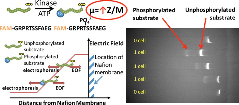
Figure 3b shows the results of single cell lysate kinase activity assay using Akt as a substrate (PKC and PKA inhibitors added to improve substrate specificity without affecting Akt activity24). In the samples corresponding to wells with no cells (negative controls), we see only one band corresponding to the unphosphorylated Akt substrate. On the other hand, in samples corresponding to wells containing one cell, we see two fluorescent bands corresponding to the phosphorylated and unphosphorylated Akt substrate. These results show conclusively that our assay can detect Akt kinase activity from single cell lysate.
One additional capability of our device is that it is possible to look at total kinase activity from multiple cells growing in the same well. Figure 3d shows the results of single, double and triple cells kinase activity assay using MK2 as a substrate. There is a clear trend indicating that total kinase activity increases with the total number of cells in the well. The measure for kinase activity, which is the substrate phosphorylation ratio, is nonzero even in wells with no cells as the image analysis algorithm identifies spurious noise in the image as a phosphorylated peak; this indicates the baseline sensitivity of the method. We observed that there are some outlier points in the activity distribution. Upon reference with the cell image taken before cell lysis, we find that these high kinase activities correlate with larger cell size (Figure S5, Supporting Information). This highlights the ability to correlate single cell phenotype (e.g., morphology, size) with cellular kinase activity in our method.
Tracking Multiple Kinase Activities Simultaneously
In studies related to the signal transduction network, investigators are often interested in understanding the functional relationship between activities of different kinases in the network. By measuring and comparing kinase activities between different signaling pathways, one could deduce how the different stimuli are integrated to produce different cellular responses. Currently, phospho-flow cytometry38 and mass cytometry39 have high degrees of multiplexing capability to measure signaling states, but would not detect small molecule binding and multiple other post-translational modification states that could potentially affect kinase activity. With the exception of the single cell capillary electrophoresis method,11 most current single cell kinase activity assays lack the capability of looking at more than one kinase activity at a time.
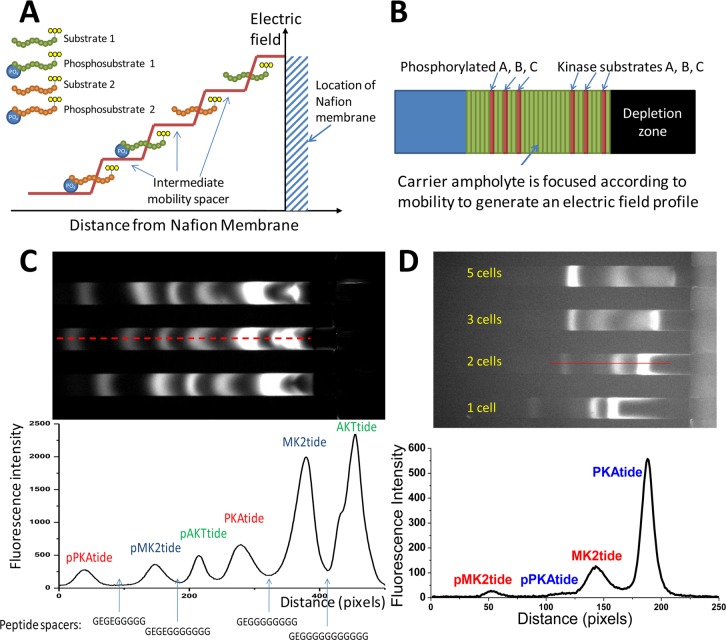
Electrophoretic separation methods such as capillary electrophoresis are powerful because they enable simultaneous measurements of several kinase activities by separating substrates of different mobility. In the previously demonstrated concentration-enhanced mobility shift assay,15 the sharp electric field gradient is essential to obtaining very high concentration factors but the same phenomenon also reduces the separation resolution. To obtain good separation for multiplexed kinase detection while maintaining high concentration factors for sensitivity enhancement, a staircase-like electric field profile, as shown in Figure 4a, is preferred. This profile is desirable because the electric field plateau can increase the separation resolution between target analytes while the individual analytes can still narrowly focus at local regions where the electric field gradient is steep. This can be achieved by concentrating various intermediate mobility species to such high concentrations that it is capable of changing the electric field profile according to the Kohlraush Regulating Function (KRF), as is commonly observed in isotachophoresis (ITP) experiments.40−42
Figure 4.
(a) Schematic showing a strategy to simultaneously separate and focus multiple kinase substrates. Upon adding suitable spacer molecules with intermediate mobilities at various concentrations, depletion zone isotachophoresis40 results in a staircase-like electric field profile that increases the separation resolution between multiple kinase substrates. (b) Compounds in the ampholyte stack in the order of their mobilities to create an extended separation zone where the equilibrium focusing position of individual fluorescent an analytes are well resolved. (c) Simultaneous detection of three kinase activities (Akt, MK2 and PKA) in a single experiment. Within a single run, one can measure the activities of three kinases by separating their substrates (AKTtide, MK2tide and PKAtide) from their phosphorylated products (pAKTtide, pMK2tide and pPKAtide) using synthetic peptide spacers. (d) Concentration-enhanced multiple kinase activity assay (for MK2 and PKA) of single and multiple adherent cells.
To provide a combination of intermediate mobility spacers, an ampholyte mixture can be used. When subjected to electric field, carrier ampholyte molecules focus at their equilibrium position, thus forming continuous bands of ampholyte species arranged in order of high mobility to low mobility as it approaches the ion-selective membrane (Figure 4b). The focusing of nonfluorescent ampholyte species in between fluorescent peptides with different mobility increases their separation resolution. However, due to the unknown identities of these proprietary carrier ampholyte molecules, it is difficult to arbitrarily increase the separation resolution between two closely spaced fluorescent peptides or decrease the distance between two distantly spaced fluorescent peptides. As an alternative to ampholytes, custom synthetic peptides can be obtained at low cost and high purity, and their mobility can be tailored by adding different amino acids (with various charge and molecular weight) during peptide synthesis. The mobilities of the peptide substrates and spacers can be approximated with the Offord Model43,44 μ = Q/M2/3 where μ = electrophoretic mobility, Q = total charge and M = total mass of the peptide.
Table S1 and Figure S1 (Supporting Information) show the predicted mobility values of 12 synthetic spacer peptides placed alongside the predicted mobility values of the kinase peptide substrates. Peptides with mobility in-between kinase substrates or phosphorylated products that we wish to resolve can be chosen as spacers. Figure 4c shows that, by rationally choosing the appropriate peptide spacers, we can simultaneously concentrate and baseline-resolve substrates and products corresponding to three kinases PKA (substrate: 5-FAM-EELGRTGRRNSI), Akt (substrate: 5-FAM-GRPRTSSFAEG-NH2) and MK2 (substrate: FITC-EEKKLNRTLSVA). This general capability would allow us to simultaneously monitor several (up to three in this study) kinase activities from the same sample and study their functional relationship to yield insights on the inner-workings of cell regulatory pathways. We also demonstrated that such multiplexed measurement (two targets, PKA and MK2) can be implemented with single cell sensitivity (Figure 4d). One point to take note is that simultaneous preconcentration of an increasing number of spacer peptides would reduce electric field gradients and therefore reduce electrokinetic preconcentration factors in multiplexed assays. The current experimental setup is sensitive enough to measure activities from two kinases from single cell. At higher applied voltages needed for high degrees of multiplexing, preconcentration bands in the current device are less stable over extended amounts of assay time needed to achieve single cell detection sensitivity. As such, more device and material optimization is needed to perform simultaneous activity assay for three or more different kinases from a single cell.
Conclusions
The presented method fills several gaps in the existing single cell kinase assay. Conventional immunohistochemical staining of single cells with phosphospecific antibodies38 has been widely used to monitor the localization of kinases in fixed cells, but the phosphorylation state of kinases does not always correlate to their biological activity, especially in the presence of other post-translational modifications, protein–protein association and drug interactions. We focus instead on directly measuring kinase activity on a target substrate, which captures the actual catalytic activity of specific kinase from single cell at the point of lysis. This is, in effect, a single cell resolution version of traditional cell lysate based kinase activity assay, where the cells are lysed prior to kinase activity assay. The cell is first rapidly lysed in this method, preserving a snapshot of the cell signaling state. As the intracellular contents of a single cell (pL volume) is then diluted into a nanoliter sized chamber, the ultrasensitive assay provided by the concentration-enhanced mobility shift assay platform is an enabling technology that allows detection of this low-abundance kinase activity. This method allows various inhibitors (for off-target kinase, protease, phosphatase) to be added to the lysate to improve specificity, reduce cross-talk, maintain stability and preserve phosphorylation states in the kinase assay, thus avoiding the complications of activating unintended cellular responses associated with incubating live cells with these inhibitors. The throughput of this platform is also scalable as all the key steps can process multiple samples in parallel. Kinase reactions are carried out in multiple chambers simultaneously; the reaction product can be frozen and analyzed at a later time. The multiplexing capability of the microfluidics chip can be further extended to process up to 128 samples in parallel, as demonstrated in a previous report.45
For biological studies of signal transduction pathway, it is important to assay for kinase activity with minimal disruption to their natural environment. Detaching and putting adherent cells into suspension, as required for flow cytometry methods, could activate many signaling pathways and seriously perturb the biochemical process to be studied. Other single cell kinase activity assays such as those involving capillary electrophoresis of injected substrates or genetically modified cells that express a reporter molecule also involve considerable perturbation to the normal physiology of the cells. The method reported here can assay for kinase activities on adherent cells, and the cells are maintained in their natural state until lysis. The rapid ultrasonic lysis method demonstrated in this paper is a simple way to lyse cells, but might not be suitable for stress responsive kinase measurement as the high forces during sonication could affect cellular response. In those cases, a more appropriate lysis method that is also compatible with this platform is pulsed laser microbeam lysis46 that would lyse cells in milliseconds and minimize perturbation to the cell signaling state.
Finally, the method developed here enables simultaneous measurement of multiple kinase activities within single cells. This general capability could allow us to study the functional relationship between different signaling pathways to yield insights on the inner-workings of cell regulatory pathways. In most assays, very good specificity can be obtained using optimized substrates and including inhibitors that prevent cross-reactivity. For very promiscuous kinases, one could potentially apply other strategies to control for any cross-reactivity in multiplexed kinase activity measurements such as deconvolution of signals using prior data on individual recombinant kinase signatures for the substrate panel.47We believe that this new platform could be a generic and powerful tool for diagnostics, drug development and systems biology research.
Acknowledgments
We gratefully acknowledge support from NIH grant P50-GM068762 (Cell Decision Processes Center). L.F.C. was supported by the A*STAR National Science Scholarship.
Supporting Information Available
Additional information as noted in text. This material is available free of charge via the Internet at http://pubs.acs.org.
The authors declare no competing financial interest.
Funding Statement
National Institutes of Health, United States
Supplementary Material
References
- Spencer S. L.; Gaudet S.; Albeck J. G.; Burke J. M.; Sorger P. K. Nature 2009, 4597245428–432. [DOI] [PMC free article] [PubMed] [Google Scholar]
- Berenbaum M. C. Cancer Chemother. Rep., Part 1 1972, 565563. [PubMed] [Google Scholar]
- Nelson D. E.; Ihekwaba A. E. C.; Elliott M.; Johnson J. R.; Gibney C. A.; Foreman B. E.; Nelson G.; See V.; Horton C. A.; Spiller D. G. Science 2004, 3065696704–708. [DOI] [PubMed] [Google Scholar]
- Nair V. D.; Yuen T.; Olanow C. W.; Sealfon S. C. J. Biol. Chem. 2004, 2792627494–27501. [DOI] [PubMed] [Google Scholar]
- Lahav G.; Rosenfeld N.; Sigal A.; Geva-Zatorsky N.; Levine A. J.; Elowitz M. B.; Alon U. Nat. Genet. 2004, 362147–150. [DOI] [PubMed] [Google Scholar]
- Johnson S. A.; Hunter T. Nat. Methods 2005, 2117–25. [DOI] [PubMed] [Google Scholar]
- Shi Q.; Qin L.; Wei W.; Geng F.; Fan R.; Shin Y. S.; Guo D.; Hood L.; Mischel P. S.; Heath J. R. Proc. Natl. Acad. Sci. U. S. A. 2012, 1092419–424. [DOI] [PMC free article] [PubMed] [Google Scholar]
- Eyer K.; Kuhn P.; Hanke C.; Dittrich P. S. Lab Chip 2012, 124765–772. [DOI] [PubMed] [Google Scholar]
- Sato M.; Ozawa T.; Inukai K.; Asano T.; Umezawa Y. Nat. Biotechnol. 2002, 203287–294. [DOI] [PubMed] [Google Scholar]
- Mehta S.; Zhang J. Annu. Rev. Biochem. 2011, 80, 375–401. [DOI] [PMC free article] [PubMed] [Google Scholar]
- Meredith G. D.; Sims C. E.; Soughayer J. S.; Allbritton N. L. Nat. Biotechnol. 2000, 183309–312. [DOI] [PubMed] [Google Scholar]
- Li H.; Sims C. E.; Kaluzova M.; Stanbridge E. J.; Allbritton N. L. Biochemistry 2004, 4361599–1608. [DOI] [PubMed] [Google Scholar]
- Li H.; Wu H. Y.; Wang Y.; Sims C. E.; Allbritton N. L. J. Chromatogr. B: Biomed. Sci. Appl. 2001, 757179–88. [DOI] [PubMed] [Google Scholar]
- Sarkar A.; Kolitz S.; Lauffenburger D. A.; Han J. Nat. Commun. 2014, 5, 3421. [DOI] [PMC free article] [PubMed] [Google Scholar]
- Cheow L. F.; Han J. Anal. Chem. 2011, 83187086–7093. [DOI] [PMC free article] [PubMed] [Google Scholar]
- Wang Y. C.; Stevens A. L.; Han J. Anal. Chem. 2005, 77144293–4299. [DOI] [PubMed] [Google Scholar]
- Wang Y. C.; Han J. Lab Chip 2008, 83392–394. [DOI] [PMC free article] [PubMed] [Google Scholar]
- Lee J. H.; Song Y. A.; Tannenbaum S. R.; Han J. Anal. Chem. 2008, 8093198–3204. [DOI] [PMC free article] [PubMed] [Google Scholar]
- Lee J. H.; Cosgrove B. D.; Lauffenburger D. A.; Han J. J. Am. Chem. Soc. 2009, 1313010340–10341. [DOI] [PMC free article] [PubMed] [Google Scholar]
- Cheow L. F.; Ko S. H.; Kim S. J.; Kang K. H.; Han J. Anal. Chem. 2010, 8283383–3388. [DOI] [PMC free article] [PubMed] [Google Scholar]
- Jung B.; Bharadwaj R.; Santiago J. G. Anal. Chem. 2006, 7872319–2327. [DOI] [PubMed] [Google Scholar]
- Ross D.; Locascio L. E. Anal. Chem. 2002, 74112556–2564. [DOI] [PubMed] [Google Scholar]
- Remenyi A.; Good M. C.; Lim W. A. Curr. Opin. Struct. Biol. 2006, 166676–685. [DOI] [PubMed] [Google Scholar]
- Shults M. D.; Janes K. A.; Lauffenburger D. A.; Imperiali B. Nat. Methods 2005, 24277–284. [DOI] [PubMed] [Google Scholar]
- Stains C. I.; Luković E.; Imperiali B. ACS Chem. Biol. 2010, 61101–105. [DOI] [PMC free article] [PubMed] [Google Scholar]
- Stains C. I.; Tedford N. C.; Walkup T. C.; Luković E.; Goguen B. N.; Griffith L. G.; Lauffenburger D. A.; Imperiali B. Chem. Biol. 2012, 192210–217. [DOI] [PMC free article] [PubMed] [Google Scholar]
- Suresh Babu C. V.; Cho S. G.; Sook Yoo Y. Electrophoresis 2005, 26193765–3772. [DOI] [PubMed] [Google Scholar]
- Schilling E. A.; Kamholz A. E.; Yager P. Anal. Chem. 2002, 7481798–1804. [DOI] [PubMed] [Google Scholar]
- Waters L. C.; Jacobson S. C.; Kroutchinina N.; Khandurina J.; Foote R. S.; Ramsey J. M. Anal. Chem. 1998, 701158–162. [DOI] [PubMed] [Google Scholar]
- Gao J.; Yin X. F.; Fang Z. L. Lab Chip 2004, 4147–52. [DOI] [PubMed] [Google Scholar]
- Lu H.; Schmidt M. A.; Jensen K. F. Lab Chip 2005, 5123–29. [DOI] [PubMed] [Google Scholar]
- Ferrell J. E. Jr. Trends Biochem. Sci. 1996, 2112460–466. [DOI] [PubMed] [Google Scholar]
- Ottesen E. A.; Hong J. W.; Quake S. R.; Leadbetter J. R. Science 2006, 31458041464–1467. [DOI] [PubMed] [Google Scholar]
- Rissin D. M.; Walt D. R. Nano Lett. 2006, 63520–523. [DOI] [PubMed] [Google Scholar]
- Li L.; Du W.; Ismagilov R. J. Am. Chem. Soc. 2009, 1321106–111. [DOI] [PMC free article] [PubMed] [Google Scholar]
- Kumar N.; Afeyan R.; Kim H. D.; Lauffenburger D. A. Mol. Pharmacol. 2008, 7361668–1678. [DOI] [PMC free article] [PubMed] [Google Scholar]
- Wynn M. L.; Ventura A. C.; Sepulchre J. A.; García H. J.; Merajver S. D. BMC Syst. Biol. 2011, 51156. [DOI] [PMC free article] [PubMed] [Google Scholar]
- Perez O. D.; Nolan G. P. Nat. Biotechnol. 2002, 202155–162. [DOI] [PubMed] [Google Scholar]
- Bodenmiller B.; Zunder E. R.; Finck R.; Chen T. J.; Savig E. S.; Bruggner R. V.; Simonds E. F.; Bendall S. C.; Sachs K.; Krutzik P. O. Nat. Biotechnol. 2012, 309858–867. [DOI] [PMC free article] [PubMed] [Google Scholar]
- Quist J.; Janssen K. G.; Vulto P.; Hankemeier T.; van der Linden H. J. Anal. Chem. 2011, 83207910–7915. [DOI] [PubMed] [Google Scholar]
- Khurana T. K.; Santiago J. G. Anal. Chem. 2008, 80166300–6307. [DOI] [PubMed] [Google Scholar]
- Bercovici M.; Kaigala G. V.; Santiago J. G. Anal. Chem. 2010, 8252134–2138. [DOI] [PubMed] [Google Scholar]
- Jalali-Heravi M.; Shen Y.; Hassanisadi M.; Khaledi M. G. Electrophoresis 2005, 26101874–1885. [DOI] [PubMed] [Google Scholar]
- Janini G. M.; Metral C. J.; Issaq H. J.; Muschik G. M. J. Chromatogr. A 1999, 8481417–433. [DOI] [PubMed] [Google Scholar]
- Ko S. H.; Kim S. J.; Cheow L. F.; Li L. D.; Kang K. H.; Han J. Lab Chip 2011, 1171351–1358. [DOI] [PubMed] [Google Scholar]
- Rau K. R.; Quinto-Su P. A.; Hellman A. N.; Venugopalan V. Biophys. J. 2006, 911317–329. [DOI] [PMC free article] [PubMed] [Google Scholar]
- Miller M. A.; Barkal L.; Jeng K.; Herrlich A.; Moss M.; Griffith L. G.; Lauffenburger D. A. Integr. Biol. 2011, 34422–438. [DOI] [PMC free article] [PubMed] [Google Scholar]
Associated Data
This section collects any data citations, data availability statements, or supplementary materials included in this article.


