Abstract
Purpose
Previous studies have demonstrated that autophagy is involved in the pathogenesis of human cytomegalovirus (HCMV) infection. However, whether autophagy is regulated by murine cytomegalovirus (MCMV) infection has not yet been investigated. The purpose of these studies was to determine how autophagy is affected by MCMV infection of the retinal pigment epithelial (RPE) cells and whether there is a functional relationship between autophagy and apoptosis; and if so, how regulation of autophagy impacts apoptosis.
Methods
RPE cells were isolated from C57BL/6 mice and infected with MCMV K181. The cells were cultured in medium containing rapamycin, chloroquine, or ammonium chloride. Green fluorescent protein–light chain 3 (GFP-LC3) plasmid was transfected to RPE cells, and the GFP-LC3 positive puncta were counted. Electron microscopic (EM) images were taken to visualize the structure of the autophagic vacuoles. Western blot was performed to detect the expression of related proteins. Trypan blue exclusion assay was used to measure the percentage of viable cells.
Results
Although the LC3B-II levels consistently increased during MCMV infection of RPE cells, administration of chloroquine or ammonium chloride increased LC3B-II expression only at the early stage of infection (6 h post-inoculation [p.i.] and 12 h p.i.), not at or after 24 h p.i. The punctate autophagic vacuoles in the GFP-LC3 transfected RPE cells were counted using light microscopy or by EM examination, the number of autophagic vacuoles was significantly increased in the MCMV-infected RPE cells compared to the uninfected controls. Compared to untreated MCMV-infected control cells, rapamycin treatment resulted in a significant decrease in the cleaved caspase 3 levels as well as a significant decrease in the ratio of phosphorylated mammalian target of rapamycin (mTOR) to total mTOR and in the ratio of phosphorylated P70S6K to total P70S6K. In contrast, chloroquine treatment resulted in a significant increase in the cleaved caspase 3 levels in the MCMV-infected RPE cells.
Conclusions
Autophagic vacuole accumulation was detected during MCMV infection of RPE cells. In contrast, autophagic flux was greatly decreased at or after 24 h p.i. The results suggest that MCMV might have a strategy for inhibiting or blocking autophagy activity by targeting a later autophagy process, such as the formation of autolysosomes or degradation of their content. Our data also suggest that there is a functional relationship between autophagy and apoptosis, which plays an important role during MCMV infection of the RPE.
Introduction
Cytomegalovirus (CMV) is a beta-herpesvirus, which is widespread in human populations and is a major cause of morbidity and mortality in individuals who are immunocompromised as a result of chemotherapy, malignancy, or acquired immunodeficiency syndrome (AIDS) [1]. CMV retinitis is the most common sight-threatening opportunistic infection observed in adult and pediatric patients who are immunosuppressed [2-5]. Although retinal necrosis is a prominent feature of CMV retinitis, apoptotic cells have been observed during microscopic examination of biopsy specimens of the eyes of patients with human cytomegalovirus (HCMV) retinitis [6,7] and in the eyes of mice with murine cytomegalovirus (MCMV) retinitis [8,9].
Autophagy, a process responsible for the transfer of intracellular components such as defective proteins, organelles [10], and viral proteins [11] into lytic vacuolar compartments for degradation, is essential to maintain the amino acid pool, to prevent neurodegradation, to suppress tumors, and to regulate innate and adaptive immunity [12-18]. Previous studies performed by Chen et al. [19] and Reme et al. [20] have shown that autophagy is a basal cellular process occurring ubiquitously in RPE cells and is critical to the health of retinal pigment epithelial (RPE) cells under normal conditions in different species. RPE cells play a central role in HCMV retinitis [21,22], and these cells are also the first target of MCMV infection in vivo as described previously in our laboratory [2]. Therefore, RPE cells are a common model for investigating pathologic conditions during HCMV retinitis [23-25].
There is a link between autophagy and virus infection. Autophagy is stimulated [26,27] or blocked [1] by virus infection, and in turn, autophagy directly or indirectly affects virus infection by regulating cellular functions through the immune response, deposition of cellular compounds, and/or cell death [28]. Recent studies have also emphasized the functional relationship between autophagy and apoptosis in a context-dependent fashion [29]. Autophagy can either be an adaption to avoid apoptosis or can lead to autophagic cell death [30]. Investigations of autophagy and apoptosis in virus-infected cells have contributed to our understanding of the role of autophagy and apoptosis in the pathogenesis of virus infection.
Chaumorcel et al. [1] have demonstrated that HCMV infection inhibits autophagy in infected fibroblasts at 24 h post-inoculation (p.i.), although accumulation of light chain 3B-II (LC3B-II) in MRC5 cells was shown with western blot at 24 h and 48 h p.i. However, the constant accumulation of LC3B-II is independent of the level of autophagy because fluorescence microscopy of cells transfected with green fluorescent protein (GFP)–LC3 and then infected with HCMV showed a clear decrease in the number of GFP-LC3-positive puncta at 24 h p.i. [31]. McFarlane et al. [27] have also demonstrated that HCMV infection induces autophagy in MRC5 cells as early as 6 h p.i. and up to 24 h p.i. However, after 24 h, HCMV infection in fibroblasts inhibits autophagy by a mechanism dependent on the interaction between the HCMV TRS1 protein and the Beclin 1 protein [31]. The HCMV TRS1 protein is the functional homolog of HSV-1 ICP34.5 [31]. However, it has not yet been determined whether autophagy is regulated by MCMV infection, in RPE cells in particular, and if so, whether there is a functional interaction between autophagy and apoptosis. Therefore, the purpose of the studies presented herein was to investigate the autophagic and apoptotic responses of MCMV-infected RPE cells as well as the functional relationship between apoptosis and autophagy during MCMV infection of RPE cells.
Methods
Virus propagation and virus titration
The original stock of MCMV (K181 strain) was a generous gift from Dr. Edward S. Mocarski (Emory University). The study was approved by the Institutional Animal Care and Use Committee of Georgia Regents University. The virus was prepared from the salivary glands of MCMV-infected BALB/c mice as described previously [32].
Stock virus was also prepared with low multiplicity of infection (MOI) passage in mouse embryonic fibroblasts (MEF) cells grown in Dulbecco’s modified Eagle's medium (DMEM; Mediatech, Manassas, VA) containing 5% fetal bovine serum (FBS; Thermoscientific, Waltham, MA) and antibiotics. The titer of the virus stock was determined with a plaque assay on the MEF cells. Aliquots of stock virus were stored at −70 °C, and a fresh aliquot was thawed and diluted for each experiment.
Preparation of RPE cells
After the neural retina was removed from C57BL/6 mice, intact sheets of RPE cells were peeled off the underlying basement (Bruch’s) membrane and transferred into a sterile 60-mm culture dish containing 5 ml of fresh RPE culture medium and then briefly triturated using a fine point Pasteur pipette. The RPE culture medium was composed of 20% FBS, 1% P/S, 1.25% L-glutamine (Life Technologies, Grand Island, NY), 1% antibiotic-antimycotic solution (Fisher Scientific, Pittsburgh, PA), and 1% HEPES buffer solution (Life Technologies, Grand Island, NY) in DMEM/F-12 50/50 (Fisher Scientific, Pittsburgh, PA). RPE cells were collected by centrifugation at 200 ×g for 5 min, resuspended in RPE culture medium, and cultured in flasks at 37 °C with 5% CO2. Cells were cultured for 7 to 10 days until the cells were confluent; the cultures showed no contamination with fibroblasts or choroidal cells based on microscopic analysis of the cell morphology. The RPE cells of passage 2 were positive when stained with an antibody specific for the RPE specific antigen, RPE 65.
Green fluorescent protein–light chain 3 transfection
The GFP-LC3 fusion plasmid was kindly provided by Dr. Zheng Dong (Georgia Regents University). RPE cells (2 × 10⁵) were plated on a coverslip and cultured to 60% confluence. Transient transfection was performed with the X-tremeGENE HP DNA transfection reagent (Roche, Basel, Switzerland) according to the manufacturer's recommendation. After 4 h, the medium was replaced with DMEM/F-12 50/50 containing 10% FBS medium, and the cells were incubated for 24 h to 48 h. Then the cells were infected with MCMV at MOI = 1; the cells were fixed in 4% paraformaldehyde for 20 min at room temperature at different times p.i., and washed three times with DPBS (Mediatech Inc, Manassas, VA). Coverslips were mounted with 4',6-diamidino-2-phenylindole (DAPI) before being analyzed with an Axioplan 2 microscope (Zeiss, Göttingen, Germany). Images were analyzed with Axiovision Rel. 4.7 software.
Western blot analysis
Proteins from uninfected, untreated cells, from MCMV-infected cells, and from rapamycin- (Selleckchem, Houston, TX) or chloroquine- (Sigma-Aldrich, St. Louis, MO) treated cells were extracted on ice with lysis buffer (Roche Diagnostics, Indianapolis, IN) supplemented with phosphatase inhibitor complex (EMD Millipore, Billerica, MA). Lysates were clarified at 13,000 × g for 10 min at 4 °C and size-fractionated with 10% or 6% sodium dodecyl sulfate–polyacrylamide gel electrophoresis (SDS–PAGE), followed by electroblotting onto a polyvinylidene difluoride (PVDF) membrane (GE Healthcare, Pittsburgh, PA). After blocking with 5% nonfat dry milk for 1 h at room temperature, the membrane was incubated overnight at 4 °C with primary antibody (rabbit anti-LC3B, cat. no. 3868; rabbit anti-cleaved caspase-3, cat. no. 9664; rabbit anti-mammalian target of rapamycin (mTOR), cat. no. 2972; rabbit anti-phospho-mTOR, cat. no. 2971; rabbit anti-p70S6K, cat. no. 2708; and rabbit anti-phospho-p70S6K, cat. no. 9234; Cell Signaling, Danvers, MA). The next day, the horseradish peroxidase (HRP)-conjugated secondary antibody was bound for 1 h at room temperature. The immune complex was visualized using a chemiluminescence detection system (Thermo Scientific, Waltham, MA) and exposure to X-ray film. The membrane was stained for β-actin to verify equal loading among the lanes. Each experiment was repeated at least once, and most were repeated three times. The density of each band was analyzed using ImageJ software.
Electron microscopy
Mock-infected or MCMV-infected RPE cells were treated with typsin for 5 min at 37 °C and centrifuged at 200 ×g for 5 min. After the supernatant was removed, the cell pellets were fixed in 2% glutaraldehyde in 0.1 M sodium cacodylate (NaCac) buffer, pH 7.4, postfixed in 2% osmium tetroxide in NaCac, stained en bloc with 2% uranyl acetate, dehydrated in a graded ethanol series, and embedded in Epon-Araldite resin. Thin sections were made using a diamond knife on a Leica EM UC6 ultramicrotome (Leica Microsystems, Wetzlar, Germany), collected on copper grids, and stained with uranyl acetate and lead citrate. Cells were observed under a JEM 1230 transmission electron microscope (JEOL USA, Peabody, MA) at 110 kV and imaged with an UltraScan 4000 CCD camera and First Light Digital Camera Controller (Gatan, Pleasanton, CA). Autophagic vacuoles were counted in individual cells from multiple fields and nonserial sections. Autophagic vacuoles were quantified by counting the number of autophagic vacuoles per cell.
Statistical analysis
Protein expression was measured with densitometry using ImageJ software and normalized to β-actin. Data are expressed as mean ± standard error of mean (SEM) reflecting the results of three independent experiments and were compared with the two-tailed Student t test or ANOVA using the GraphPad Prism 5 Analysis tool. A p value of p<0.05 was considered significant [33].
Results
Autophagic flux during murine cytomegalovirus infection of RPE cells
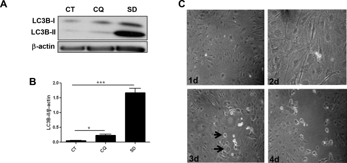
A commonly used biochemical marker of autophagy is lipidation of LC3B-I in the membrane of the autophagosome that leads to the formation of LC3B-II that migrates more rapidly in SDS-polyacrylamide gels than LC3B-I [34]. Serum deprivation (SD) and chloroquine (CQ) were used to modulate autophagy. Serum deprivation increases autophagy, which protects cells against apoptosis [35]. In contrast, chloroquine, a negative regulator of autophagy that localizes to acidic vesicles, impairs lysosomal acidification and suppresses protease activity, resulting in accumulation of autophagosomes, and consequently increases the expression of LC3B-II [36]. The results showed that the intensity of the characteristic faster-migrating form of LC3B (LC3B-II) was increased in uninfected RPE cells under serum-free conditions or following chloroquine treatment compared to normal untreated cells, suggesting that RPE cells are sensitive to negative and positive regulation of autophagy (Figure 1A,B).
Figure 1.
Autophagic response to different stimuli. A: Retinal pigment epithelial (RPE) cells were cultured in normal medium (CT) and treated with chloroquine (CQ, 10−6 M) or serum deprivation (SD) for 24 h. Expression of processed light-chain 3B (LC3B) was monitored. B: Semi-quantitative analysis of western blot for LC3B protein expression in the RPE cells from the control, CQ-treated, and SD-treated groups. C: Representative images (×200) of murine cytomegalovirus (MCMV)-infected RPE cells. RPE cells were infected with MCMV at multiplicity of infection (MOI) = 1 for 1, 2, 3, and 4 days. Black arrow: infected cell. 1d: 1 day postinfection; 2d: 2 days postinfection; 3d: 3 days postinfection; 4d: 4 days postinfection. *p<0.05, ***p<0.001, ANOVA. Data are shown as mean±SEM (n=3).
RPE cells infected with MCMV at MOI = 1 were characterized by cytoplasmic stranding, cell rounding in a focal pattern, cell enlargement, cell fusion, and refraction, and some exhibited an irregular outline (Figure 1C).
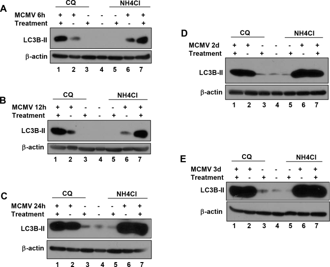
Autophagic flux was assessed in the MCMV-infected RPE cells. RPE cells were mock-infected or infected with MCMV followed by treatment with chloroquine or ammonium chloride. Ammonium chloride, a commonly used negative regulator of autophagy, localizes to acidic vesicles and impairs lysosomal acidification and protease activity, resulting in impaired fusion of autophagosomes and lysosomes, and accumulation of autophagosomes [27,37]. The LC3B-II level constantly increased during MCMV infection (lane 2 and lane 6 in Figure 2A–E). At early stages of infection, such as 6 h or 12 h p.i., adding chloroquine or ammonium chloride resulted in a large increase in LC3B-II in MCMV-infected RPE cells (compare lane 1 to lane 2 or lane 7 to lane 6 in Figure 2A,B), suggesting that the increase in LC3B-II at this stage of infection reflects increased autophagic flux rather than impaired function of autolysosome formation or degradation. However, at or after 24 h p.i., chloroquine or ammonium chloride treatment did not have an additive increase in the LC3B-II level (compare lane 1 to lane 2 or lane 7 to lane 6 in Figure 2C–E), indicating that increased LC3B-II at this stage of infection does not represent increased autophagic flux, but instead might represent inhibition or blocking of autophagy activity at or after autolysosomes are formed.
Figure 2.
Autophagic flux during murine cytomegalovirus infection of retinal pigment epithelial (RPE) cells. RPE cells were infected with murine cytomegalovirus (MCMV) at low multiplicity of infection (MOI) = 1 in normal medium or in medium containing chloroquine (CQ, 10−6 M) or ammonium chloride (NH4Cl, 10−6 M) to block autophagic flux for 6 h (A), 12 h (B), 24 h (C), 2 days (D), and 3 days (E). Expression of processed light-chain 3B (LC3B) was monitored.
The accumulation of autophagic vacuoles during the late stage of MCMV infection of RPE cells
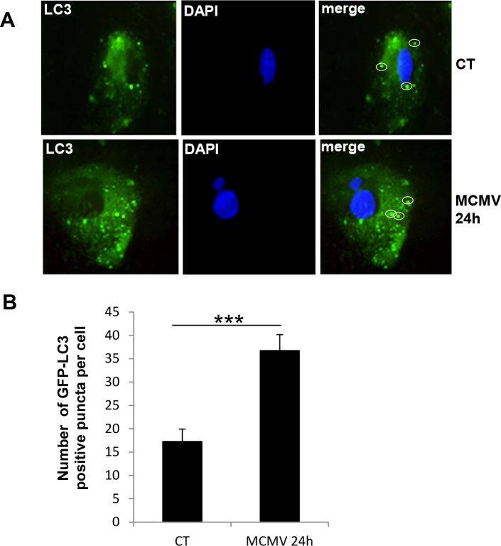
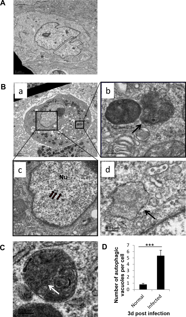
Since the autophagic flux results suggest that MCMV might inhibit autophagy activity during a later autophagy process such as formation of autolysosomes or degradation of their contents on or after 24 h p.i., we hypothesized that the number of autophagic vacuoles increased during the late stage of MCMV infection. To confirm this hypothesis, the RPE cells were transfected with GFP-LC3 and then infected with MCMV. Then the GFP-LC3 positive puncta were counted under a fluorescent microscope. The results showed that the number of GFP-LC3 positive puncta increased significantly in MCMV-infected RPE cells compared to uninfected control cells at 24 h p.i. (Figure 3). Additional evidence of the increased accumulation of autophagic vacuoles during MCMV infection was obtained by comparing the electron microscopic appearance of uninfected RPE cells (Figure 4A) with RPE cells infected with MCMV for 3 days (Figure 4B). At 3 days p.i., autophagic vacuoles, characterized by the presence of double-membrane vesicles (immature and degradative) containing cytoplasmic components or degrading mitochondria [11,38], were observed in the RPE cells (Figure 4B, panel b). The vacuoles we observed were different from phagosomes, which were seen as electron-dense structures in the cytoplasm surrounded by a single membrane in an in vivo model [39]. In this model, the author observed that RPE cells have the ability to phagocytize retinal photoreceptor outer segments (ROS). However, in our in vitro RPE culture, most of the vacuoles we observed had double-membrane autophagic characteristics. We occasionally observed phagosomes in the cytoplasm of the RPE cells (Figure 4B, panel d).
Figure 3.
Representative images of retinal pigment epithelial (RPE) cells with green fluorescent protein-light chain 3 (GFP-LC3) puncta. RPE cells cultured in normal medium were transiently transfected with green fluorescent protein–light chain 3 (GFP-LC3) plasmid (CT) or infected with murine cytomegalovirus (MCMV) at low multiplicity of infection (MOI) = 1 for 24 h. A: Representative images (×630) of RPE cells with GFP-LC3 puncta. White circle: GFP-LC3 positive puncta. B: Quantification of autophagy in RPE cells transiently transfected with GFP-LC3 plasmid in normal medium or infected with MCMV at MOI = 1 for 24 h. ***p<0.001, two-tailed Student t test. Data are shown as mean±SEM (n=3).
Figure 4.
Representative electron microscopic images of autophagic vacuoles in murine cytomegalovirus-infected and -uninfected retinal pigment epithelial (RPE) cells. RPE cells were cultured in normal medium or infected with murine cytomegalovirus (MCMV) at low multiplicity of infection (MOI) = 1 for 3 days and then fixed and processed for electron microscopy. A: Representative view of normal RPE cells. B: Higher magnification view of autophagic vacuoles in MCMV-infected cells. Panel b is the enlarged view of the rectangle in panel a. Panel c is the enlarged view of the rectangle in panel a. Black arrow in panel b: autophagic vacuole; black arrows in panel c: viral particles; Nu: nucleus; black arrow in panel d: phagosome. C: Higher magnification view of autophagic vacuoles containing viral particle. White arrow: viral particle. D: Quantification of autophagic vacuoles for a minimum of 20 MCMV-infected or uninfected cells. The number of autophagic vacuoles per cell was determined in electron micrographs. ***p<0.001, two-tailed Student t test. Data are shown as mean±SEM (n=3).
Because we observed viral particles inside autophagic vacuoles (Figure 4C), autophagy herein is referred to as xenophagy, which involves recognition of an intracellular pathogen and targeting of the pathogen to autophagic machinery for degradation [40]. When the autophagic vacuoles were counted, RPE cells infected with MCMV had more autophagic vacuoles than were observed in normal uninfected RPE cells (Figure 4D). These results suggest that during the late stage of infection, MCMV induces the accumulation of autophagic vacuoles in RPE cells. These results also support the idea that the decrease in autophagy during the late stage of MCMV infection is not because of the blockade in the initiation of autophagosome but because of the blockade in the late step of the autophagy process.
Rapamycin induces autophagy and decreases apoptosis during MCMV infection
Virus infection induces cell death through the caspase 3-dependent pathway [41,42]. Since recent reports suggest that autophagy may be an adaption to avoid cell death [29,43], we hypothesized that increased autophagy in infected cells might contribute to inhibition of apoptosis. To test whether there is a functional relationship between autophagy and apoptosis during MCMV infection, the RPE cells were infected with MCMV and were cultured in medium containing rapamycin, which positively regulates autophagy through inhibition of mTOR. mTOR is the mammalian target of rapamycin and an important protein kinase that regulates cellular functions, such as cell growth, cell proliferation, protein synthesis, and transcription [44]. One way to regulate autophagy is by mTOR signaling through phosphorylation of its downstream target, P70S6K [45-48]. mTOR inhibits the initiation of the phagophore [49,50], which blocks the early step of autophagy leading to decreased formation of autophagosome and expression of LC3B-II.
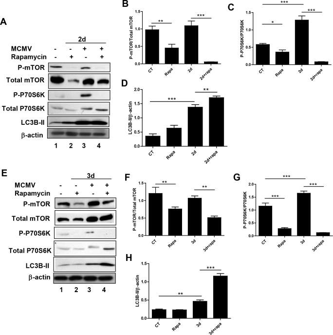
First, we wanted to know whether autophagy can be regulated by rapamycin during MCMV infection of RPE cells. Decreased ratios of phosphorylated mTOR and total mTOR, phosphorylated P70S6K, and total P70S6K in rapamycin-treated, infected RPE cells (Figure 5A,B, lane 4) were observed compared to infected, untreated RPE cells (Figure 5A,B, lane 3), suggesting that rapamycin treatment inhibits mTOR activity during MCMV infection of RPE cells. Moreover, rapamycin treatment increased the LC3B-II levels in the virus-infected cells (Figure 5A,B, lane 4) compared to the infected, untreated cells (Figure 5A,B, lane 3). Taken together, these results suggest that regulation of autophagy by rapamycin during MCMV infection occurs through the inhibition of mTOR.
Figure 5.
Murine cytomegalovirus infection, autophagy, and mammalian target of rapamycin pathway. Retinal pigment epithelial (RPE) cells were infected with murine cytomegalovirus (MCMV) at low multiplicity of infection (MOI) = 1 in normal medium or in medium containing rapamycin (10−6 M). Samples were collected at day 2 (A) and 3 p.i. (E). Expression of processed light-chain 3B (LC3B), P-mammalian target of rapamycin (mTOR), mTOR, P-P70S6K, and P70S6K was monitored and quantified at day 2 (B–D) and at day 3 (F–H). Rapa: rapamycin; 2d: 2 days postinfection; 3d: 3 days postinfection. *p<0.05, **p<0.01, ***p<0.001, ANOVA. Data are shown as mean±SEM (n=3).
We also observed that MCMV infection increased the ratios of phosphorylated P70S6K and total P70S6K (Figure 5A,B, lane 3) compared to the normal control (Figure 5A,B, lane 1), although the ratios of phosphorylated mTOR and total mTOR remained unchanged (Figure 5A,B, lanes 1 and 3). These results suggest that MCMV infection activates mTOR signaling in RPE cells.
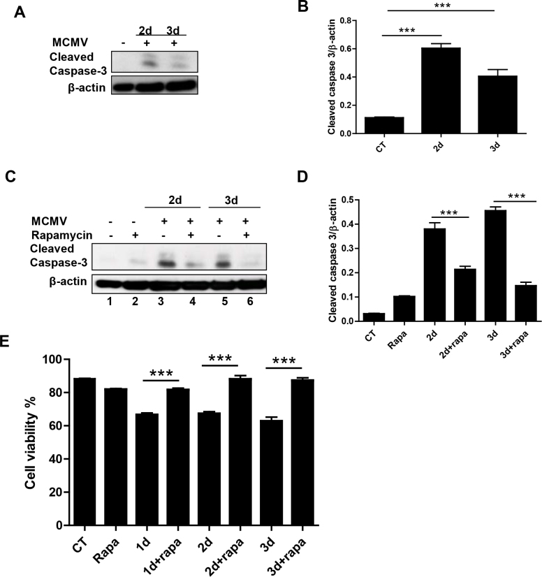
Next, we investigated the effect of rapamycin treatment on caspase 3-dependent apoptosis during the MCMV infection of the RPE cells. The MCMV infection in the RPE cells induced cell death through activation of caspase 3 (Figure 6A). Rapamycin treatment (Figure 6C, lanes 4 and 6) decreased MCMV infection–induced cleavage of caspase 3 compared with infected, untreated cells (Figure 6C, lanes 3 and 5), suggesting that activation of autophagy by rapamycin results in a concomitant decrease in apoptosis during MCMV infection.
Figure 6.
Effect of rapamycin treatment on apoptosis during murine cytomegalovirus infection. Retinal pigment epithelial (RPE) cells were infected with murine cytomegalovirus (MCMV) at low multiplicity of infection (MOI) = 1 in normal medium (A) or in medium containing rapamycin (10−6 M; C) for 2 and 3 days. B and D: Expression of cleaved caspase 3 was monitored and quantified. E: Collected cells were diluted to 1:1 using a 0.4% trypan blue solution. The stained cells and unstained cells were counted under a microscope. The calculated percentage of unstained cells represents the percentage of viable cells. Rapa: rapamycin; 2d: 2 days postinfection; 3d: 3 days postinfection. **p<0.01, ***p<0.001, ANOVA. Data are shown as mean±SEM (n=3).
To further explore whether activation of autophagy by rapamycin during MCMV infection is cytoprotective, the number of viable cells in a cell suspension infected with MCMV and treated with a non-toxic dose of rapamycin was determined with a trypan blue exclusion assay. Not surprisingly, the number of viable cells decreased in the MCMV-infected samples compared with the uninfected controls (Figure 6E). Moreover, the percentage of viable cells in the MCMV-infected, rapamycin-treated samples increased compared to that of the infected, untreated samples (Figure 6E). Taken together, these results suggest that activation of autophagy by rapamycin through blocking mTOR plays a role in protecting cells from apoptosis during MCMV infection.
Chloroquine blocks autophagy and increases apoptosis during MCMV infection
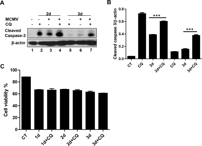
To further confirm the functional relationship between autophagy and apoptosis, cells were infected with MCMV and treated with chloroquine, which blocks the late step of autophagy and, consequently, results in increased levels of LC3B-II. Chloroquine-treated infected cells had increased expression of cleaved caspase 3 (Figure 7A, lanes 4 and 7) compared to infected RPE cells not treated with chloroquine (Figure 7A, lanes 3 and 6) suggesting that chloroquine treatment inhibits autophagy but increases apoptosis during MCMV infection of RPE cells. The trypan blue exclusion assay revealed that more than 80% cells were viable in the chloroquine-treated cells and the control cells. After MCMV infection, similar percentages of viable cells were observed in the chloroquine-treated cells and the control cells (Figure 7C). These results provide additional support for the idea that there is a functional relationship between autophagy and apoptosis and that this relationship plays an important role during MCMV infection of RPE cells.
Figure 7.
Effect of chloroquine treatment on apoptosis during murine cytomegalovirus infection. Retinal pigment epithelial (RPE) cells were infected with murine cytomegalovirus (MCMV) at low multiplicity of infection (MOI) = 1 in normal medium or in medium containing chloroquine (10−6 M) for 2 and 3 days. A and B: Expression of cleaved caspase 3 was monitored and quantified. C: Collected cells were diluted to 1:1 using a 0.4% trypan blue solution. The stained cells and unstained cells were counted under a microscope. The calculated percentage of unstained cells represents the percentage of viable cells. CQ: chloroquine; 2d: 2 days postinfection; 3d: 3 days postinfection. ***p<0.001, ANOVA. Data are shown as mean±SEM (n=3).
Discussion
In this study, we first demonstrated that MCMV infection of RPE cells induces autophagy during the early stage of infection. MCMV is a double-stranded DNA virus, and a broad spectrum of DNA viruses induces de novo formation and subsequent accumulation of autophagy-related vesicles [28]. Our results showing decreased autophagic flux plus increased expression of LC3B and an increased number of autophagic vacuoles suggest that during the late stage of MCMV infection, a late step of autophagy is inhibited. Previous studies have shown the induction of autophagy in human fibroblasts during the early stage of HCMV infection [27]; however, the inhibition of autophagy in HCMV-infected MRC5 fibroblasts during the late stage of infection is through the interaction between HCMV protein TRS1 and Beclin 1 [1,31], which blocks the early step of the autophagy process.
The possible reasons why the inhibition of autophagy during the late stage of MCMV infection is through the blockade of the late step of the autophagy process are as follows. First, the viral protein may interfere with the fusion of the autophagosome with the lysosome. For example, the matrix 2 protein of influenza A virus blocks the fusion of the autophagosome with the lysosome, resulting in accumulation of autophagosomes [51]. Second, enhanced virus replication and egress may inhibit the degradation of autolysosomes [52]. In vitro [53] and in vivo [54] studies have demonstrated that coxsackievirus B3 (CVB3) induces autophagic signaling to promote virus replication but prevents autophagic degradation [55]. Third, use of different cell types (RPE versus MRC5) and different viruses (MCMV versus HCMV) may contribute to apparent disparities between publications.
We observed activation of mTOR and the accumulation of autophagic vacuoles. mTOR has been reported to interfere with initiation of the phagophore [49,50] to block formation of the autophagosome, which leads to decreased expression of LC3B and a decreased number of autophagic vacuoles. Thus, even though MCMV infection activates mTOR signaling, activation of mTOR does not inhibit autophagy, or, perhaps, inhibition of autophagy by mTOR activation is impaired during MCMV infection since our results showed that MCMV inhibited autophagy after the autophagosome was formed. MCMV may use activation of mTOR to promote cell viability because during MCMV infection the cells are not likely to divide [56].
RPE cells are important in maintaining vision through the phagocytic function. The burst of RPE phagocytosis in the early morning rapidly clears ROS from the retina [57]. Using knockout mice with Atg5-deficient RPE cells, Kim et al. [39] observed that phagocytosis of the RPE coincided with the conversion of autophagy protein LC3B-I to its lipidated form, LC3B-II, and that the phagosomes had only a single membrane. In our in vitro RPE culture, most of the vacuoles we observed had the double membranes characteristic of autophagy. We occasionally observed phagosomes in the cytoplasm of RPE cells, perhaps because phagocytosis does not participate in clearing virus in intro. In addition, several passages of murine RPE cells might alter their phagocytotic functions, since a previous report showed that passage 4 human-induced pluripotent stem RPE (hiPSC-RPE) cells failed to form monolayers and possessed altered morphological and functional characteristics and gene expression levels [58].
As an important mechanism of host defense after viral infection, apoptosis is always considered to limit viral replication. However, even large and slowly replicating viruses may cause persistent infections. To ensure survival, these viruses have evolved mechanisms to keep infected cells alive by expressing cell death suppressors such as Bcl-2 and Bcl-xL [59]. These viruses, for example, MCMV, depend on the expression of cell death suppressors for effective replication and pathogenesis. Recently, MCMV genes m36 (gene ID AM237574.1) [60], m45 (gene ID DQ978788.1) [61], m38.5 (gene ID AM237292.1) [62], and m41.1 (gene ID FJ477245.1) [63] have been reported to encode cell death suppressors. When these genes are disrupted, the endogenous cellular defenses are ineffective, particularly during infections in the mouse. Although cytomegalovirus has the ability to block apoptosis to the benefit of viral replication [59-63], other data suggest that CMV may also activate apoptosis pathways resulting in the death of infected cells [64,65]. Recent studies have shown that autophagy and apoptosis are closely linked [30]. Depending on the conditions within the cell, autophagy can either be an adaptation to avoid apoptosis or may eventually lead to autophagic cell death. Autophagy may therefore be another mechanism for protecting cells from apoptosis since evidence from in vitro and in vivo studies has shown that autophagy limits caspase-dependent cell death and mortality after virus infection, including chikungunya (CHKV) virus [66], dengue virus [67], and CVB3 [68].
The results of the studies reported herein support the idea that there is cross talk between autophagy and apoptosis during MCMV infection of RPE cells. MCMV induces apoptosis. However, autophagy induced by rapamycin precluded apoptosis. Several mechanisms may explain why autophagy limits apoptotic cell death. One possible mechanism is that the degradation of protein aggregates and the sequestration of damaged mitochondria resulting from viral infection may delay induction of apoptosis by preventing released cytochrome c from forming a functional apoptosome [69]. Another possible mechanism is that the Atg3 protein complex releases Bcl-2 and Fas-associated death domain-like interleukin-1beta-converting enzyme (FLICE)-like inhibitory protein (FLIP) to block apoptotic pathways [30,69]. Our studies herein showed that caspase 3-dependent apoptosis is induced during RPE cell infection. Whether inhibition of autophagy by MCMV during the late stage of infection constitutes one mechanism of MCMV-induced apoptosis remains to be elucidated. In summary, these studies provide evidence for the early induction of autophagy as well as for the late inhibition of autophagy and support the idea of a functional relationship between autophagy and apoptosis during MCMV infection of RPE cells.
Acknowledgments
The authors are grateful to Dr. Zheng Dong for providing the GFP-LC3 plasmid. The authors thank Robert Smith for technical support for electron microscopy. The research was supported by NIH grant: RO1EY009169.
References
- 1.Chaumorcel M, Souquere S, Pierron G, Codogno P, Esclatine A. Human cytomegalovirus controls a new autophagy-dependent cellular antiviral defense mechanism. Autophagy. 2008;4:46–53. doi: 10.4161/auto.5184. [DOI] [PubMed] [Google Scholar]
- 2.Zhang M, Xin H, Roon P, Atherton SS. Infection of retinal neurons during murine cytomegalovirus retinitis. Invest Ophthalmol Vis Sci. 2005;46:2047–55. doi: 10.1167/iovs.05-0005. [DOI] [PubMed] [Google Scholar]
- 3.Thorne JE, Jabs DA, Kempen JH, Holbrook JT, Nichols C, Meinert CL. Incidence of and risk factors for visual acuity loss among patients with AIDS and cytomegalovirus retinitis in the era of highly active antiretroviral therapy. Ophthalmology. 2006;113:1432–40. doi: 10.1016/j.ophtha.2006.03.021. [DOI] [PubMed] [Google Scholar]
- 4.Thorne JE, Jabs DA, Kempen JH, Holbrook JT, Nichols C, Meinert CL. Causes of visual acuity loss among patients with AIDS and cytomegalovirus retinitis in the era of highly active antiretroviral therapy. Ophthalmology. 2006;113:1441–5. doi: 10.1016/j.ophtha.2006.03.022. [DOI] [PubMed] [Google Scholar]
- 5.Jabs DA, Holbrook JT, Van Natta ML, Clark R, Jacobson MA, Kempen JH, Murphy RL. Risk factors for mortality in patients with AIDS in the era of highly active antiretroviral therapy. Ophthalmology. 2005;112:771–9. doi: 10.1016/j.ophtha.2004.10.049. [DOI] [PubMed] [Google Scholar]
- 6.Chiou SH, Liu JH, Hsu WM, Chen SS, Chang SY, Juan LJ, Lin JC, Yang YT, Wong WW, Liu CY, Lin YS, Liu WT, Wu CW. Up-regulation of Fas ligand expression by human cytomegalovirus immediate-early gene product 2: a novel mechanism in cytomegalovirus-induced apoptosis in human retina. J Immunol. 2001;167:4098–103. doi: 10.4049/jimmunol.167.7.4098. [DOI] [PubMed] [Google Scholar]
- 7.Buggage RR, Chan CC, Matteson DM, Reed GF, Whitcup SM. Apoptosis in cytomegalovirus retinitis associated with AIDS. Curr Eye Res. 2000;21:721–9. [PubMed] [Google Scholar]
- 8.Bigger JE, Tanigawa M, Zhang M, Atherton SS. Murine cytomegalovirus infection causes apoptosis of uninfected retinal cells. Invest Ophthalmol Vis Sci. 2000;41:2248–54. [PubMed] [Google Scholar]
- 9.Zhang M, Marshall B, Atherton SS. Murine cytomegalovirus infection and apoptosis in organotypic retinal cultures. Invest Ophthalmol Vis Sci. 2008;49:295–303. doi: 10.1167/iovs.07-0612. [DOI] [PMC free article] [PubMed] [Google Scholar]
- 10.Virgin HW, Levine B. Autophagy genes in immunity. Nat Immunol. 2009;10:461–70. doi: 10.1038/ni.1726. [DOI] [PMC free article] [PubMed] [Google Scholar]
- 11.English L, Chemali M, Duron J, Rondeau C, Laplante A, Gingras D, Alexander D, Leib D, Norbury C, Lippe R, Desjardins M. Autophagy enhances the presentation of endogenous viral antigens on MHC class I molecules during HSV-1 infection. Nat Immunol. 2009;10:480–7. doi: 10.1038/ni.1720. [DOI] [PMC free article] [PubMed] [Google Scholar]
- 12.Deretic V. Autophagy in innate and adaptive immunity. Trends Immunol. 2005;26:523–8. doi: 10.1016/j.it.2005.08.003. [DOI] [PubMed] [Google Scholar]
- 13.Levine B, Deretic V. Unveiling the roles of autophagy in innate and adaptive immunity. Nat Rev Immunol. 2007;7:767–77. doi: 10.1038/nri2161. [DOI] [PMC free article] [PubMed] [Google Scholar]
- 14.Schmid D, Munz C. Innate and adaptive immunity through autophagy. Immunity. 2007;27:11–21. doi: 10.1016/j.immuni.2007.07.004. [DOI] [PMC free article] [PubMed] [Google Scholar]
- 15.Cecconi F, Levine B. The role of autophagy in mammalian development: cell makeover rather than cell death. Dev Cell. 2008;15:344–57. doi: 10.1016/j.devcel.2008.08.012. [DOI] [PMC free article] [PubMed] [Google Scholar]
- 16.Levine B, Kroemer G. Autophagy in the pathogenesis of disease. Cell. 2008;132:27–42. doi: 10.1016/j.cell.2007.12.018. [DOI] [PMC free article] [PubMed] [Google Scholar]
- 17.Mizushima N, Levine B, Cuervo AM, Klionsky DJ. Autophagy fights disease through cellular self-digestion. Nature. 2008;451:1069–75. doi: 10.1038/nature06639. [DOI] [PMC free article] [PubMed] [Google Scholar]
- 18.Rubinsztein DC. The roles of intracellular protein-degradation pathways in neurodegeneration. Nature. 2006;443:780–6. doi: 10.1038/nature05291. [DOI] [PubMed] [Google Scholar]
- 19.Chen Y, Sawada O, Kohno H, Le YZ, Subauste C, Maeda T, Maeda A. Autophagy protects the retina from light-induced degeneration. J Biol Chem. 2013;288:7506–18. doi: 10.1074/jbc.M112.439935. [DOI] [PMC free article] [PubMed] [Google Scholar]
- 20.Remé CE, Young RW. The effects of hibernation on cone visual cells in the ground squirrel. Invest Ophthalmol Vis Sci. 1977;16:815–40. [PubMed] [Google Scholar]
- 21.Scholz M, Doerr HW, Cinatl J. Human cytomegalovirus retinitis: pathogenicity, immune evasion and persistence. Trends Microbiol. 2003;11:171–8. doi: 10.1016/s0966-842x(03)00066-0. [DOI] [PubMed] [Google Scholar]
- 22.Chiou SH, Liu JH, Chen SS, Liu WT, Lin JC, Wong WW, Tseng WS, Chou CK, Liu CY, Ho LL, Hsu WM. Apoptosis of human retina and retinal pigment cells induced by human cytomegalovirus infection. Ophthalmic Res. 2002;34:77–82. doi: 10.1159/000048332. [DOI] [PubMed] [Google Scholar]
- 23.Michaelis M, Suhan T, Reinisch A, Reisenauer A, Fleckenstein C, Eikel D, Gumbel H, Doerr HW, Nau H, Cinatl J., Jr Increased replication of human cytomegalovirus in retinal pigment epithelial cells by valproic acid depends on histone deacetylase inhibition. Invest Ophthalmol Vis Sci. 2005;46:3451–7. doi: 10.1167/iovs.05-0369. [DOI] [PubMed] [Google Scholar]
- 24.Allart S, Lule J, Serres B, Jones T, Davignon JL, Malecaze F, Davrinche C. Impaired killing of HCMV-infected retinal pigment epithelial cells by anti-pp65 CD8(+) cytotoxic T cells. Invest Ophthalmol Vis Sci. 2003;44:665–71. doi: 10.1167/iovs.02-0547. [DOI] [PubMed] [Google Scholar]
- 25.Momma Y, Nagineni CN, Chin MS, Srinivasan K, Detrick B, Hooks JJ. Differential expression of chemokines by human retinal pigment epithelial cells infected with cytomegalovirus. Invest Ophthalmol Vis Sci. 2003;44:2026–33. doi: 10.1167/iovs.02-0980. [DOI] [PubMed] [Google Scholar]
- 26.Wang X, Gao Y, Tan J, Devadas K, Ragupathy V, Takeda K, Zhao J, Hewlett I. HIV-1 and HIV-2 infections induce autophagy in Jurkat and CD4+ T cells. Cell Signal. 2012;24:1414–9. doi: 10.1016/j.cellsig.2012.02.016. [DOI] [PubMed] [Google Scholar]
- 27.McFarlane S, Aitken J, Sutherland JS, Nicholl MJ, Preston VG, Preston CM. Early induction of autophagy in human fibroblasts after infection with human cytomegalovirus or herpes simplex virus 1. J Virol. 2011;85:4212–21. doi: 10.1128/JVI.02435-10. [DOI] [PMC free article] [PubMed] [Google Scholar]
- 28.Dreux M, Chisari FV. Viruses and the autophagy machinery. Cell Cycle. 2010;9:1295–307. doi: 10.4161/cc.9.7.11109. [DOI] [PubMed] [Google Scholar]
- 29.Maiuri MC, Zalckvar E, Kimchi A, Kroemer G. Self-eating and self-killing: crosstalk between autophagy and apoptosis. Nat Rev Mol Cell Biol. 2007;8:741–52. doi: 10.1038/nrm2239. [DOI] [PubMed] [Google Scholar]
- 30.Thorburn A. Apoptosis and autophagy: regulatory connections between two supposedly different processes. Apoptosis. 2008;13:1–9. doi: 10.1007/s10495-007-0154-9. [DOI] [PMC free article] [PubMed] [Google Scholar]
- 31.Chaumorcel M, Lussignol M, Mouna L, Cavignac Y, Fahie K, Cotte-Laffitte J, Geballe A, Brune W, Beau I, Codogno P, Esclatine A. The human cytomegalovirus protein TRS1 inhibits autophagy via its interaction with Beclin 1. J Virol. 2012;86:2571–84. doi: 10.1128/JVI.05746-11. [DOI] [PMC free article] [PubMed] [Google Scholar]
- 32.Atherton SS, Newell CK, Kanter MY, Cousins SW. T cell depletion increases susceptibility to murine cytomegalovirus retinitis. Invest Ophthalmol Vis Sci. 1992;33:3353–60. [PubMed] [Google Scholar]
- 33.Nuzzo R. Scientific method: statistical errors. Nature. 2014;506:150–2. doi: 10.1038/506150a. [DOI] [PubMed] [Google Scholar]
- 34.McLeland CB, Rodriguez J, Stern ST. Autophagy monitoring assay: qualitative analysis of MAP LC3-I to II conversion by immunoblot. Methods Mol Biol. 2011;697:199–206. doi: 10.1007/978-1-60327-198-1_21. [DOI] [PubMed] [Google Scholar]
- 35.Boya P, Gonzalez-Polo RA, Casares N, Perfettini JL, Dessen P, Larochette N, Metivier D, Meley D, Souquere S, Yoshimori T, Pierron G, Codogno P, Kroemer G. Inhibition of macroautophagy triggers apoptosis. Mol Cell Biol. 2005;25:1025–40. doi: 10.1128/MCB.25.3.1025-1040.2005. [DOI] [PMC free article] [PubMed] [Google Scholar]
- 36.Wu YT, Tan HL, Shui G, Bauvy C, Huang Q, Wenk MR, Ong CN, Codogno P, Shen HM. Dual role of 3-methyladenine in modulation of autophagy via different temporal patterns of inhibition on class I and III phosphoinositide 3-kinase. J Biol Chem. 2010;285:10850–61. doi: 10.1074/jbc.M109.080796. [DOI] [PMC free article] [PubMed] [Google Scholar]
- 37.Calvo-Garrido J, Carilla-Latorre S, Mesquita A, Escalante R. A proteolytic cleavage assay to monitor autophagy in Dictyostelium discoideum. Autophagy. 2011;7:1063–8. doi: 10.4161/auto.7.9.16629. [DOI] [PubMed] [Google Scholar]
- 38.Shelly S, Lukinova N, Bambina S, Berman A, Cherry S. Autophagy is an essential component of Drosophila immunity against vesicular stomatitis virus. Immunity. 2009;30:588–98. doi: 10.1016/j.immuni.2009.02.009. [DOI] [PMC free article] [PubMed] [Google Scholar]
- 39.Kim JY, Zhao H, Martinez J, Doggett TA, Kolesnikov AV, Tang PH, Ablonczy Z, Chan CC, Zhou Z, Green DR, Ferguson TA. Noncanonical autophagy promotes the visual cycle. Cell. 2013;154:365–76. doi: 10.1016/j.cell.2013.06.012. [DOI] [PMC free article] [PubMed] [Google Scholar]
- 40.Dong X, Levine B. Autophagy and viruses: adversaries or allies? J Innate Immun. 2013;5:480–93. doi: 10.1159/000346388. [DOI] [PMC free article] [PubMed] [Google Scholar]
- 41.Roulston A, Marcellus RC, Branton PE. Viruses and apoptosis. Annu Rev Microbiol. 1999;53:577–628. doi: 10.1146/annurev.micro.53.1.577. [DOI] [PubMed] [Google Scholar]
- 42.Barber GN. Host defense, viruses and apoptosis. Cell Death Differ. 2001;8:113–26. doi: 10.1038/sj.cdd.4400823. [DOI] [PubMed] [Google Scholar]
- 43.Baehrecke EH. Autophagy: dual roles in life and death? Nat Rev Mol Cell Biol. 2005;6:505–10. doi: 10.1038/nrm1666. [DOI] [PubMed] [Google Scholar]
- 44.Shrivastava S, Bhanja Chowdhury J, Steele R, Ray R, Ray RB. Hepatitis C virus upregulates Beclin1 for induction of autophagy and activates mTOR signaling. J Virol. 2012;86:8705–12. doi: 10.1128/JVI.00616-12. [DOI] [PMC free article] [PubMed] [Google Scholar]
- 45.Ravikumar B, Sarkar S, Davies JE, Futter M, Garcia-Arencibia M, Green-Thompson ZW, Jimenez-Sanchez M, Korolchuk VI, Lichtenberg M, Luo S, Massey DC, Menzies FM, Moreau K, Narayanan U, Renna M, Siddiqi FH, Underwood BR, Winslow AR, Rubinsztein DC. Regulation of mammalian autophagy in physiology and pathophysiology. Physiol Rev. 2010;90:1383–435. doi: 10.1152/physrev.00030.2009. [DOI] [PubMed] [Google Scholar]
- 46.Inoki K, Zhu T, Guan KL. TSC2 mediates cellular energy response to control cell growth and survival. Cell. 2003;115:577–90. doi: 10.1016/s0092-8674(03)00929-2. [DOI] [PubMed] [Google Scholar]
- 47.Narita M, Young AR, Arakawa S, Samarajiwa SA, Nakashima T, Yoshida S, Hong S, Berry LS, Reichelt S, Ferreira M, Tavare S, Inoki K, Shimizu S, Narita M. Spatial coupling of mTOR and autophagy augments secretory phenotypes. Science. 2011;332:966–70. doi: 10.1126/science.1205407. [DOI] [PMC free article] [PubMed] [Google Scholar]
- 48.Settembre C, Di Malta C, Polito VA, Garcia Arencibia M, Vetrini F, Erdin S, Erdin SU, Huynh T, Medina D, Colella P, Sardiello M, Rubinsztein DC, Ballabio A. TFEB links autophagy to lysosomal biogenesis. Science. 2011;332:1429–33. doi: 10.1126/science.1204592. [DOI] [PMC free article] [PubMed] [Google Scholar]
- 49.Liu EY, Ryan KM. Autophagy and cancer–issues we need to digest. J Cell Sci. 2012;125:2349–58. doi: 10.1242/jcs.093708. [DOI] [PubMed] [Google Scholar]
- 50.Nair S, Ren J. Autophagy and cardiovascular aging: lesson learned from rapamycin. Cell Cycle. 2012;11:2092–9. doi: 10.4161/cc.20317. [DOI] [PMC free article] [PubMed] [Google Scholar]
- 51.Gannagé M, Dormann D, Albrecht R, Dengjel J, Torossi T, Ramer PC, Lee M, Strowig T, Arrey F, Conenello G, Pypaert M, Andersen J, Garcia-Sastre A, Munz C. Matrix protein 2 of influenza A virus blocks autophagosome fusion with lysosomes. Cell Host Microbe. 2009;6:367–80. doi: 10.1016/j.chom.2009.09.005. [DOI] [PMC free article] [PubMed] [Google Scholar]
- 52.Silva LM, Jung JU. Modulation of the autophagy pathway by human tumor viruses. Semin Cancer Biol. 2013;23:323–8. doi: 10.1016/j.semcancer.2013.05.005. [DOI] [PMC free article] [PubMed] [Google Scholar]
- 53.Wong J, Zhang J, Si X, Gao G, Mao I, McManus BM, Luo H. Autophagosome supports coxsackievirus B3 replication in host cells. J Virol. 2008;82:9143–53. doi: 10.1128/JVI.00641-08. [DOI] [PMC free article] [PubMed] [Google Scholar]
- 54.Kemball CC, Alirezaei M, Flynn CT, Wood MR, Harkins S, Kiosses WB, Whitton JL. Coxsackievirus infection induces autophagy-like vesicles and megaphagosomes in pancreatic acinar cells in vivo. J Virol. 2010;84:12110–24. doi: 10.1128/JVI.01417-10. [DOI] [PMC free article] [PubMed] [Google Scholar]
- 55.Klein KA, Jackson WT. Picornavirus subversion of the autophagy pathway. Viruses. 2011;3:1549–61. doi: 10.3390/v3091549. [DOI] [PMC free article] [PubMed] [Google Scholar]
- 56.Kosugi I, Shinmura Y, Kawasaki H, Arai Y, Li RY, Baba S, Tsutsui Y. Cytomegalovirus infection of the central nervous system stem cells from mouse embryo: a model for developmental brain disorders induced by cytomegalovirus. Laboratory. 2000;80:1373–83. doi: 10.1038/labinvest.3780145. [DOI] [PubMed] [Google Scholar]
- 57.Strauss O. The retinal pigment epithelium in visual function. Physiol Rev. 2005;85:845–81. doi: 10.1152/physrev.00021.2004. [DOI] [PubMed] [Google Scholar]
- 58.Singh R, Phillips MJ, Kuai D, Meyer J, Martin JM, Smith MA, Perez ET, Shen W, Wallace KA, Capowski EE, Wright LS, Gamm DM. Functional analysis of serially expanded human iPS cell-derived RPE cultures. Invest Ophthalmol Vis Sci. 2013;54:6767–78. doi: 10.1167/iovs.13-11943. [DOI] [PMC free article] [PubMed] [Google Scholar]
- 59.Brune W. Inhibition of programmed cell death by cytomegaloviruses. Virus Res. 2011;157:144–50. doi: 10.1016/j.virusres.2010.10.012. [DOI] [PubMed] [Google Scholar]
- 60.McCormick AL, Skaletskaya A, Barry PA, Mocarski ES, Goldmacher VS. Differential function and expression of the viral inhibitor of caspase 8-induced apoptosis (vICA) and the viral mitochondria-localized inhibitor of apoptosis (vMIA) cell death suppressors conserved in primate and rodent cytomegaloviruses. Virology. 2003;316:221–33. doi: 10.1016/j.virol.2003.07.003. [DOI] [PubMed] [Google Scholar]
- 61.Upton JW, Kaiser WJ, Mocarski ES. Cytomegalovirus M45 cell death suppression requires receptor-interacting protein (RIP) homotypic interaction motif (RHIM)-dependent interaction with RIP1. J Biol Chem. 2008;283:16966–70. doi: 10.1074/jbc.C800051200. [DOI] [PMC free article] [PubMed] [Google Scholar]
- 62.Jurak I, Schumacher U, Simic H, Voigt S, Brune W. Murine cytomegalovirus m38.5 protein inhibits Bax-mediated cell death. J Virol. 2008;82:4812–22. doi: 10.1128/JVI.02570-07. [DOI] [PMC free article] [PubMed] [Google Scholar]
- 63.Crosby LN, McCormick AL, Mocarski ES. Gene products of the embedded m41/m41.1 locus of murine cytomegalovirus differentially influence replication and pathogenesis. Virology. 2013;436:274–83. doi: 10.1016/j.virol.2012.12.002. [DOI] [PMC free article] [PubMed] [Google Scholar]
- 64.Lee GC, Lee JH, Kim BY, Lee CH. Mitochondria-targeted apoptosis in human cytomegalovirus-infected cells. J Microbiol Biotechnol. 2013;23:1627–35. doi: 10.4014/jmb.1306.06023. [DOI] [PubMed] [Google Scholar]
- 65.McCormick AL, Roback L, Wynn G, Mocarski ES. Multiplicity-dependent activation of a serine protease-dependent cytomegalovirus-associated programmed cell death pathway. Virology. 2013;435:250–7. doi: 10.1016/j.virol.2012.08.042. [DOI] [PMC free article] [PubMed] [Google Scholar]
- 66.Joubert PE, Werneke SW, de la Calle C, Guivel-Benhassine F, Giodini A, Peduto L, Levine B, Schwartz O, Lenschow DJ, Albert ML. Chikungunya virus-induced autophagy delays caspase-dependent cell death. J Exp Med. 2012;209:1029–47. doi: 10.1084/jem.20110996. [DOI] [PMC free article] [PubMed] [Google Scholar]
- 67.Green AM, Beatty PR, Hadjilaou A, Harris E. Innate Immunity to Dengue Virus Infection and Subversion of Antiviral Responses. J Mol Biol. 2014 doi: 10.1016/j.jmb.2013.11.023. [DOI] [PMC free article] [PubMed] [Google Scholar]
- 68.Li M, Wang X, Yu Y, Yu Y, Xie Y, Zou Y, Ge J, Peng T, Chen R. Coxsackievirus B3-induced calpain activation facilitates the progeny virus replication via a likely mechanism related with both autophagy enhancement and apoptosis inhibition in the early phase of infection: An in vitro study in H9c2 cells. Virus Res. 2014 doi: 10.1016/j.virusres.2013.10.014. [DOI] [PubMed] [Google Scholar]
- 69.Kroemer G, Marino G, Levine B. Autophagy and the integrated stress response. Mol Cell. 2010;40:280–93. doi: 10.1016/j.molcel.2010.09.023. [DOI] [PMC free article] [PubMed] [Google Scholar]


