Abstract
Drug treatment, electric stimulation and decimeter wave therapy have been shown to promote the repair and regeneration of the peripheral nerves at the injured site. This study prepared a Mackinnon's model of rat sciatic nerve compression. Electric stimulation was given immediately after neurolysis, and decimeter wave radiation was performed at 1 and 12 weeks post-operation. Histological observation revealed that intraoperative electric stimulation and decimeter wave therapy could improve the local blood circulation of repaired sites, alleviate hypoxia of compressed nerves, and lessen adhesion of compressed nerves, thereby decreasing the formation of new entrapments and enhancing compressed nerve regeneration through an improved microenvironment for regeneration. Immunohistochemical staining results revealed that intraoperative electric stimulation and decimeter wave could promote the expression of S-100 protein. Motor nerve conduction velocity and amplitude, the number and diameter of myelinated nerve fibers, and sciatic functional index were significantly increased in the treated rats. These results verified that intraoperative electric stimulation and decimeter wave therapy contributed to the regeneration and the recovery of the functions in the compressed nerves.
Keywords: neural regeneration, peripheral nerve injury, physical therapy, electric stimulation, sciatic nerve compression, Schwann cells, functional recovery, neuroregeneration
Research Highlights
-
(1)
This study concluded that decimeter wave therapy promoted the proliferation of Schwann cells and elevated S-100 protein expression in injured sciatic nerves, and contributed to neural regeneration and functional recovery at cellular and molecular levels.
-
(2)
Decimeter wave therapy promoted axonal regeneration and remyelination, delayed myatrophy and facilitated neural regeneration and functional recovery.
-
(3)
Decimeter wave therapy inhibited the inflammatory reaction and anticoagulation, improved the local blood circulation, reduced the formation of scar and effectively prevented nerve adhesion and re-entrapment after repairing.
INTRODUCTION
Nerve compression syndromes have long been a common clinical problem in hand surgery. Sophisticated microsurgery skills have much improved neurorrhaphy and a number of biologically active factors and gene transfer have been used to promote nerve regeneration. However, the functional recovery of an injured nerve is often unsatisfactory. Peripheral nerve regeneration involves a complex process of biochemical and cellular events, and the microenvironment of the regenerating nerve very much influences its repair. How to protect the nerve to avoid the secondary damage is currently an unresolved problem. Moreover, mechanisms underlying neural repair and regeneration are very complicated. Some scholars treated patients with flexor tendons injury after repairing and patients with peripheral nerve diseases of diabetes by decimeter wave, they found that decimeter wave could accelerate tendon intrinsic healing and inhibit extrinsic healing after tendon repairing and then reduce tendon adhesion[1,2,3,4,5,6,7,8,9,10,11,12,13]. They also deduced that decimeter wave could improve local blood circulation of the repairing site, alleviate hypoxia of regenerating nerves and increase the expression of immunologic reaction to S-100 protein in Schwann cells[14,15,16,17,18,19,20,21]. Some bioactive substances and relevant drugs that are effective in promoting neural regeneration have been reported in recent studies[22,33]. Decimeter wave, a long-wavelength electromagnetic radiation with effects of improving local blood circulation, accelerating metabolism, reducing local inflammation and alleviating pain, has attracted the attention of many researchers and clinical workers. Some scholars[7,12,13] gave decimeter treatment to patients recovering from flexor tendons injury and patients with peripheral nerve diseases of diabetes, and found that the excellent and good rate of tendon function recovery was 97% according to TAM standard and the whole effective rate of treatment to the pathological nerves was 83%. He deduced that decimeter waves could accelerate intrinsic tendon healing and inhibit extrinsic healing after tendon repairing and then reduce tendon adhesion. This could alleviate pain and be advantageous for patients’ earlier rehabilitation exercises. Physical therapy is a type of natural therapy, has no side effects and is relatively cheap. Previous studies have shown that physical therapy promotes nerve regeneration; however, mechanisms underlying the improvement have not been investigated.
This study aims to investigate the mechanism of the effects of electric stimulation and decimeter waves on peripheral nerve regeneration, and then to provide a theoretical basis for clinical applications. It was investigated by anatomical observation, light microscopy, electron microscopy, immunochemistry, and morphometric analysis in a Mackinnon's model of sciatic nerve compression in adult Sprague-Dawley rats.
RESULTS
Quantitative analysis of experimental animals
Under anesthesia, the right sciatic nerves of the 90 healthy adult Sprague-Dawley rats were each compressed by a silicone tube similar to the model of Mackinnon. A total of 90 healthy adult Sprague-Dawley rats were randomly divided into two groups as follows (n = 45 in each group): the intraoperative electric stimulation and decimeter wave group and the control group. At 4 weeks post-operation, three rats in each group were randomly sacrificed; the original wounds were reopened and simple decompression was applied, such that the silicone tube was simply moved and the epineurium of the entrapped segment was not decompressed. The animals in the intraoperative electric stimulation and decimeter wave group were treated with electric stimulation. After thorough hemostasis, the intermuscular space was closed and the skin was sutured. A DXZ-1 polymorphism wave therapy instrument was used in the present study. The rats of the intraoperative electric stimulation and decimeter wave group were fixed on a table prostrated and the right posterior thigh was exposed to decimeter waves every day from 1 day post-operation to the sacrifice day. The rats of the control group were also fixed on a table prostrated at the same time, but were not exposed to decimeter waves. At 1, 2, 3, 4 and 12 weeks post-operation, the samples were observed by anatomical, light and electron microscope observation, morphometric analysis and S-100 protein immunochemical staining. At 12 weeks post-operation, the latency, nerve conduction velocity and amplitude of compound muscle action potentials were measured electrophysiologically. At 4, 8 and 12 weeks post- operation, the sciatic functional indices were analyzed. All data were analyzed statistically using two-sample t-test. Ninety rats were involved in the final analysis.
Effects of electric stimulation and decimeter wave on general morphology of sciatic nerves in rats with sciatic nerve compression
Stage I healing was observed in each group, without infection, plantar ulceration or autophagocytic phenomena. At each time point, hyperemia and edema were observed in the subcutaneous tissues and tissues surrounding the nerves 1 week post-operation. Signs of entrapment were also clearly visible. The entrapped segment became thin with an enlarged neuroma-like appearance at both ends. These changes were smaller in the intraoperative electric stimulation and decimeter wave group than those in the control group. Hyperemia and edema of the tissues surrounding the neurons abated gradually and the entrapped neural segments thickened in both groups. By the 8th week post-operation, neither hyperemia nor edema was observed in the tissues surrounding the neurons in both groups, whereas the entrapped segments were significantly thickened in both groups. Neuroma-like changes in both ends of the entrapped segment had significantly abated in both groups. At 12 weeks post-operation, the surface of the entrapped nerve was smooth in the intraoperative electric stimulation and decimeter wave group, and there were mostly thin-membrane adhesions. The neural tissue surrounding adhesions, which could be readily blunt-separated, was looser in the intraoperative electric stimulation and decimeter wave group than in the control group. The entrapped nerves in both groups were almost normal.
Effects of electric stimulation and decimeter wave on the structure of the sciatic nerves in rats with sciatic nerve compression
Examination by light microscope, 2 weeks post-operation, showed that the inflammatory cells had infiltrated tissues surrounding the nerve in both groups. In the intraoperative electric stimulation and decimeter wave group, the inflammatory cells were limited to the superficial layer of the epineurium, while in the control group, many inflammatory cells and interspersed multinucleated giant cells had infiltrated around the nerves (Figure 1A, B). The epineuria in both groups were intact; however, the myelin sheaths were uneven in thickness and irregular in shape, and different degrees of demyelination were noticeable.
Figure 1.
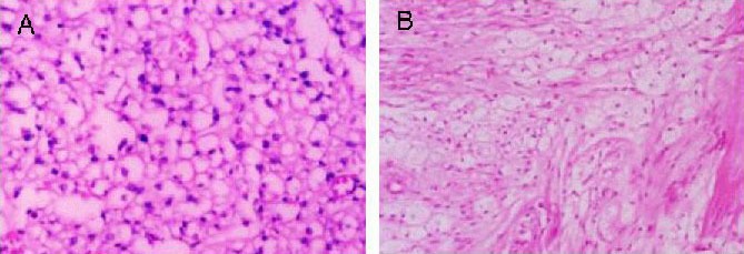
Effects of electric stimulation and decimeter wave on morphology of the sciatic nerves in rats with sciatic nerve compression at the 2nd week post-operation (hematoxylin-eosin staining, × 400).
Many inflammatory cells and interspersed multinucleated giant cells infiltrated around the nerves in the control group (A). The inflammatory cells were limited to the superficial layer of the epineurium in the intraoperative electric stimulation and decimeter wave group (B). Inflammatory cells had infiltrated the tissues surrounding the nerve in both groups.
By the 4th week post-operation, the inflammatory responses were significantly reduced and swelling of the myelin sheaths had lessened in both groups. In the intraoperative electric stimulation and decimeter wave group, the inflammatory cells were rarely observed, whereas they were still accumulating in the control group Figure 2A, B).
Figure 2.
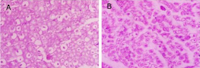
Effects of electric stimulation and decimeter wave on morphology of the sciatic nerves in rats with sciatic nerve compression at the 4th week post-operation (hematoxylin-eosin staining, × 400).
The inflammatory cells were still accumulating in the control group (A). Inflammatory cells were rarely observed in the intraoperative electric stimulation and decimeter wave group (B). Inflammatory responses were significantly reduced and myelin swelling began to fade in both groups.
At 8 weeks post-operation, the demyelination of the two groups had significantly been alleviated. In the intraoperative electric stimulation and decimeter wave group, the swelling of the axons had almost disappeared, detached myelin sheaths were gradually decreasing, and regenerated neural fibers were observed in the entrapped segment and at the distal end. However, the regenerated axons were of fine diameter and their myelin sheaths were thin. In the control group, axon swelling had abated despite the increased demyelination. There were few regenerated nerve fibers in both the entrapped segment and its distal end. The development of these fibers was still immature (Figure 3A, B).
Figure 3.
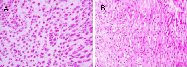
Effects of electric stimulation and decimeter wave on morphology of the sciatic nerves in rats with sciatic nerve compression at the 8th week post-operation (hematoxylin-eosin staining, × 200).
Axon swelling was abating despite increased demyelination in the control group (A). The swelling of the axons almost disappeared, detached myelin sheaths were gradually decreasing in the intraoperative electric stimulation and decimeter wave group (B).
At 12 weeks post-operation, the surfaces of the nerves in the intraoperative electric stimulation and decimeter wave group were smooth and showed no adhesion to their surrounding tissues, or only had slight filamentous adhesions. In the control group, the surfaces of the nerves were rough, and many adhesions were observed. In both groups, the myelin sheath fragments had significantly decreased, neovascularization was noted, and regenerated axons and myelin sheaths had formed. The damaged neural fibers, with fine diameters and thin myelin sheaths, were mostly adhering together and arranged orderly. In conclusion, the intraoperative electric stimulation and decimeter wave group recovered better than the control group (Figure 4A, B).
Figure 4.
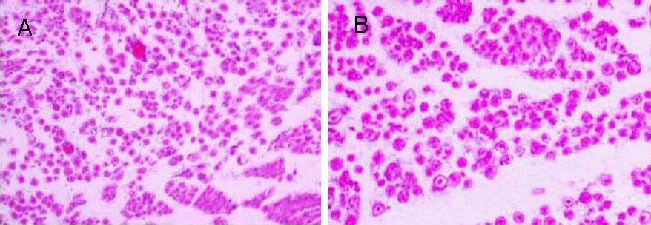
Effects of electric stimulation and decimeter wave on morphology of the sciatic nerves in rats with sciatic nerve compression at the 12th week post-operation (hematoxylin-eosin staining, × 200).
The surfaces of the nerves were rough, and many adhesions were observed in the control group (A). The surfaces of the nerves were smooth and showed no adhesion with their surrounding tissues, or only had slight filamentous adhesions in the intraoperative electric stimulation and decimeter wave group (B).
Effects of electric stimulation and decimeter wave on ultrastructure of the sciatic nerves in rats with sciatic nerve compression
At 2 weeks post-operation, a novel axon bud, encapsulated by the epineurium, was present in the proximal end of the entrapped segment in a rat of the intraoperative electric stimulation and decimeter wave group. Organelle such as mitochondria, microfilament, microtubule and vesicle within the neonatal axon bud was few and sparsely distributed. In the control group, the cellular membrane of the myelin sheath epineurium was detached from the axon, the myelin sheath was swollen unevenly, and the mitochondria had necrotic vacuoles and many were disintegrating (Figure 5A, B).
Figure 5.
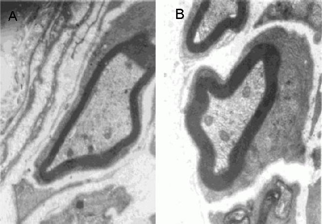
Effects of electric stimulation and decimeter wave on ultrastructure of the sciatic nerves in rats with sciatic nerve compression at the 2nd week post-operation (transmission electron microscopy, × 15 000).
The cellular membrane of myelin sheath epineurium was detached from the axon, the myelin sheath was unevenly swollen, and the mitochondria appeared vacuolar necrotic and disintegrated in the control group (A). A novel axon bud, which was encapsulated by the epineurium, was present in the proximal end of the entrapped segment in the intraoperative electric stimulation and decimeter wave group (B).
In the intraoperative electric stimulation and decimeter wave group, 4 weeks post-operation, the myelin sheaths were relatively thick and the regenerated axons appeared normal. A few nearly normal organelles were noted in sparse but arranged axons. In the control group, there were a few myelinated fibers that had fine axons and thin myelin sheaths but they were arranged irregularly. In addition, parts of the sheaths were empty, and oval-like bodies associated with myelin sheath degeneration were occasionally noticed (Figure 6A, B).
Figure 6.
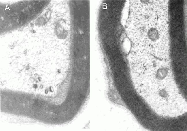
Effects of electric stimulation and decimeter wave on ultrastructure of the sciatic nerves in rats with sciatic nerve compression at the 4th week post-operation (transmission electron microscopy, × 30 000).
In the control group (A), there were few irregularly arranged myelinated fibers. In the intraoperative electric stimulation and decimeter wave group (B), the myelin sheath was relatively thick and the regenerated axon had a normal appearance.
However, by the 8th week post-operation, there was a large quantity of myelinated fibers seen in the control group. They had small diameters and were arranged orderly. Furthermore, the myelin sheath was thick with connective tissue of low hyperplasia (Figure 7A). In the intraoperative electric stimulation and decimeter wave group, the regenerated myelinated fibers were arranged orderly, the fasciculated structure was fine and the endoneurium was well developed. Connective tissue hyperplasia was not visible (Figure 7B).
Figure 7.

Effects of electric stimulation and decimeter wave on ultrastructure of the sciatic nerves in rats with sciatic nerve compression at the 8th week post-operation (transmission electron microscopy, × 30 000).
In the control group (A), there was a large quantity of orderly arranged myelinated fibers with small diameters. In the intraoperative electric stimulation and decimeter wave group (B), regenerated myelinated fibers were orderly arranged. The fasciculated structure was fine and the endoneurium was well developed.
Effects of electric stimulation and decimeter waves on S-100 protein expression in rats with sciatic nerve compression
There were fewer S-100-positive particles in the proximal end of the entrapped segment in both groups 2 weeks post-operation. In contrast, the positive particles in the Schwann cells increased with time, and were approaching normal both in quantity and distribution by the 4th week post-operation (Figure 8A, B).
Figure 8.
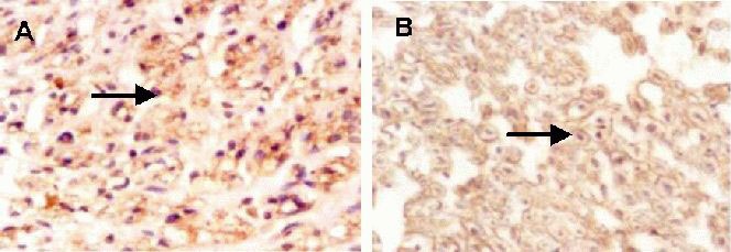
Effects of electric stimulation and decimeter wave on S-100 expression (arrows) in compressed sciatic nerve (immunohistochemistry, × 400).
In the control group (A), nerve fibers in the proximal end of the entrapped segment were wrapped with normal Schwann cells. In the intraoperative electric stimulation and decimeter wave group (B), nerve fibers were wrapped with Schwann cells and the positive particles in Schwann cells had increased with time.
Effects of electric stimulation and decimeter wave on morphometric analysis and electrophysiological assessment of the sciatic nerves
At 12 weeks post-operation, distal nerve segments in the intraoperative electric stimulation and decimeter wave group had more myelinated axon counts and a larger mean axon diameter than those in the control group. The parameters (latency, nerve conduction velocity and amplitude of compound muscle action potentials) revealed a better recovery in the intraoperative electric stimulation and decimeter wave group compared with that in the control group (P < 0.01; Table 1).
Table 1.
Effects of electric stimulation and decimeter wave on morphometric and electrophysiological changes of rat sciatic nerves

Effects of electric stimulation and decimeter wave on the recovery of sciatic nerve function
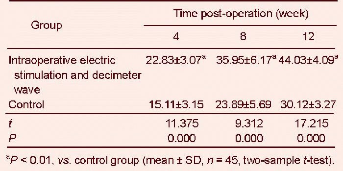
Sciatic functional index recovery rate in the intraoperative electric stimulation and decimeter wave group was significantly higher than that in the control group (P < 0.01; Table 2).
Table 2.
Effects of electric stimulation and decimeter wave on sciatic functional index at 4th, 8th and 12th weeks post-operation
DISCUSSION
Chronic peripheral nerve compression is a functional disorder of the peripheral nerves that results from chronic entrapment of specific parts of the nerve.
There are three basic pathological changes for chronic peripheral nerve compression: chronic ischemia; blood-neuron barrier changes; and severe Wallerian degeneration[34,35,36,37,38,39]. In the initial phase of entrapment, endoneurial fluid pressure increases, leading to edema in the endoneurium and perineurium, followed by progressive thickening of the epineurium, and finally, segmental demyelination in local nerve fibers[40,41,42,43,44,45,46]. Moreover, there are antegrade and retrograde axoplasmic flows in neural fibers, both of which can be blocked by entrapment[5,39,41]. Decompression, in releasing entrapment, eliminates the above-mentioned adverse factors, reduces neural compression, improves neural microcirculation, facilitates myelin sheath regeneration and modifies electrolyte concentration and distribution, all of which contribute to restoring neural functions[42,43,44,45,46].
It has been shown that electric stimulation promotes the regeneration of capillaries and leakage of certain elements, and then provides a good microenvironment for peripheral nerve repair and regeneration[47,48,49,50,51]. Electric stimulation could accelerate the Waller degeneration of distal nerve injury to promote the functional recovery of nerve cells. Studies have shown that electric stimulation begins shortly after nerve injury to promote nerve generation[47,48,49,50,51]. Technically, intraoperative electric stimulation during surgery is advantageous because of its accurate stimulation of the selected site. Moreover, it does not induce complications, such as infection and poor compliance, and patients suffer less pain, as compared with transcutaneous electric stimulation[52,53,54,55].
Decimeter wave therapy has been shown to inhibit inflammatory reaction and improve local circulation of the affected site; thus, the treatment enhances the metabolism of the affected area and reduces scar formation and adhesion[5,6,56]. In the present study, in the rats without decimeter wave therapy, extensive infiltration of inflammatory cells around the nerves and dense adhesion around tissues surrounding the nerve were observed. In contrast, at 1 week post-operation, decimeter wave therapy significantly reduced the inflammation and adhesion, and the inflammation was hardly observed by the 4th week after the operation. These findings demonstrate that decimeter waves could effectively inhibit the inflammatory reaction after injury, as well as improve the local circulation of the injured nerve and consequently reduce adhesion between the injured nerves. Furthermore, the treatment facilitated neural degeneration[57,58,59,60,61].
Here, we used S-100 protein as a Schwann cell marker to compare the effect of decimeter wave therapy on the proliferation of Schwann cells after nerve injury[62,63,64]. Previous immunohistochemical and immunocytochemical studies have shown that S-100 protein expression was limited in Schwann cells in the peripheral nervous system, and its expression was absent in axons[65,66,67,68,69]. However, S-100 protein expression increases when Schwann cells proliferate. Therefore, high levels of S-100 protein indicate active proliferation of Schwann cells, which has been shown to promote nerve regeneration[25,52,53,54,55]. Thus, treatment strategies that up- regulate S-100 protein expression may be beneficial for the enhancement of neural regeneration[70,71,72]. Results of the present study showed that S-100 protein expression levels in the compressed nerve from the rats with decimeter wave therapy were significantly higher than those in the rats without the treatment in various phases (data not shown), suggesting that decimeter waves induced S-100 protein expression in the regenerated nerves[73,74,75,76]. In conclusion, the decimeter wave therapy was effective in promoting the proliferation of Schwann cells and increasing S-100 protein expression levels, which might have been beneficial for nerve regeneration and neural repair.
The process of repair and regeneration of the peripheral nerves is characterized by the growth of the proximal end into the distal end. Therefore, regeneration status can be reflected by the quantity of regenerated axons and the degree of maturation. Our results from transmission electron microscopy showed that at 2 weeks post- operation, in the nerves from the rats treated with decimeter wave, many neonatal axon buds containing different types of organelles extended from the proximal end of the entrapped nerve. With time, regenerated myelin sheaths became thick, regenerated axons looked normal, and organelle structure was nearly normal at the proximal end of the axon. In contrast, there were no neonatal axon buds in the nerves from rats without the treatment. Quantitative analysis results further revealed that the number of myelinated nerve fibers and the diameter and thickness of myelin sheath in the decimeter wave therapy group were significantly greater than those in the group without treatment. These results indicated that the regenerated nerves in the treatment group were more mature than those in the rats without treatment. The nerves in the treatment group were characterized by a shorter latent period, faster conductivity and higher wave amplitude, suggesting that nerve function recovered better in the treatment group than in the non-treatment group.
This study illustrates the mechanism underlying electrical stimulation and decimeter wave therapy in the improvement of peripheral nerve regeneration. Our findings will provide a theoretical basis for clinical application.
MATERIALS AND METHODS
Design
A randomized, controlled animal experiment.
Time and setting
Experiments were performed at the Experimental Center of the First Hospital of Hebei Medical University, China, from January 2009 to February 2012.
Materials
A total of 90 healthy adult Sprague-Dawley rats weighing 200–250 g were purchased from the Animal Experiment Center of Hebei Medical University, China (license No. DK0408-0089). The protocols were conducted in accordance with the Guidance Suggestions for the Care and Use of Laboratory Animals, formulated by the Ministry of Science and Technology of China[77].
Methods
Preparation of chronically compressed nerve model of Mackinnon
Rats were fasted for 8 hours before operation, and then anesthetized with 1% pentobarbital sodium (30 mg/kg) (the Experimental Center of the First Hospital of Hebei Medical University, China) by intra-abdominal injection. The posterior median line of the left thigh was cut and the biceps femoris and semitendinosus muscle were separated. The sciatic nerves were exposed in the intermuscular space of the semi-membranous muscle. The diameter of the nerve was measured with a vernier caliper to be 1.0 mm. A silicone tube with a diameter of 1.0 mm and length of 6.0 mm was cut longitudinally and fixed to the nerve in the lower margin (10 mm) of the piriformis. The silicone tube was sutured with three stitches of non-invasive suture with an operating microscope. Thorough hemostasis and prophylactic therapy with 0.4 mL gentamicin was completed. Finally, the intermuscular space was closed and the skin was sutured (Figure 9A, B).
Figure 9.
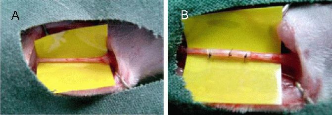
Morphology of sciatic nerves of normal rats (A) and model of Mackinnon (B).
Three experimental animals in each group were randomly selected at the 4th week post-operation. The original wound was opened and the silicone tube was observed to be wrapped with connective tissue without adhesion. Both ends of the entrapped segment became thickened with a fibroneuroma-like appearance, and the surface of the entrapped segment appeared pale. Conductivity was decreased below 1/6 of the normal value by electrophysiological recording. Moderate-to-severe demyelination was considered as pathological phenonmenon of the entrapped segment, indicating a successful operation[78] (Figure 10A, B).
Figure 10.
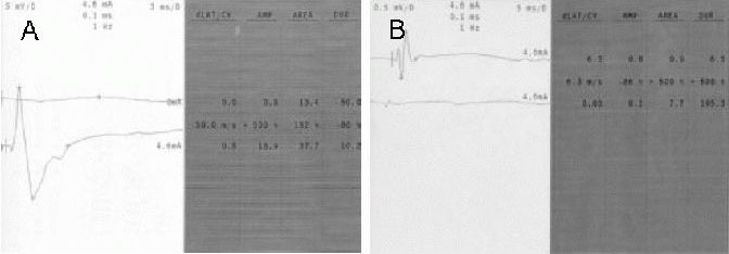
Electrophysiological indexes of normal rats (A) and model of Mackinnon (B).
Screenshot of electrophysiology recorder system. Left: Electrophysiological waves; right: the detected data.
Nerve release and electric stimulation
The original wounds were reopened and simple decompression was applied, such that the silicone tube was simply moved and the epineurium of the entrapped segment was not decompressed. The animals in the intraoperative electric stimulation and decimeter wave group were treated with electric stimulation. After thorough hemostasis, the intermuscular space was closed and the skin was sutured. A DXZ-1 polymorphism wave therapy instrument (Medical Equipment Technology Development Co., Ltd., Tianjin, China) was used in the present study. Parameters of frequency, intensity and treatment time were set to 2 Hz, 2 mA and 30 seconds, respectively.
Postoperative treatment of decimeter wave
Male and female animals were fed separately, and free movement was allowed. Rats of the intraoperative electric stimulation and decimeter wave group were fixed on a table prostrated and their right posterior thighs were exposed to decimeter wave every day from 1 day post-operation until sacrifice. The TMA-A double-frequency microwave hyperthermia therapeutic instrument (Beijing Electronic Medical Instrument Factory, Beijing, China) was used in the present study. Parameters of frequency, power and radiating distance were set to 915 MHz, 5 W and 10 cm, respectively. The treatment was performed once a day with duration of 10 minutes until sacrifice. Rats of the control group were also fixed on a table prostrated, but were not exposed to decimeter wave therapy.
Morphological observation of the sciatic nerves
Postoperative observations of wound healing included ulceration of the plantar and/or auto-phagocytic phenomenon. At the 1st, 2nd, 4th, 8th and 12th weeks post-operation, the neural segments in three animals from each group were observed. The entrapped segment of the sciatic nerve was exposed, and the lesions of the sciatic nerves and surrounding tissues and neuronal adhesion were grossly observed. Neural segments (2 mm) were obtained from the proximal and distal end of the entrapped segment, followed by 10% formalin fixation and hematoxylin-eosin staining. The nerve structure was observed under a light microscope (Shanghai Optical Instrument Factory, Shanghai, China).
Ultrastructural observation of the sciatic nerves under an electron microscope
At 2, 4 and 8 weeks post-operation, samples of the sciatic nerve from three animals from each group were selected. The samples were then fixed with 3% glutaraldehyde and embedded with resin. Thereafter, ultrathin sections were obtained to observe the neural regeneration conditions under an electron microscope (Japanese Electronics Co., Ltd., Tokyo, Japan).
S-100 expression in regenerating tissues as detected by immunohistochemistry
At the 3rd day and 1st, 2nd, 4th and 8th weeks post-operation, the samples from three animals in each group were selected to be cut into 5 μm-thick paraffin sections and stained by streptavidin peroxidase method. Paraffin sections were de-waxed, and then incubated in 3% H2O2 for 10 minutes to eliminate endogenous peroxidase. The sections were rinsed with water and soaked in PBS for 5 minutes. They were then blocked in 10% normal goat serum (diluted in PBS), rinsed, and incubated for 10 minutes at room temperature in a rabbit anti-rat S-100 antibody (Boster, Wuhan, Hubei Province, China) 1:100 for 1 hour at 37°C. The sections were washed in PBS three times for 5 minutes each, followed by incubation in biotinylated-secondary goat anti-rabbit antibody (1:100; Boster; diluted in 1% bovine serum albumin-PBS solution) at 37°C for 30 minutes. After washing in PBS three times, the sections were incubated in streptavidin-horseradish peroxidase at room temperature for 30 minutes. After washing in PBS, they were stained with 3,3′-diaminobenzidine and counterstained with hematoxylin. S-100 expression in regenerating tissues was observed under a light microscope (Shanghai Optical Instrument Factory, Shanghai, China).
Morphometric analysis of the sciatic nerves
At the 12th week post-operation, three animals were selected from each group to analyze the osmium tetroxide-stained distal end of the entrapped nerve; the number of axons, axon diameter, and myelin thickness were analyzed by image analyzer (Quantimet 970, Cambridge Instrument Co., UK). A total of 200–400 nerves in each field were chosen for analysis.
Electrophysiological assessment of the sciatic nerves
At 12 weeks post-operation, six animals were selected from each group to analyze muscle action potentials (EMG evoked instrument DISA-1500, Aalborg, Denmark). The maximum stimulation was 18 V, the stimulation phase was 0.1 ms, and the frequency was 1 Hz. Profiles of six animals were recorded and the latency, nerve conduction velocity and amplitude were measured.
Determination of sciatic functional index
A trough that opened at both ends was made; it was 60 cm long, 10 cm wide and 15 cm high. A sheet of white paper was spread into the bottom of trough. At the 4th, 8th and 12th weeks post-operation, the posterior limbs of rats were pigmented, and then the rats were put in at one end of the trough, induced to walk to the other end of the trough to a food reward. Each side of the posterior limbs left a 4–5 podogram, and a clear podogram was selected to measure three indicators of the normal foot (N) and the exceptional foot (E): (1) print length (PL): the greatest distance from the point of foot to the calcar pedis; (2) toes spread (TS): the distance from the first toe to the fifth toe; (3) intermediate toe (IT): the distance from the second toe to the fourth toe. Results were accurate to 0.1 mm. The data were put into the Bain formula to calculate sciatic functional index. Sciatic functional index = 0 for normal, −100 for complete injury. Bain formula is[79]:
Sciatic functional index = −38.3[(EPL-NPL)/NPL] + 109.5[(ETS–NTS)/NTS] + 13.3[(EIT–NIT)/NIT]−8.8.
Statistical analysis
All data were analyzed using SPSS 16.0 software (SPSS, Chicago, IL, USA). Measurement data were expressed as mean ± SD and a two-sample t-test was used for intergroup comparison. A value of P < 0.05 was considered statistically significant.
Acknowledgments
We received help and support from a teacher during the laboratory experiment and appreciated the hard work and enthusiasm.
Footnotes
Conflict of Interest: None declared.
Ethical approval: This study was approved by the Animal Ethics Committee of the Hebei Medical University, China.
(Reviewed by Dawes E, Rave W, Lv GM, Chen TY)
(Edited by Wang J, Qiu Y, Li CH, Song LP, Liu WJ, Zhao M)
REFERENCES
- [1].Yu KL, Li XM, Tian DH, et al. The mechanism of the effect of decimeter wave irradiation on the repair of acute injured peripheral nerves. Zhongguo Kangfu Yixue Zazhi. 2008;23(12):1089–1091. [Google Scholar]
- [2].Yao SQ, Zhang YZ, Tian DH, et al. The effect of decineter wave on endothelial cell and intinal hyperplasia of the vein graft. Zhongguo Kangfu Yixue Zazhi. 2006;21(1):11–14. [Google Scholar]
- [3].Tian DH, Li XM, Zhang Q, et al. The influence of decimeter wave on the regeneration of motor end plate after nerve injury. Zhongguo Kangfu Yixue Zazhi. 2008;23(11):976–978. [Google Scholar]
- [4].Tian DH, Luo J, Zhang Q, et al. Effects of decimeter wave and sodium hyaluronate product on postoperative adhesions in flexor tendon. Zhongguo Xiu Fu Chong Jian Wai Ke Za Zhi. 2008;22(11):1318–1322. [PubMed] [Google Scholar]
- [5].Tian DH, Zhao M, Wang LM, et al. Effect of enhancing peripheral nerve regeneration with combined physical factors. Zhongguo Kangfu Yixue Zazhi. 2007;22(2):100–102. [Google Scholar]
- [6].Zhang YL, Tian DH, Zhang YZ, et al. Observation of the therapeutic effects of comprehensive rehabilitation on peripheral nerve complete impairment. Zhongguo Kangfu Yixue Zazhi. 2008;23(11):1001–1003. [Google Scholar]
- [7].Tian DH, Zhang YZ, Zhao F, et al. Mechanisms of decimeter wave on the regeneration of peripheral nerves. Zhongguo Kangfu Yixue Zazhi. 2005;20(4):261–263. [Google Scholar]
- [8].Zhang LX, Tong XJ, Sun XH, et al. Experimental study of low dose ultrashortwave promoting nerve regeneration after acellular nerve allografts repairing the sciatic nerve gap of rats. Cell Mol Neurobiol. 2008;28(4):501–509. doi: 10.1007/s10571-007-9226-1. [DOI] [PMC free article] [PubMed] [Google Scholar]
- [9].Sun XH, Che YQ, Tong XJ, et al. Improving nerve regeneration of acellular nerve allografts seeded with SCs bridging the sciatic nerve defects of rat. Cell Mol Neurobiol. 2009;29(3):347–353. doi: 10.1007/s10571-008-9326-6. [DOI] [PMC free article] [PubMed] [Google Scholar]
- [10].Zhang LX, Tong XJ, Yuan XH, et al. Effects of 660-nm gallium-aluminum-arsenide low-energy laser on nerve regeneration after acellular nerve allograft in rats. Synapse. 2010;64(2):152–160. doi: 10.1002/syn.20724. [DOI] [PubMed] [Google Scholar]
- [11].Tian DH, Zhao F, Zhang YZ. Research progress of peripheral nerve compression. Zhongguo Kangfu Yixue Zazhi. 2007;22(2):85–87. [Google Scholar]
- [12].Li GF, Tian DH, Zhang YZ, et al. Rehabilitation treatment in peripheral nerve compression. Zhongguo Kangfu Yixue Zazhi. 2007;22(2):178–179. [Google Scholar]
- [13].Tian DH. Peripheral nerve injury and rehabilitation. Zhongguo Kangfu Yixue Zazhi. 2007;22(2):99. [Google Scholar]
- [14].Yao CH, Chang RL, Chang SL, et al. Electrical stimulation improves peripheral nerve regeneration in streptozotocin-induced diabetic rats. J Trauma Acute Care Surg. 2012;72(1):199–205. doi: 10.1097/TA.0b013e31822d233c. [DOI] [PubMed] [Google Scholar]
- [15].Inoue M, Katsumi Y, Itoi M, et al. Direct current electrical stimulation of acupuncture needles for peripheral nerve regeneration: an exploratory case series. Acupunct Med. 2011;29(2):88–93. doi: 10.1136/aim.2010.003046. [DOI] [PubMed] [Google Scholar]
- [16].Inoue M, Hojo T, Nakajima M, et al. The effect of electrical stimulation of the pudendal nerve on sciatic nerve blood flow in animals. Acupunct Med. 2008;26(3):145–148. doi: 10.1136/aim.26.3.145. [DOI] [PubMed] [Google Scholar]
- [17].Inoue M, Hojo T, Yano T, et al. Effects of lumbar acupuncture stimulation on blood flow to the sciatic nerve trunk--an exploratory study. Acupunct Med. 2005;23(4):166–170. doi: 10.1136/aim.23.4.166. [DOI] [PubMed] [Google Scholar]
- [18].Inoue M, Hojo T, Yano T, et al. Electroacupuncture direct to spinal nerves as an alternative to selective spinal nerve block in patients with radicular sciatica--a cohort study. Acupunct Med. 2005;23(1):27–30. doi: 10.1136/aim.23.1.27. [DOI] [PubMed] [Google Scholar]
- [19].Hausner T, Pajer K, Halat G, et al. Improved rate of peripheral nerve regeneration induced by extracorporeal shock wave treatment in the rat. Exp Neurol. 2012;236(2):363–370. doi: 10.1016/j.expneurol.2012.04.019. [DOI] [PubMed] [Google Scholar]
- [20].Shen CC, Yang YC, Liu BS. Effects of large-area irradiated laser phototherapy on peripheral nerve regeneration across a large gap in a biomaterial conduit. J Biomed Mater Res A. 2013;101(1):239–252. doi: 10.1002/jbm.a.34314. [DOI] [PubMed] [Google Scholar]
- [21].Tian DH, Zhang YZ, Mi LX, et al. Effects of decimeter wave on the expression of immunologic reaction to s-100 protein in Schwann's cell after peripheral nerve injury. Zhongguo Kangfu Yixue Zazhi. 2004;19(4):269–271. [Google Scholar]
- [22].Sun H, Yang T, Li Q, et al. Dexamethasone and vitamin B(12) synergistically promote peripheral nerve regeneration in rats by upregulating the expression of brain-derived neurotrophic factor. Arch Med Sci. 2012;8(5):924–930. doi: 10.5114/aoms.2012.31623. [DOI] [PMC free article] [PubMed] [Google Scholar]
- [23].Liu YR, Liu Q. Meta-analysis of mNGF therapy for peripheral nerve injury: a systematic review. Chin J Traumatol. 2012;15(2):86–91. [PubMed] [Google Scholar]
- [24].Young C, Miller E, Nicklous DM, et al. Nerve growth factor and neurotrophin-3 affect functional recovery following peripheral nerve injury differently. Restor Neurol Neurosci. 2001;18(4):167–175. [PubMed] [Google Scholar]
- [25].Patodia S, Raivich G. Role of transcription factors in peripheral nerve regeneration. Front Mol Neurosci. 2012;5(10):8. doi: 10.3389/fnmol.2012.00008. [DOI] [PMC free article] [PubMed] [Google Scholar]
- [26].Takagi T, Kimura Y, Shibata S, et al. Sustained bFGF- release tubes for peripheral nerve regeneration: comparison with autograft. Plast Reconstr Surg. 2012;130(4):866–876. doi: 10.1097/PRS.0b013e318262f36e. [DOI] [PubMed] [Google Scholar]
- [27].Saceda J, Isla A, Santiago S, et al. Effect of recombinant human growth hormone on peripheral nerve regeneration: experimental work on the ulnar nerve of the rat. Neurosci Lett. 2011;504(2):146–150. doi: 10.1016/j.neulet.2011.09.020. [DOI] [PubMed] [Google Scholar]
- [28].Sorensen J, Haase G, Krarup C, et al. Gene transfor to Schwann cells after peripheral nerve injury: a delivery systerm for therapeatic agents. Ann Neurol. 1998;43(2):205–211. doi: 10.1002/ana.410430210. [DOI] [PubMed] [Google Scholar]
- [29].Turgut M, Kaplan S. Effects of melatonin on peripheral nerve regeneration. Recent Pat Endocr Metab Immune Drug Discov. 2011;5(2):100–108. doi: 10.2174/187221411799015336. [DOI] [PubMed] [Google Scholar]
- [30].Hsiang SW, Lee HC, Tsai FJ, et al. Puerarin accelerates peripheral nerve regeneration. Am J Chin Med. 2011;39(6):1207–1217. doi: 10.1142/S0192415X11009500. [DOI] [PubMed] [Google Scholar]
- [31].Wu D, Raafat A, Pak E, et al. Dicer-microRNA pathway is critical for peripheral nerve regeneration and functional recovery in vivo and regenerative axonogenesis in vitro. Exp Neurol. 2012;233(1):555–565. doi: 10.1016/j.expneurol.2011.11.041. [DOI] [PMC free article] [PubMed] [Google Scholar]
- [32].Weidner N, Blesch A, Grill RJ, et al. Nerve growth factor-hypersecreting Schwann cell grafts augment and guide spinal cord axonal growth and remyelinate central nervous system axons in a phenotypically appropriate manner that correlates with expression of L1. J Comp Neurol. 1999;413(4):495–506. doi: 10.1002/(sici)1096-9861(19991101)413:4<495::aid-cne1>3.0.co;2-z. [DOI] [PubMed] [Google Scholar]
- [33].Mandel RJ, Snyder RO, Leff SE. Recombinant adenoassociated viral vector-mediated glial cell line-derived neurotrophic factor gene transfer protects nigral dopamine neurons after onset of progressive degeneration in a rat model of Parkinson's disease. Exp Neurol. 1999;160(1):205–214. doi: 10.1006/exnr.1999.7203. [DOI] [PubMed] [Google Scholar]
- [34].Ahmed MR, Basha SH, Gopinath D, et al. Initial upregulation of growth factors and inflammatory mediators during nerve regeneration in the presence of cell adhesive peptide-incorporated collagen tubes. J Peripher Nerv Syst. 2005;10(1):17–30. doi: 10.1111/j.1085-9489.2005.10105.x. [DOI] [PubMed] [Google Scholar]
- [35].Ikeda M, Oka Y. The relationship between nerve conduction velocity and fiber morphology during peripheral nerve regeneration. Brain Behav. 2012;2(4):382–390. doi: 10.1002/brb3.61. [DOI] [PMC free article] [PubMed] [Google Scholar]
- [36].Maffei L, Carmignoto G, Perry VH, et al. Schwann cells promote the survival of rat retinal ganglion cells after optic nerve section. Proc Natl Acad Sci USA. 1990;87(5):1855–1859. doi: 10.1073/pnas.87.5.1855. [DOI] [PMC free article] [PubMed] [Google Scholar]
- [37].Terenghi G. Peripheral nerve injury and regeneration. Histol Histopathol. 1995;10(3):709–718. [PubMed] [Google Scholar]
- [38].Yan Y, Sun HH, Hunter DA, et al. Efficacy of short-term FK506 administration on accelerating nerve regeneration. Neurorehabil Neural Repair. 2012;26(6):570–580. doi: 10.1177/1545968311431965. [DOI] [PubMed] [Google Scholar]
- [39].Wang Y, Zhang P, Yin X, et al. Characteristics of peripheral nerve regeneration following a second nerve injury and repair. Artif Cells Blood Substit Immobil Biotechnol. 2012;40(4):296–302. doi: 10.3109/10731199.2011.652259. [DOI] [PubMed] [Google Scholar]
- [40].Avellino AM, Hart D, Dailey AT, et al. Differential macrophage responses in the peripheral and central nervous system during wallerian degeneration of axons. Exp Neurol. 1995;136(2):183–198. doi: 10.1006/exnr.1995.1095. [DOI] [PubMed] [Google Scholar]
- [41].Podhajsky RJ, Myers RR. A diffusion-reaction model of nerve regeneration. J Neurosci Methods. 1995;60(1-2):79–88. doi: 10.1016/0165-0270(94)00222-3. [DOI] [PubMed] [Google Scholar]
- [42].Torigoe K, Tanaka HF, Takahashi A, et al. Basic behavior of migratory Schwann cells in peripheral nerve regeneration. Exp Neurol. 1996;137(2):301–308. doi: 10.1006/exnr.1996.0030. [DOI] [PubMed] [Google Scholar]
- [43].Torigoe K, Hashimoto K, Lundhorg G. A role of migratory Schwann cells in a conditioning effect of peripheral nerve regeneration. Exp Neurol. 1999;160(1):99–108. doi: 10.1006/exnr.1999.7202. [DOI] [PubMed] [Google Scholar]
- [44].Bryan DJ, Wang KK, Summerhayes C. Migration of Schwann cells in peripheral nerve regeneration. J Reconstr Microsurg. 1999;15(8):591–596. doi: 10.1055/s-2007-1000143. [DOI] [PubMed] [Google Scholar]
- [45].Kater SB, Mills LR. Regulation of growth cone behavior by calcium. J Neurosci. 1991;11(4):891–899. doi: 10.1523/JNEUROSCI.11-04-00891.1991. [DOI] [PMC free article] [PubMed] [Google Scholar]
- [46].Doherty P, Ashton SV, Moore SE, et al. Morphoregulatory activities of NCAM and N-cadherin can be accounted by G protein-dependent activation of L-and N-type neuronal Ca2+ channels. Cell. 1991;67(1):21–33. doi: 10.1016/0092-8674(91)90569-k. [DOI] [PubMed] [Google Scholar]
- [47].Ansselin AD, Pollard JD. Immunopathological factors in peripheral nerve allograft rejection: quantification of lymphocyte invasion and major histocompatility complex expression. J Neurol Sci. 1990;96(1):75–88. doi: 10.1016/0022-510x(90)90058-u. [DOI] [PubMed] [Google Scholar]
- [48].Hoang NS, Sar C, Valmier J, et al. Electro-acupuncture on functional peripheral nerve regeneration in mice: a behavioural study. BMC Complement Altern Med. 2012;31(12):141. doi: 10.1186/1472-6882-12-141. [DOI] [PMC free article] [PubMed] [Google Scholar]
- [49].Zochodne DW, Cheng C. Neurotrophins and other growth factors in the regenerative milieu of proximal nerve stump tips. J Anat. 2000;196(2):279–283. doi: 10.1046/j.1469-7580.2000.19620279.x. [DOI] [PMC free article] [PubMed] [Google Scholar]
- [50].Chernousov MA, Carey DJ. Schwann cell extracellular matrix molecules and their receptors. Histol Histopathol. 2000;15(2):593–601. doi: 10.14670/HH-15.593. [DOI] [PubMed] [Google Scholar]
- [51].Liu X, Wang J, Dai K. Research advance of treatment of peripheral nerve injury with neuromuscular electric stimulation. Zhongguo Xiu Fu Chong Jian Wai Ke Za Zhi. 2010;24(5):622–627. [PubMed] [Google Scholar]
- [52].Schmidt A. Nerve compression syndromes in the area of the elbow joint in patients with chronic polyarthritis. Review of the literature. Handchir Mikrochir Plast Chir. 1993;25(2):75–79. [PubMed] [Google Scholar]
- [53].Kerns JM, Fakhouri AJ, Weinrib HP, et al. Electrical stimulation of nerve regeneration in the rat: the early effects evaluated by vibrating probe and electron microscopy. Neuroscience. 1991;40(1):93–107. doi: 10.1016/0306-4522(91)90177-p. [DOI] [PubMed] [Google Scholar]
- [54].Zanakis MF. Differential effects of various electrical parameters on peripheral and central nerve regeneration. Acupunct Electrother Res. 1990;15(3-4):185–191. doi: 10.3727/036012990816358199. [DOI] [PubMed] [Google Scholar]
- [55].Rajaram A, Chen XB, Schreyer DJ. Strategic design and recent fabrication techniques for bioengineered tissue scaffolds to improve peripheral nerve regeneration. Tissue Eng Part B Rev. 2012;18(6):454–467. doi: 10.1089/ten.TEB.2012.0006. [DOI] [PubMed] [Google Scholar]
- [56].Gupta R, Rowshan K, Chao T, et al. Chronic nerve compression induces local demyelination and remyelination in a rat model of carpal tunnel syndrome. Exp Neurol. 2004;187(2):500–508. doi: 10.1016/j.expneurol.2004.02.009. [DOI] [PubMed] [Google Scholar]
- [57].Mendonca AC, Barbieri CH, Mazzer N. Directly applied low intensity direct electric current enhances peripheral nerve regeneration in rats. J Neurosci Methods. 2003;129(2):183–190. doi: 10.1016/s0165-0270(03)00207-3. [DOI] [PubMed] [Google Scholar]
- [58].Popović M, Bresjanac M, Sketelj J. Role of axon-deprived schwann cells in perineurial regeneration in the ratsciatic nerve. Neuropathol Appl Neurohiol. 2000;26(3):221–231. doi: 10.1046/j.1365-2990.2000.00238.x. [DOI] [PubMed] [Google Scholar]
- [59].Hare GM, Evans PJ, Mackinnon SE, et al. Walking track analysis: a long-term assessment of periphreal nerve recovery. Plast Reconstr Surg. 1992;89(2):251–258. [PubMed] [Google Scholar]
- [60].Badalamente MA, Hurst LC, Paul SB, et al. Enhancement of neuromuscular recovery after nerve repair in primates. J Hand Surg. 1987;12(2):211–217. doi: 10.1016/0266-7681_87_90015-5. [DOI] [PubMed] [Google Scholar]
- [61].Tian DH, Zhang YZ, Zhao F, et al. Effect of decameter wave on the expression of NGF mRNA in regenerated nerve. Zhonghua Wuli Yixue yu Kangfu Zazhi. 2005;27(3):141–144. [Google Scholar]
- [62].Shakhbazau A, Kawasoe J, Hoyng SA, et al. Early regenerative effects of NGF-transduced Schwann cells in peripheral nerve repair. Mol Cell Neurosci. 2012;50(1):103–112. doi: 10.1016/j.mcn.2012.04.004. [DOI] [PubMed] [Google Scholar]
- [63].Dubový P, Svízenská I, Klusáková I, et al. Laminin molecules in freeze-treated nerve segments are associated with migrating Schwann cells that display the corresponding alpha6beta1 integrin receptor. Glia. 2001;33(1):36–44. doi: 10.1002/1098-1136(20010101)33:1<36::aid-glia1004>3.3.co;2-2. [DOI] [PubMed] [Google Scholar]
- [64].Dezawa M, Adachi-Usami E. Role of Schwann cells in retinal ganglion cell axon regeneration. Prog Retin Eye Res. 2000;19(2):171–204. doi: 10.1016/s1350-9462(99)00010-5. [DOI] [PubMed] [Google Scholar]
- [65].Kobayashi M, Ishibashi S, Tomimitsu H, et al. Proliferating immature Schwann cells contribute to nerve regeneration after ischemic peripheral nerve injury. J Neuropathol Exp Neurol. 2012;71(6):511–519. doi: 10.1097/NEN.0b013e318257fe7b. [DOI] [PubMed] [Google Scholar]
- [66].McGrath AM, Novikova LN, Novikov LN, et al. BDTM PuraMatrixTM peptide hydrogel seeded with Schwann cells for peripheral nerve regeneration. Brain Res Bull. 2010;83(5):207–213. doi: 10.1016/j.brainresbull.2010.07.001. [DOI] [PubMed] [Google Scholar]
- [67].Matsuse D, Kitada M, Kohama M, et al. Human umbilical cord-derived mesenchymal stromal cells differentiate into functional Schwann cells that sustain peripheral nerve regeneration. J Neuropathol Exp Neurol. 2010;69(9):973–985. doi: 10.1097/NEN.0b013e3181eff6dc. [DOI] [PubMed] [Google Scholar]
- [68].Liu H, Kim Y, Chattopadhyay S, et al. Matrix metalloproteinase inhibition enhances the rate of nerve regeneration in vivo by promoting dedifferentiation and mitosis of supporting schwann cells. J Neuropathol Exp Neurol. 2010;69(4):386–395. doi: 10.1097/NEN.0b013e3181d68d12. [DOI] [PMC free article] [PubMed] [Google Scholar]
- [69].Torigoe K, Hashimoto K, Lundborg G. A role of migratory Schwann cells in a conditioning effect of peripheral nerve regeneration. Exp Neurol. 1999;160(1):99–108. doi: 10.1006/exnr.1999.7202. [DOI] [PubMed] [Google Scholar]
- [70].Bryan DJ, Wang KK, Summerhayes C. Migration of Schwann cells in peripheral-nerve regeneration. J Reconstr Microsurg. 1999;15(8):591–596. doi: 10.1055/s-2007-1000143. [DOI] [PubMed] [Google Scholar]
- [71].Blottner D, Baumgarten HG. Neurotrophy and regeneration in vivo. Acta Anat (Basel) 1994;150(4):235–245. doi: 10.1159/000147626. [DOI] [PubMed] [Google Scholar]
- [72].Bajrovic F, Bresjanac M, Sketelj J. Long-term effects of deprivation of cell support in the distal stump on peripheral nerve regeneration. J Neurosci Res. 1994;39(1):23–30. doi: 10.1002/jnr.490390104. [DOI] [PubMed] [Google Scholar]
- [73].Miyauchi A, Kanje M, Danielsen N, et al. Role of macrophages in the stimulation and regeneration of sensory nerves by transposed granulation tissue and temporal aspects of the response. Scand J Plast Reconstr Surg Hand Surg. 1997;31(1):17–23. doi: 10.3109/02844319709010501. [DOI] [PubMed] [Google Scholar]
- [74].Krieglstein K, Richter S, Farkas L, et al. Reduction of endogenous transforming growth factors beta prevents ontogenetic neuron death. Nat Neurosci. 2000;3(11):1085–1090. doi: 10.1038/80598. [DOI] [PubMed] [Google Scholar]
- [75].Einheber S, Hannocks MJ, Metz CN, et al. Transforming growth factor-beta 1 regulates axon/Schwann cell interactions. J Cell Biol. 1995;129(2):443–458. doi: 10.1083/jcb.129.2.443. [DOI] [PMC free article] [PubMed] [Google Scholar]
- [76].Nath RK, Kwon B, Mackinnon SE, et al. Antibody to transforming growth factor beta reduces collagen production in injured peripheral nerve. Plast Reconstr Surg. 1998;102(4):1100–1108. [PubMed] [Google Scholar]
- [77].The Ministry of Science and Technology of the People's Republic of China. Guidance Suggestions for the Care and Use of Laboratory Animals. 2006 Sep 30; [Google Scholar]
- [78].Mackinnon SE, Dellon AL, Hudson AR, et al. A primate model for chronic nerve compression. J Reconstr Mcrosurg. 1985;1(3):185–195. doi: 10.1055/s-2007-1007073. [DOI] [PubMed] [Google Scholar]
- [79].Hare GM, Evans PJ, Mackinnon SE, et al. Walking track analysis: utilization of individual footprint parameters. Ann Plast Surg. 1993;30(2):147–153. doi: 10.1097/00000637-199302000-00009. [DOI] [PubMed] [Google Scholar]


