

Keywords: neural regeneration, transient receptor potential channel A1, calcitonin gene-related peptide, dorsal root ganglion neurons, pain, hyperalgesia, noxious stimuli, sensory neuron, grants-supported paper, neuroregeneration
Abstract
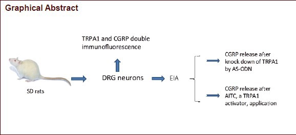
Transient receptor potential channel A1 is one of the important transducers of noxious stimuli in the primary afferents, which may contribute to generation of neurogenic inflammation and hyperalgesia. The present study was designed to investigate if activation of transient receptor potential channel A1 may induce calcitonin gene-related peptide release from the primary afferent neurons. We found that application of allyl isothiocyanate, a transient receptor potential channel A1 activator, caused calcitonin gene-related peptide release from the cultured rat dorsal root ganglion neurons. Knockdown of transient receptor potential channel A1 with an antisense oligodeoxynucleotide prevented calcitonin gene-related peptide release by allyl isothiocyanate application in cultured dorsal root ganglion neurons. Thus, we concluded that transient receptor potential channel A1 activation caused calcitonin gene-related peptide release in sensory neurons.
INTRODUCTION
The transient receptor potential (TRP) channels constitute a large and diverse family of channel proteins that are expressed in many tissues and cell types in both vertebrates and invertebrates. Transient receptor potential channel A1 (TRPA1) is a member of branch A of the TRP family of cation channels[1]. Transient receptor potential channel A1 has been reported to form channels activated by icilin, a chemical that induces a cooling sensation, and by temperatures ≤ 17°C[2]. This channel was also reported to be activated by some pungent chemicals, such as horseradish, mustard oil, cinnamon oil and allicin[1,2,3,4,5]. A recent study using knockout mice demonstrated that transient receptor potential channel A1 is an important component of the transduction machinery through which environmental irritants and endogenous proalgesic agents depolarize nociceptors to elicit inflammatory pain[6,7,8,9,10,11,12,13,14,15,16]. Taking the above into account, it is clear that this channel is one of the important transducers of noxious stimuli in the primary afferents.
Activation of channels (such as transient receptor potential V1, a member of branch V of the transient receptor potential family) elicits acute pain accompanied by vasodilation, vascular leakage, and inflammation due to peripheral release of peptides (such as substance P and calcitonin gene-related peptide [CGRP] from the activated nerve terminals[17,18,19,20,21,22,23,24]. However, there is no direct evidence that activation of transient receptor potential channel A1 contributes to neuropeptide release from primary afferent neurons. Calcitonin gene-related peptide as a neurotransmitter in transient receptor potential channel A1-mediated pain sensation (such as migraine) has been suggested to be dominant over substance P[25,26]. In the present study, we demonstrated for the first time that allyl isothiocyanate (AITC; also known as mustard oil) activates transient receptor potential channel A1, resulting in calcitonin gene-related peptide release from the cultured rat dorsal root ganglion (DRG) neurons.
RESULTS
Calcitonin gene-related peptide was co-expressed with transient receptor potential channel A1 in dorsal root ganglion neurons and released after transient receptor potential channel A1 activation
To investigate if transient receptor potential channel A1 activation can induce calcitonin gene-related peptide release in dorsal root ganglion neurons, we first examined the co-localization of transient receptor potential channel A1 and calcitonin gene-related peptide, using double immunofluorescence. We found that 39.9 % of transient receptor potential channel A1-labeled neurons were also labeled with calcitonin gene-related peptide. Further, 38.3% of calcitonin gene-related peptide-positive neurons also contained transient receptor potential channel A1 (Figure 1A, B). This indicates a possibility for calcitonin gene-related peptide release from transient receptor potential channel A1-labeled neurons. Then we applied a transient receptor potential channel A1 activator, AITC to the cultured dorsal root ganglion neurons, and measured calcitonin gene-related peptide release by calcitonin gene-related peptide enzyme-linked immunoassay (CGRP-EIA) kit from the culture supernatant. The content of calcitonin gene- related peptide was increased within 3 minutes after AITC application. A significant and concentration-dependent increase of calcitonin gene-related peptide-induced by AITC was observed (Figure 1C).
Figure 1.

Calcitonin gene-related peptide (CGRP) is co-expressed with transient receptor potential channel A1 (TRPA1) and released by allyl isothiocyanate (AITC) application.
(A) Double immunofluorescence of TRPA1 (green) and CGRP (red) shows that a large population of TRPA1 is co-localized with CGRP in the rat DRG neurons. Yellow indicates double labeling. Scale bar: 100 μm. (B) Percentages of colocalization of CGRP and TRPA1 in the dorsal root ganglion (DRG) neurons (A1: TRPA1). Data are expressed as mean ± SEM from four rats in each group. (C) AITC (50 μmol/L and 500 μmol/L) induced a significant increase in CGRP release. Data were collected from six rats in each group. CGRP releases were determined in parallel by enzyme-linked immunoassay from the same incubation fluid samples. aP < 0.05, bP < 0.01, vs. control. One-way analysis of variance followed by Fisher's protected least significant difference (PLSD) was used.
Knockdown of transient receptor potential channel A1 decreased calcitonin gene-related peptide release from cultured dorsal root ganglion neurons
To further confirm the AITC-induced calcitonin gene-related peptide release is through transient receptor potential channel A1 activation indeed, we knocked down transient receptor potential channel A1 (see method) and tested calcitonin gene-related peptide release from the transient receptor potential channel A1-knockdowned neurons.
The antisense oligodeoxynucleotide (AS-ODN) targeting to transient receptor potential channel A1 has been used in our previous studies and has been demonstrated to be able to inhibit transient receptor potential channel A1 mRNA synthesis in vivo[27,28]. In the current study, we tested the effect of AS-ODN (100 μmol/L) on protein synthesis of transient receptor potential channel A1 in cultured dorsal root ganglion neurons. Results of western blot analysis indicated that AS-ODN significantly decreased transient receptor potential channel A1 expression in cultured dorsal root ganglion neurons, compared with control or the mismatch oligodeoxynucleotide (MM-ODN; 100 μmol/L) treated neurons (Figure 2A, B). As expected, knockdown of transient receptor potential channel A1 significantly suppressed AITC-evoked calcitonin gene-related peptide release in cultured dorsal root ganglion neurons (Figure 2C).
Figure 2.

Knockdown of transient receptor potential channel A1 (TRPA1) by antisense oligodeoxynucleotide (AS-ODN) decreased calcitonin gene-related peptide (CGRP) release from cultured dorsal root ganglion (DRG) neurons.
(A, B) The raised TRPA1 antibody recognized an expected band (~128 kDa) in the western blot from the naive and AS-ODN (or mismatch oligodeoxynucleotide [MM-ODN])-treated rat DRG lysate (A). AS-ODN but not MM-ODN (100 μmol/L) significantly decreased TRPA1 expression (B). aP < 0.05, vs. naive. Data are expressed as mean ± SEM from three rats in each group. The experiment was repeated at least twice. (C) AS-ODN (100 μmol/L) treatment significantly decreased allyl isothiocyanate (AITC; 50 μmol/L)-induced CGRP release. Data were collected from six rats in each group. The experiment was repeated at least twice. CGRP releases were determined in parallel by enzyme-linked immunoassay from the same incubation fluid samples. The data were normalized to that without AITC application. aP < 0.05, vs. control. One-way analysis of variance followed by Fisher's protected least significant difference was used.
DISCUSSION
In this study, we investigated if activation of transient receptor potential channel A1 may induce calcitonin gene-related peptide release from primary afferent. We found that transient receptor potential channel A1 and calcitonin gene-related peptide were highly co-expressed in rat DGR neurons. AITC could induce calcitonin gene-related peptide release from the cultured rat dorsal root ganglion neurons and knockdown of transient receptor potential channel A1 prevented calcitonin gene-related peptide release. These data provide a critical role of transient receptor potential channel A1 in neurotransmission.
Morphological evidence shows that calcitonin gene-related peptide is contained in populations of dorsal root ganglion neurons that overlap with populations of nociceptive primary afferent neurons[29,30,31]. Calcitonin gene-related peptide immunoreactivity is found in approximately 60% of the small neurons that are believed to give rise to C-fibers[32]. Transient receptor potential channel A1 is also expressed in a subset of small-sized dorsal root ganglions or trigeminal ganglia neurons in adult rats or mice[4,27,33]. We showed that 39.9% of transient receptor potential channel A1 positive neurons were also labeled with calcitonin gene-related peptide, which was lower than that in a previous study[7], indicating that 97% of transient receptor potential channel A1-positive neurons were calcitonin gene-related peptide -positive in adult mice. The difference of the results may be due to the different methodologies that were used in two studies (immunofluorescence versus in situ hybridization). In any case, co-localization of these two molecules gave the morphological evidence for transient receptor potential channel A1 activation induced calcitonin gene-related peptide release.
AITC has been used for activating transient receptor potential channel A1 in many previous reports, but its specificity for transient receptor potential channel A1 activation remained unclear. A calcium imaging study revealed that AITC still induced calcium entry in a part of dorsal root ganglion neurons of transient receptor potential channel A1 null mice[6]. To make sure that AITC-evoked calcitonin gene-related peptide release was due to the transient receptor potential channel A1 activation, we knocked down transient receptor potential channel A1 using the AS-ODN, which has been demonstrated to be able to significantly decrease transient receptor potential channel A1 mRNA in our previous studies[27,28], and also inhibit transient receptor potential channel A1 protein expression. Knockdown of transient receptor potential channel A1 significantly reduced the calcitonin gene-related peptide release in cultured dorsal root ganglion neurons. These data demonstrated that the AITC-evoked calcitonin gene-related peptide release from dorsal root ganglion neurons was mediated by transient receptor potential channel A1 activation. In our experiment, the content of calcitonin gene-related peptide was increased within 3 minutes after AITC application. The AITC-induced activation of transient receptor potential channel A1 may induce a significant extracellular calcium influx, and then the increased intracellular calcium can evoke calcitonin gene- related peptide release rapidly[34].
Nociceptor activation may release peptides, such as substance P and calcitonin gene-related peptide peripherally to produce vascular leakage and vasodilation, leading to inflammation and tenderness at the site of irritant application[35,36,37,38]. Besides the peripheral effects, calcitonin gene-related peptide may contribute to the neurotransmission of nociceptive primary afferent neurons in the spinal cord[19,39]. Considering the various effects of calcitonin gene-related peptide in peripheral and central nerve system, the transient receptor potential channel A1-mediated calcitonin gene-related peptide release in the present study provides a critical evidence for realizing the function of transient receptor potential channel A1 channel.
MATERIALS AND METHODS
Materials
Adult male Sprague-Dawley rats (4–5 weeks of age, 100 –150 g body weight; Japanese animals, Shizuoka, Japan) were used in this study. Rats were housed at a temperature of 22°C, and were fed food and water ad libitum.
Methods
Immunohistochemistry study
Rats were deeply anesthetized with sodium pentobarbital and perfused transcardially with 1% paraformaldehyde in 0.1 mol/L phosphate-buffer (PB) (pH 7.4), followed by 4% paraformaldehyde in 0.1 mol/L PB. The L4−5 dorsal root ganglions were dissected out and processed for transient receptor potential channel A1 and calcitonin gene-related peptide double immunofluorescence according to the procedure of our previous study[40]. The tyramide signal amplification (TSA) (NEN Life Science Products, Boston, MA, USA) fluorescence procedures were used for transient receptor potential channel A1 (1:10 000) staining. Then, the rabbit polyclonal primary antibody for transient receptor potential channel A1 at 1:10 000 was combined with rabbit polyclonal calcitonin gene-related peptide antibody (1:2 000; Amersham International plc, Buckinghamshire, England). The preparation of the polyclonal transient receptor potential channel A1 antibody was performed according to our previous study[41]. Goat anti-rabbit-Alexa 488 (1:1 000; Molecular Probes, Eugene, OR, USA) for transient receptor potential channel A1 and goat anti-rabbit-Alexa 594 (1:1 000; Molecular Probes) for calcitonin gene-related peptide were used as second antibodies. Non-specific double labeling was not observed in the present study. In control single labeling using indirect labeled immunofluorescence, we were unable to visualize the transient receptor potential channel A1 antiserum at the dilutions used for the TSA procedure. For quantification, 8–12 sections of the L4−5 dorsal root ganglion were selected randomly in each rat. The number of transient receptor potential channel A1 or calcitonin gene-related peptide immunopositive neurons per section was counted. An averaged percentage of transient receptor potential channel A1-labeled neurons relative to calcitonin gene-related peptide-labeled neurons were determined.
Primary culture of rat dorsal root ganglion neurons
Dorsal root ganglions were collected using sterile techniques, and placed in ice-cold Earle's balanced salt solution (EBSS; Sigma, St. Louis, MO, USA). Adhering fat and connective tissue were removed and each dorsal root ganglion was minced with scissors and placed immediately in a medium consisting of 2 mL of EBSS containing 1.25 mg/mL of collagenase P (Roche Diagnostics GmbH, Mannheim, Germany) and kept at 37°C for 60 minutes with occasional agitation. After dissociation of the dorsal root ganglion cells, this cell suspension was centrifuged for 5 minutes at 200 × g and the cell pellets were resuspended in EBSS supplemented with 10% fetal bovine serum (FBS), 2 mmol/L glutamax, penicillin/ streptomycin and recombinant rat NGF (100 ng/mL; R& D Systems, Inc, Minneapolis, MN, USA). Neurons were counted using a haemocytometer and suspension was diluted to get a final cell density of 30 000 neurons/mL. Cells in 500 μL suspension were plated onto poly-L-lysine-coated 24-well plates for enzyme immunoassay study. Cells in 2 mL suspension were plated onto a 35 mm dish for western blot analysis. The neurons were then kept in a 5% CO2 incubator at 37°C overnight. An AS-ODN (5′-TCTATGCGGTTATGTTGG-3′) targeting transient receptor potential channel A1 and an MM-ODN (5′-ACTACTACACTAGACTAC-3′) were designed and manufactured by BIOGNOSTIK (Gottingen, Lower Saxony, Germany). In some case, the AS-ODN or MM-ODN was applied onto the cultures at a concentration of 100 μmol/L to obtain the transient receptor potential channel A1-knockdowned neurons.
Western blot analysis
Western blot analysis was performed 24 hours after AS-ODN or MM-ODN application to culture dorsal root ganglion neurons. In accordance with the procedure of our previous report[41], the cultured cells were washed with ice-cold phosphate-buffered saline (PBS) and lysed using cell lysis buffer containing 1% NP40, 150 mmol/L NaCl, 1 mmol/L ethylenediamine tetraacetic acid, 10% glycerol, 0.1% 2-mercaptoethanal, 0.5 mmol/L dithiothreitol, and a mixture of proteinase inhibitors. The cell lysates were kept on ice for 1 hour with intermittent vortex mixing and centrifuged at 10 000 × g for 30 minutes at 4°C. Equal amount of protein samples from each dorsal root ganglion culture was denatured, loaded, electrophoresed in a 10–20 SDS-polyacrylamide gel (Bio-Rad, Richmond, CA, USA) and blotted onto hybond P membranes (Amersham Biosciences, Arlington Heights, IL, USA) using multiphore II (Amersham Biosciences, Uppsala, Sweden). Membranes were incubated with 1% bovine serum albumin in Tris buffer saline containing Tween 20 (10 mmol/L Tris-HCl, pH 8.0, 150 mmol/L NaCl, and 0.2% Tween 20) for at least 10 minutes at room temperature and incubated with the raised rabbit polyclonal primary antibody for TRPA1 (1:500) at 4°C overnight. A monoclonal mouse anti β-actin antibody (1:1 000; Sigma, St. Louis, MO, USA) was added as the loading control. The membranes were then incubated with an alkaline phosphatase-conjugated goat anti-rabbit IgG (1:5 000; Jackson Immuno-Research Lab, West Grove, PA, USA) for TRPA1, and/or an alkaline phosphatase-conjugated goat anti-mouse IgG (1: 5 000; Chemicon, Temecula, CA, USA) for β-actin for 2 hours at room temperature. Finally, membranes were washed several times with Tris buffer saline to remove unbound secondary antibodies and visualized using a BCIP-NBT Solution Kit for alkaline phosphatase stain (Nacalai Tesque, Kyoto, Japan). The density of specific bands was measured using a computerized image analysis system (NIH image, version 1.62, W. Rasband, National Institutes of Health, Bethesda, MD, USA).
Calcitonin gene-related peptide release assay
AS-ODN or MM-ODN 100 μmol/L was applied to culture dorsal root ganglion neurons for 24 hours. Then the neurons were washed twice with Hanks’ buffered salt solution (HBSS; in mmol/L, NaCl, 136; KCl, 5.36; MgCl2, 0.49; CaCl2, 1.27; glucose, 5.5; MgSO4, 0.40; KH2PO4, 0.44; Na2HPO4, 0.33; pH 7.4) and was applied with AITC (at 50 μmol/L or 500 μmol/L) for 3 minutes at 37°C. The buffers from the wells were collected and kept at 4°C until analysis. Their calcitonin gene-related peptide content was determined with a commercially available EIA kit utilizing the double antibody sandwich technique (SPI-BIO, Massy Cedex, France), and by a SPECTRAmax microplate reader equipped with a 414-nm filter (Molecular device, Sunnyvale, CA, USA). For normalization, the actual value was divided by the value of control which was not treated with any drug.
Statistical analysis
All results were expressed as mean ± SEM, and processed with statview 5.0 software (SAS Institute Inc., Cary, NC, USA). One-way analysis of variance followed by Fisher's protected least significant difference was used. A level of P < 0.05 was considered statistically significant.
Research background: TRPA1 is a well-known pain sensor for environmental irritants, cold and mechanical stimuli. It is an important component of the transduction machinery which activates nociceptors to elicit inflammatory pain and neuropathic pain.
Research frontiers: There has been no direct evidence that activation of TRPA1 contributes to neuropeptide release from primary afferent neurons. We demonstrated for the first time that AITC; also known as mustard oil) activates TRPA1, resulting in CGRP release from the cultured rat DRG neurons.
Clinical significance: Pharmacological inhibition of TRPA1 significantly attenuated mechanical hyper-sensitivity in inflammation and neuropathy.
Academic terminology: Antisense oligonucleotide is the oligonucleotide fragment expressed by artificially synthesized or reconstructed antisense expression vector, with a length of 15–30 nucleotides. It regulates cell growth and differentiation by interfering gene unwinding, replication, transcription, mRNA splicing, output and translation based on the principle of base complementrity. It is often used for gene knockdown
Peer review: Considering the various effects of CGRP in peripheral and central nerve system, the TRPA1-mediated CGRP release in the present study provides a critical evidence for realizing the function of the TRPA1 channel.
Acknowledgments:
We would like to thank Ms. Kusumoto N from Department of Anatomy and Neuroscience at Hyogo College of Medicine, Japan for her help and support with experiments.
Footnotes
Funding: This work was supported by the Research Basis Formation Supporting Project for Private University.
Conflicts of interest: None declared.
Ethical approval: The study was approved by the Animal Ethics Committee of Hyogo College of Medicine, Japan.
(Reviewed by Mowa H, Reen P, Zhang N, Wang LS)
(Edited by Li CH, Song LP, Liu WJ, Zhao M)
REFERENCES
- [1].Jaquemar D, Schenker T, Trueb B. An ankyrin-like protein with transmembrane domains is specifically lost after oncogenic transformation of human fibroblasts. J Biol Chem. 1999;274:7325–7333. doi: 10.1074/jbc.274.11.7325. [DOI] [PubMed] [Google Scholar]
- [2].Story GM, Peier AM, Reeve AJ, et al. ANKTM1, a TRP- like channel expressed in nociceptive neurons, is activated by cold temperatures. Cell. 2003;112:819–829. doi: 10.1016/s0092-8674(03)00158-2. [DOI] [PubMed] [Google Scholar]
- [3].Bandell M, Story GM, Hwang SW, et al. Noxious cold ion channel TRPA1 is activated by pungent compounds and bradykinin. Neuron. 2004;41:849–857. doi: 10.1016/s0896-6273(04)00150-3. [DOI] [PubMed] [Google Scholar]
- [4].Jordt SE, Bautista DM, Chuang HH, et al. Mustard oils and cannabinoids excite sensory nerve fibres through the TRP channel ANKTM1. Nature. 2004;427:260–265. doi: 10.1038/nature02282. [DOI] [PubMed] [Google Scholar]
- [5].Macpherson LJ, Geierstanger BH, Viswanath V, et al. The pungency of garlic: activation of TRPA1 and TRPV1 in response to allicin. Curr Biol. 2005;15:929–934. doi: 10.1016/j.cub.2005.04.018. [DOI] [PubMed] [Google Scholar]
- [6].Kwan KY, Allchorne AJ, Vollrath MA, et al. TRPA1 contributes to cold, mechanical, and chemical nociception but is not essential for hair-cell transduction. Neuron. 2006;50:277–289. doi: 10.1016/j.neuron.2006.03.042. [DOI] [PubMed] [Google Scholar]
- [7].Bautista DM, Jordt SE, Nikai T, et al. TRPA1 mediates the inflammatory actions of environmental irritants and proalgesic agents. Cell. 2006;124:1269–1282. doi: 10.1016/j.cell.2006.02.023. [DOI] [PubMed] [Google Scholar]
- [8].Ohkawara S, Tanaka-Kagawa T, Furukawa Y, et al. Methylglyoxal activates the human transient receptor potential ankyrin 1 channel. J Toxicol Sci. 2012;37:831–835. doi: 10.2131/jts.37.831. [DOI] [PubMed] [Google Scholar]
- [9].Komatsu T, Uchida K, Fujita F, et al. Primary alcohols activate human TRPA1 channel in a carbon chain length- dependent manner. Pflugers Arch. 2012;463:549–459. doi: 10.1007/s00424-011-1069-4. [DOI] [PubMed] [Google Scholar]
- [10].Taylor-Clark TE, Undem BJ, Macglashan DW, Jr, et al. Prostaglandin-induced activation of nociceptive neurons via direct interaction with transient receptor potential A1 (TRPA1) Mol Pharmacol. 2008;73:274–281. doi: 10.1124/mol.107.040832. [DOI] [PubMed] [Google Scholar]
- [11].Lennertz RC, Kossyreva EA, Smith AK, et al. TRPA1 mediates mechanical sensitization in nociceptors during inflammation. PLoS One. 2012;7:e43597. doi: 10.1371/journal.pone.0043597. [DOI] [PMC free article] [PubMed] [Google Scholar]
- [12].Lapointe TK, Altier C. The role of TRPA1 in visceral inflammation and pain. Channels (Austin) 2011;5:525–529. doi: 10.4161/chan.5.6.18016. [DOI] [PMC free article] [PubMed] [Google Scholar]
- [13].Materazzi S, Nassini R, Andrè E, et al. Cox-dependent fatty acid metabolites cause pain through activation of the irritant receptor TRPA1. Proc Natl Acad Sci U S A. 2008;105:12045–12050. doi: 10.1073/pnas.0802354105. [DOI] [PMC free article] [PubMed] [Google Scholar]
- [14].Maher M, Ao H, Banke T, et al. Activation of TRPA1 by farnesyl thiosalicylic acid. Mol Pharmacol. 2008;73:1225–1234. doi: 10.1124/mol.107.042663. [DOI] [PubMed] [Google Scholar]
- [15].Fujita F, Uchida K, Moriyama T, et al. Intracellular alkalization causes pain sensation through activation of TRPA1 in mice. J Clin Invest. 2008;118:4049–4057. doi: 10.1172/JCI35957. [DOI] [PMC free article] [PubMed] [Google Scholar]
- [16].Cruz-Orengo L, Dhaka A, Heuermann RJ, et al. Cutaneous nociception evoked by 15-delta PGJ2 via activation of ion channel TRPA1. Mol Pain. 2008;4:30. doi: 10.1186/1744-8069-4-30. [DOI] [PMC free article] [PubMed] [Google Scholar]
- [17].Gamse R, Holzer P, Lembeck F. Decrease of substance P in primary afferent neurones and impairment of neurogenic plasma extravasation by capsaicin. Br J Pharmacol. 1980;68:207–213. doi: 10.1111/j.1476-5381.1980.tb10409.x. [DOI] [PMC free article] [PubMed] [Google Scholar]
- [18].Julius D, Basbaum AI. Molecular mechanisms of nociception. Nature. 2001;413:203–210. doi: 10.1038/35093019. [DOI] [PubMed] [Google Scholar]
- [19].Garry MG, Walton LP, Davis MA. Capsaicin-evoked release of immunoreactive calcitonin gene-related peptide from the spinal cord is mediated by nitric oxide but not by cyclic GMP. Brain Res. 2000;861:208–219. doi: 10.1016/s0006-8993(99)02448-8. [DOI] [PubMed] [Google Scholar]
- [20].Sauer SK, Reeh PW, Bove GM. Noxious heat-induced CGRP release from rat sciatic nerve axons in vitro. Eur J Neurosci. 2001;14:1203–1208. doi: 10.1046/j.0953-816x.2001.01741.x. [DOI] [PubMed] [Google Scholar]
- [21].Hsieh YL, Lin CL, Chiang H, et al. Role of peptidergic nerve terminals in the skin: reversal of thermal sensation by calcitonin gene-related peptide in TRPV1-depleted neuropathy. PLoS One. 2012;7:e50805. doi: 10.1371/journal.pone.0050805. [DOI] [PMC free article] [PubMed] [Google Scholar]
- [22].Chatchaisak D, Srikiatkhachorn A, Maneesri-le Grand S, et al. The role of calcitonin gene-related peptide on the increase in transient receptor potential vanilloid-1 levels in trigeminal ganglion and trigeminal nucleus caudalis activation of rat. J Chem Neuroanat. 2013;47:50–56. doi: 10.1016/j.jchemneu.2012.09.005. [DOI] [PubMed] [Google Scholar]
- [23].Pan XQ, Gonzalez JA, Chang S, et al. Experimental colitis triggers the release of substance P and calcitonin gene-related peptide in the urinary bladder via TRPV1 signaling pathways. Exp Neurol. 2010;225:262–273. doi: 10.1016/j.expneurol.2010.05.012. [DOI] [PMC free article] [PubMed] [Google Scholar]
- [24].Gong H, Liu Q, Yang X, et al. Effects of selective alpha 2-adrenoreceptor stimulation on capsaicin-evoked substance P release from primary cultured dorsal root ganglion neurons. Pharmazie. 2010;65:202–205. [PubMed] [Google Scholar]
- [25].Kunkler PE, Ballard CJ, Oxford GS, et al. TRPA1 receptors mediate environmental irritant-induced meningeal vasodilatation. Pain. 2011;152:38–44. doi: 10.1016/j.pain.2010.08.021. [DOI] [PMC free article] [PubMed] [Google Scholar]
- [26].Pozsgai G, Hajna Z, Bagoly T, et al. The role of transient receptor potential ankyrin 1 (TRPA1) receptor activation in hydrogen-sulphide-induced CGRP-release and vasodilation. Eur J Pharmacol. 2012;689:56–64. doi: 10.1016/j.ejphar.2012.05.053. [DOI] [PubMed] [Google Scholar]
- [27].Kobayashi K, Fukuoka T, Obata K, et al. Distinct expression of TRPM8, TRPA1, and TRPV1 mRNAs in rat primary afferent neurons with adelta/c-fibers and colocalization with trk receptors. J Comp Neurol. 2005;493:596–606. doi: 10.1002/cne.20794. [DOI] [PubMed] [Google Scholar]
- [28].Katsura H, Obata K, Mizushima T, et al. Antisense knock down of TRPA1, but not TRPM8, alleviates cold hyperalgesia after spinal nerve ligation in rats. Exp Neurol. 2006;200:112–123. doi: 10.1016/j.expneurol.2006.01.031. [DOI] [PubMed] [Google Scholar]
- [29].Noguchi K, Senba E, Morita Y, et al. Co-expression of alpha-CGRP and beta-CGRP mRNAs in the rat dorsal root ganglion cells. Neurosci Lett. 1990;108:1–5. doi: 10.1016/0304-3940(90)90696-7. [DOI] [PubMed] [Google Scholar]
- [30].Ryu PD, Gerber G, Murase K, et al. Calcitonin gene-related peptide enhances calcium current of rat dorsal root ganglion neurons and spinal excitatory synaptic transmission. Neurosci Lett. 1988;89:305–312. doi: 10.1016/0304-3940(88)90544-7. [DOI] [PubMed] [Google Scholar]
- [31].Ryu PD, Murase K, Gerber G, et al. Actions of calcitonin gene-related peptide on rat sensory ganglion neurones. Physiol Bohemoslov. 1988;37:259–265. [PubMed] [Google Scholar]
- [32].McCarthy PW, Lawson SN. Cell type and conduction velocity of rat primary sensory neurons with calcitonin gene-related peptide-like immunoreactivity. Neuroscience. 1990;34:623–632. doi: 10.1016/0306-4522(90)90169-5. [DOI] [PubMed] [Google Scholar]
- [33].Nagata K, Duggan A, Kumar G, et al. Nociceptor and hair cell transducer properties of TRPA1, a channel for pain and hearing. J Neurosci. 2005;25:4052–4061. doi: 10.1523/JNEUROSCI.0013-05.2005. [DOI] [PMC free article] [PubMed] [Google Scholar]
- [34].Sanada M, Yasuda H, Omatsu-Kanbe M, et al. Increase in intracellular Ca(2+) and calcitonin gene-related peptide release through metabotropic P2Y receptors in rat dorsal root ganglion neurons. Neuroscience. 2002;111:413–422. doi: 10.1016/s0306-4522(02)00005-2. [DOI] [PubMed] [Google Scholar]
- [35].Lembeck F, Holzer P. Substance P as neurogenic mediator of antidromic vasodilation and neurogenic plasma extravasation. Naunyn Schmiedebergs Arch Pharmacol. 1979;310:175–183. doi: 10.1007/BF00500282. [DOI] [PubMed] [Google Scholar]
- [36].Louis SM, Jamieson A, Russell NJ, et al. The role of substance P and calcitonin gene-related peptide in neurogenic plasma extravasation and vasodilatation in the rat. Neuroscience. 1989;32:581–586. doi: 10.1016/0306-4522(89)90281-9. [DOI] [PubMed] [Google Scholar]
- [37].Hoffmann J, Wecker S, Neeb L, et al. Primary trigeminal afferents are the main source for stimulus-induced CGRP release into jugular vein blood and CSF. Cephalalgia. 2012;32:659–667. doi: 10.1177/0333102412447701. [DOI] [PubMed] [Google Scholar]
- [38].Engel MA, Khalil M, Mueller-Tribbensee SM, et al. The proximodistal aggravation of colitis depends on substance P released from TRPV1-expressing sensory neurons. J Gastroenterol. 2012;47:256–265. doi: 10.1007/s00535-011-0495-6. [DOI] [PubMed] [Google Scholar]
- [39].Seybold VS, Galeazza MT, Garry MG, et al. Plasticity of calcitonin gene related peptide neurotransmission in the spinal cord during peripheral inflammation. Can J Physiol Pharmacol. 1995;73:1007–1014. doi: 10.1139/y95-141. [DOI] [PubMed] [Google Scholar]
- [40].Dai Y, Iwata K, Fukuoka T, et al. Phosphorylation of extracellular signal-regulated kinase in primary afferent neurons by noxious stimuli and its involvement in peripheral sensitization. J Neurosci. 2002;22:7737–7745. doi: 10.1523/JNEUROSCI.22-17-07737.2002. [DOI] [PMC free article] [PubMed] [Google Scholar]
- [41].Dai Y, Wang S, Tominaga M, et al. Sensitization of TRPA1 by PAR2 contributes to the sensation of inflammatory pain. J Clin Invest. 2007;117:1979–1987. doi: 10.1172/JCI30951. [DOI] [PMC free article] [PubMed] [Google Scholar]