Abstract
Hepatic abscess due to perforation of the gastrointestinal tract caused by ingested foreign bodies is uncommon. Pre-operative diagnosis is difficult as patients are often unaware of the foreign body ingestion and symptoms and imagiology are usually non-specific. The authors report a case of 62-year-old woman who was admitted with fever and abdominal pain. Further investigation revealed hepatic abscess, without resolution despite antibiotic therapy. A liver abscess resulting from perforation and intra-hepatic migration of a bone coming from the pilorum was diagnosed by surgery. The literature concerning foreign body-induced perforation of the gastrointestinal tract complicated by liver abscess is reviewed.
Keywords: Liver abscess, Foreign body, Gastrointestinal perforation
INTRODUCTION
Perforation of the gastrointestinal tract caused by ingested foreign bodies is uncommon and formation of posterior hepatic abscess is even more rare[1-5]. In the majority of cases an early diagnosis is difficult to make by laparotomy due to the variability of clinical presentation and non specificity of complementary examinations. The authors report a rare case of gastric perforation induced by a chicken bone with hepatic perforation and abscess formation. Despite computed tomography scan (CT) showed possible perforation, laparotomy established the diagnosis.
CASE REPORT
A 62-year old woman presented in March 2005 to our emergency room with abdominal pain, fever and asthenia. She had a history of hypertension, gastro-oesophageal disease and hemorrhoids and was treated with ramipril and lansoprazole.
She had a 6-wk history of intermittent epigastric pain that progressively worsened, asthenia, anorexia and more recently developed mild fever. There was no history of chills, nausea, vomiting, thoracic pain, jaundice, respiratory or urinary complaints.
Physical examination revealed stable vital signs. And lung examination was unremarkable. Her abdomen was soft and tender to palpation but the liver was mildly tender and enlarged.
Laboratory investigations revealed a haemoglobin level of 10 g/dL, leukocytosis with granulocytosis (16 600/mm3 and 87%), C-reactive protein 24 mg/dL, elevated aspartate aminotransferase and alanine aminotransferase (43 and 35 IU/mL; normal < 31), γ-glutamil transferase 93 UI/L (N < 55), with normal bilirrubin and alkaline phosphatase. Plain radiographs of the chest and abdomen were normal. Abdomen ultrasound (US) revealed a hypoechoic lesion in the left lobe containing both gas and fluid. Contrast enhanced CT scan showed a large collection, measuring approximately 8.5 cm × 7.0 cm, consistent with left-sided intra-hepatic abscess extending up to the gastric antrum, that presented parietal thickening (Figure 1). An abdominal RM did not rule out a liver tumor, but failed to show continuity with the gastric antrum (Figure 2).
Figure 1.
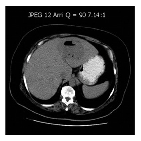
Contrast en-hanced CT scan showing a low-density area with gas and fluid, measuring approximately 8.5 cm x 7.0 cm, consistent with left-sided intra-hepatic abscess.
Figure 2.
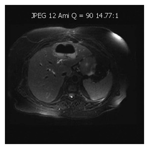
Abdominal RM demonstrating a large collection with gas and fluid.
Using CT guidance, the hepatic abscess was drained percutaneously and pus and blood cultures were obtained. Microbiological examination of the drained fluid was negative and biopsies taken only revealed inflammatory process (Figure 3). Upper GI endoscopy revealed a pre-pyloric thickened fold (Figure 4), with normal histological evaluation. Entamoeba histolytica serology was negative.
Figure 3.
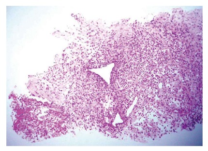
Biopsy of the liver abscess showing fibrosis, fibrin and acute inflammatory cells, consistent with abscess wall (HE).
Figure 4.
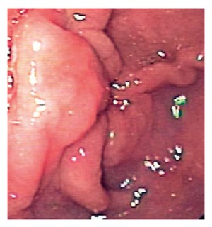
Upper GI endoscopy revealing a thickened gastric fold (pre-pyloric).
The patient started on antibiotherapy (ampicilin, gentamicin and metronidazole) with clinical improvement. Four weeks later abdominal ultrasonography showed abscess size reduction (3 cm) and the patient was discharged and maintained antibiotic therapy.
Three weeks later the patient presented with fever, abdominal pain and elevated C-reactive protein. Abdominal ultrasonography and CT scan showed enlargement of the abscess cavity (8.4 cm × 5.3 cm), which extended to the gastric antrum. Laparotomy was then performed and a foreign body (bone) was found embedded in the left lobe of the liver, resulting in a gastric antrum perforation (Figure 5). The bone was removed, the abscess drained, the stomach defect closed and a drain placed. The post-operative course was uneventful.
Figure 5.
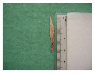
Removed foreign body (chicken bone, with 3.3 cm x 0.5 cm).
DISCUSSION
About 80%-90% of ingested foreign bodies pass trough the gut without discovery within 1 wk[1,2,4]. When symptoms arise they are usually secondary to obstruction[1,2]. Gastrointestinal perforation has been reported in less than 1% of patients[3-5] and the most commonly affected areas are the ileocecal and rectosigmoidal regions[4,5] and duodenum[2]. Development of hepatic abscess due to penetration induced by a foreign body is even more rare, the first case was published in 1898[6]. Since then, the world literature has been increased, with 46 cases reported until now. The most common sites of perforation of the gut are stomach and duodenum[5] which can induced by sharp foreign bodies like fish bones, chicken bones, needles or toothpicks[2,4,5,6] although pens or dental plates have also been reported[6,7].
It is difficult to establish the time until the onset of symptoms as patients rarely recall the episode of ingestion[1,3,4] and the migrating foreign body may remain silent until an abscess formation[5].
Most patients have non specific symptoms such as abdominal pain, fever, vomiting, anorexia or weight loss[4,5,8] which are features of a systemic response against an infection or abscess formation[4]. Furthermore, the classical presentation of hepatic abscess (fever, abdominal pain and jaundice) is only present in a few cases[5].
The results of routine laboratory studies are also non specific and unless the foreign body is radio-opaque it will not be identified on plain radiography[3,4].
An abdominal US or CT scan is preferred techniques for the diagnosis, the latter is excellent in detection of foreign bodies due to its high resolution and accuracy[1,2,4]. Endoscopy may be helpful when performed early, before the foreign body migration and mucosal healing[2,9] (which happened in our patient). In addition, endoscopy does not allow examination of the mid-gut[2]. Therefore, pre-operative diagnosis is difficult and a high degree of suspicion is required[1,3].
We reviewed the world literature, and summarized it in Table 1. We found that fish bones were the most common foreign body and the stomach was the principal site of perforation. Abscess formation occurs more often on the left lobe. Microorganisms isolated on abscess or fluid cultures are usually part of the normal flora of human oropharynx[4,5,6,10-12]. Prognosis depends on a quick diagnosis, not only for morbidity but also for mortality[5,6].
Table 1.
World literature review of hepatic abscess induced by foreign bodies
| Ref | Year | Author | Symptoms | Suffering period | Foreign body | Size (cm) | Penetration | Liver | Bacteria | Laparo tomy | Treatment | Mortality |
| [1] | 2003 | Kanazana | Epigastralgia | 1 mo | Toothpick | 5.5 | Stomach | Left lobe | Unknown | Yes | Abscess drained and removal of a small part of the liver | No |
| [2] | 2000 | Cheung | Epigastralgia, fever | 3 mo | Toothpick | - | Stomach | Left lobe | Unknown | Yes | removal of the toothpick and a small part of the liver | No |
| [3] | 2000 | Broome | Epigastralgia, anorexia, fever | 7 d | Chicken bone | 4.0 | Stomach | Left lobe | Unknown | Yes | Removal of the chicken bone and abscess drainage | No |
| [4] | 1999 | Horii | Fever, vomiting | 2 wk | Fish bone | 2.8 | Unknown | Left lobe | Streptococcus constellatus | No | Percutaneous abscess drainage | No |
| [5] | 2003 | Chintamani | Fever, vomiting | 1 yr | Needle | 3.0 | Unknown | Right lobe | Streptococcus pyogenes, E. coli | Yes | Removal of the needle and abscess drainage | No |
| [6] | 2001 | La Veja | Abdominal pain, vomiting | Unknown | Fish bone | 2.5 | Unknown | Right lobe | - | Autopsy | Yes | |
| [7] | 1999 | Perkins | Fever, anaemia | 2 wk | Pen | - | Duodenum | Right lobe | Streptococcus malleri (group C), Sreptococcus malleri | No | Removal of the pen and abscess drainage | No |
| [8] | 1983 | Shaw | Fever | Dental plate | - | Descending colon | Unknown | |||||
| [9] | 1997 | Tsui | Clothespin, Tooth pick | - | Duodenum Stomach | Unknown | ||||||
| [10] | 1993 | Chen | Epigastralgia, fever, weight loss | 3 mo | Chicken bone | 4.0 | Duodenum | Left lobe | Unknown | Yes | Removal of the chicken bone and abscess drainage | No |
| [11] | 2003 | Bilimoria | Right upper abdominal pain, fever | Unknown | Toothpick | - | Sigmoid colon | Right lobe | Estreptococcus | Yes | Removal of the toothpick and abscess drainage | No |
| [12] | 2004 | Tomimori | Epigastralgia | 4 wk | Fish bone | 1.0 | Stomach | Left lobe | Sreptococcus constellatus | Yes | Removal of the fish bone and abscess drainage | No |
| [13] | 2001 | Kessler | Abdominal pain | 4 wk | Fish bone | Unknown | Duodenum | Left lobe | Eikenella corrodens | Yes | Removal of the fish bone and abscess drainage | No |
| [14] | 2000 | Paraskeva | Abdominal pain | 4 mo | Fish bone | 3.7 | Sigmoid colon | Right lobe | Sreptococcus malleri | No | Removal of the fish bone | No |
| [15] | 1999 | Drnovsek | Abdominal pain, vomiting | 1 d | Toothpick | Unknown | Duodenum | Both | Streptococcus viridens | Yes | Removal of the toothpick | No |
| [16] | 1999 | Guglielminet ti | Toothpick | - | Stomach | Left lobe | Unknown | No | Endoscopic toothpick removal and percutaneous abscess drainage | |||
| [17] | 2002 | Theodoropo ulou | Right upper abdominal pain, fever, jaundice | 3 d | Fish bone | 5.5 | Stomach | Left lobe | Unknown | Autopsy | Yes | |
| [18] | 1981 | Wood | Fever, diarrhea | 9 mo | Needle | - | Retrocecal appendix | Right lobe | Streptococcus viridens | Yes | Removal of the needle and abscess drainage | |
| [19] | 2005 | Starakis | Right upper abdominal pain, fever | 3 wk | Chicken bone | - | Duodenum | Left lobe | Sreptococcus viridans, Eikenella corrodens | Yes | Removal of the chiken bone and abscess drainage | No |
| [20] | 2003 | Houli | Right upper abdominal pain, fever | 2 wk | Chicken bone | 3.5 | Transverse colon | Right lobe | Streptococcus angiosus and mixed anaerobic flora | Yes | Abscess drainage, removal of the chicken bone and a small part of the liver | No |
| [21] | 2001 | Byard | Abdominal pain, fever | Several years | Chicken bone | 3.8 | Duodenum | Both | E. coli, mixed anaerobes and Candida albicans | Autopsy | Yes | |
| [22] | 1999 | Chan | Abdominal pain, fever | Unknown | Fish bone | - | Stomach | Unknown | Yes | Removal of the fish bone, abscess drainage and parcial gastrectomy | No | |
| [23] | 1999 | Tsai | Abdominal pain, fever | Fish bone | 3.7 | Stomach | Left lobe | Unknown | No | Abscess drainage and simple closure of the perforated hole | No | |
| [24] | 1992 | Shuldais | Fish bone | - | Stomach | Unknown | ||||||
| [25] | 1991 | Masunaga | Abdominal pain, fever, vomiting | 1wk | Fish bone | 4.0 | Stomach | Left lobe | Unknown | Yes | Percutaneous abscess drainage, parcial gastrectomy and lateral segmentectomy | |
| [26] | 1990 | Allimant | Fever, astenia | 3 wk | Toothpick | - | Stomach | Left lobe | Unknown | Yes | Drainage and removal of the tooth pick and a small part of the liver | No |
| [27] | 1986 | Penderson | Abdominal pain, shock | Unknown | Toothpick | 3.5 | Stomach | Left lobe | Unknown | Yes | Removal of the toothpick and abscess drainage | No |
| [28] | 1988 | Gonzalez | Abdominal pain, fever, jaundice, nausea | 1 mo | Fish bone | Unknown | Stomach | Left lobe | Unknown | Yes | Removal of the fish bone and abscess drainage | No |
| [29] | 1981 | Rafizadeth | Low-grade fever | 10 d | Toothpick | 4.2 | Duodenum | Left lobe | Estreptococcus | Yes | Removal of the toothpick and abscess drainage | No |
| [30] | 1966 | Aron | Astenia, fever, jaundice | 3 mo | Fish bone | 2.2 | Stomach | Right lobe | E. coli, Proteus | Yes | Removal of the toothpick, abscess drainage and piloroplasty | No |
| [31] | 1971 | Berk | Right upper abdominal pain | Several weeks | Chicken bone | 4.0 | Stomach | Left lobe | Unknown | Yes | Removal of the chicken bone, abscess drainage and parcial gastrectomy | |
| [32] | 1996 | Acosta | Needle | - | Appendix | Unknown | ||||||
| [33] | 1971 | Abel | None | Unknown | Needle | 2.5 | Stomach | Left lobe | Unknown | Yes | Removal of the needle and segmentectomy | No |
| [34] | 1981 | Tsuboi | Epigastralgia, weight loss | 1 mo | Fish bone | 4.7 | Stomach | Left lobe | Unknown | Yes | Removal of the fish bone and abscess drainage | No |
| [35] | 1984 | Bloch | Fever, myalgia | 2 wk | toothpick | 4.5 | Stomach or Duodenum | Left lobe | Estreptococcus | Yes | Removal of the toothpick and abscess drainage | |
| [36] | 1955 | Griffiths | Septic shock | Unknown | Needle | 4.0 | Stomach | Right lobe | Unknown | Autopsy | Yes | |
| 1955 | Griffiths | Fever , vomiting | 1 mo | Toothpick | 6.0 | Duodenum | Right lobe | Unknown | Autopsy | Yes | ||
| [37] | 1990 | Dugger | Fever, right upper abdominal pain | 3 wk | Fish bone or Chicken bone | 3.8 | Stomach | Right lobe | E. coli, Proteus | Autopsy | ||
| [38] | 2005 | Lee | Epigastralgia | 5 d | Body piercing | 5.0 | Stomach | Left lobe | Klebsiella spp, Streptococcus milleri | Yes | Removal of the piercing, closure of the perforated hole and abscess drainage | No |
| 2005 | Lee | Fever, epigastralgia, nausea, vomiting | 1 wk | Fish bone | 3.5 | Stomach | Left lobe | Streptococcus milleri | Yes | Removal of the fish bone, closure of the perforated hole and abscess drainage | No | |
| 2005 | Lee | Epigastralgia | 10 d | - | - | Stomach | Left lobe | Streptococcus milleri | Yes | Closure of the perforated hole | No | |
| [39] | 2005 | Goh | Fever | 5 d | Fish bone | 3.0 | Duodenum | Left lobe | Streptococcus milleri | Yes | Removal of the fish bone and abscess drainage | No |
| [40] | 2006 | Chiang | Right upper abdominal pain, fever | 3 d | Toohpick | 6.7 | Duodenum | Right lobe | Staphylococcus aureus | No | Antibiotics (refused surgery) | No |
Our clinical report is similar to the world literature and enhances the difficulty of diagnosing such an entity. Our patient who did not recall the ingestion, had non specific symptoms and laboratory results as well as US and CT showed a hepatic abscess on the left lobe and its fistulous track. The diagnosis was obtained after exploratory laparotomy. Considering all issues we suppose that the chicken bone perforated through the pylorus.
Hepatic abscess treatment includes aspiration and antibiotic therapy[4]. Nevertheless if we suspect perforation of the gut caused by a foreign body or it is detected by radiography, US or CT, surgery is the option[13], although there are some descriptions of endoscopic[4,12,15] or percutaneous[4,14] removal. In our case surgery not only allowed to make a diagnosis but also treated it.
In conclusion, hepatic abscess diagnosis based on perforation of the gastrointestinal tract caused by a foreign body is difficult due to a variety of non specific symptoms and because patients are often unaware of the ingestion. In a hepatic abscess that does not respond to aspiration and antibiotic therapy we should look for an aetiology. Despite its rarity we should consider a foreign body and surgical therapy. Surgery still has a major role in the diagnosis and treatment of hepatic abscess induced by a foreign body although US and CT may establish it in some cases.
Footnotes
S- Editor Liu Y L- Editor Wang XL E- Editor Zhou T
References
- 1.Kanazawa S, Ishigaki K, Miyake T, Ishida A, Tabuchi A, Tanemoto K, Tsunoda T. A granulomatous liver abscess which developed after a toothpick penetrated the gastrointestinal tract: report of a case. Surg Today. 2003;33:312–314. doi: 10.1007/s005950300071. [DOI] [PubMed] [Google Scholar]
- 2.Cheung YC, Ng SH, Tan CF, Ng KK, Wan YL. Hepatic inflammatory mass secondary to toothpick perforation of the stomach: triphasic CT appearances. Clin Imaging. 2000;24:93–95. doi: 10.1016/s0899-7071(00)00182-0. [DOI] [PubMed] [Google Scholar]
- 3.Broome CJ, Peck RJ. Hepatic abscess complicating foreign body perforation of the gastric antrum: an ultrasound diagnosis. Clin Radiol. 2000;55:242–243. doi: 10.1053/crad.1999.0339. [DOI] [PubMed] [Google Scholar]
- 4.Horii K, Yamazaki O, Matsuyama M, Higaki I, Kawai S, Sakaue Y. Successful treatment of a hepatic abscess that formed secondary to fish bone penetration by percutaneous transhepatic removal of the foreign body: report of a case. Surg Today. 1999;29:922–926. doi: 10.1007/BF02482788. [DOI] [PubMed] [Google Scholar]
- 5.Chintamani V, Lubhana P, Durkhere R, Bhandari S. Liver abscess secondary to a broken needle migration--a case report. BMC Surg. 2003;3:8. doi: 10.1186/1471-2482-3-8. [DOI] [PMC free article] [PubMed] [Google Scholar]
- 6.de la Vega M, Rivero JC, Ruíz L, Suárez S. A fish bone in the liver. Lancet. 2001;358:982. doi: 10.1016/s0140-6736(01)06106-2. [DOI] [PubMed] [Google Scholar]
- 7.Shaw PJ, Freeman JG. The antemortem diagnosis of pyogenic liver abscess due to perforation of the gut by a foreign body. Postgrad Med J. 1983;59:455–456. doi: 10.1136/pgmj.59.693.455. [DOI] [PMC free article] [PubMed] [Google Scholar]
- 8.Tsui BC, Mossey J. Occult liver abscess following clinically unsuspected ingestion of foreign bodies. Can J Gastroenterol. 1997;11:445–448. doi: 10.1155/1997/815876. [DOI] [PubMed] [Google Scholar]
- 9.Bilimoria KY, Eagan RK, Rex DK. Colonoscopic identification of a foreign body causing an hepatic abscess. J Clin Gastroenterol. 2003;37:82–85. doi: 10.1097/00004836-200307000-00021. [DOI] [PubMed] [Google Scholar]
- 10.Tomimori K, Nakasone H, Hokama A, Nakayoshi T, Sakugawa H, Kinjo F, Shiraishi M, Nishimaki T, Saito A. Liver abscess. Gastrointest Endosc. 2004;59:397–398. doi: 10.1016/s0016-5107(03)02590-2. [DOI] [PubMed] [Google Scholar]
- 11.Kessler AT, Kourtis AP. Images in clinical medicine. Liver abscess due to Eikenella corrodens from a fishbone. N Engl J Med. 2001;345:e5. doi: 10.1056/ENEJMicm010433. [DOI] [PubMed] [Google Scholar]
- 12.Paraskeva KD, Bury RW, Isaacs P. Streptococcus milleri liver abscesses: an unusual complication after colonoscopic removal of an impacted fish bone. Gastrointest Endosc. 2000;51:357–358. doi: 10.1016/s0016-5107(00)70372-5. [DOI] [PubMed] [Google Scholar]
- 13.Drnovsek V, Fontanez-Garcia D, Wakabayashi MN, Plavsic BM. Gastrointestinal case of the day. Pyogenic liver abscess caused by perforation by a swallowed wooden toothpick. Radiographics. 1999;19:820–822. doi: 10.1148/radiographics.19.3.g99ma20820. [DOI] [PubMed] [Google Scholar]
- 14.Guglielminetti D, Poddie DB. Liver abscess secondary to ingestion of foreign body. A case report. G Chir. 1999;20:453–455. [PubMed] [Google Scholar]
- 15.Theodoropoulou A, Roussomoustakaki M, Michalodimitrakis MN, Kanaki C, Kouroumalis EA. Fatal hepatic abscess caused by a fish bone. Lancet. 2002;359:977. doi: 10.1016/S0140-6736(02)07999-0. [DOI] [PubMed] [Google Scholar]
- 16.Wood MK, Harrison MR. 'A-pin-dicitis' and liver abscess. JAMA. 1981;246:940. [PubMed] [Google Scholar]
- 17.Starakis I, Karavias D, Marangos M, Psoni E, Bassaris H. A rooster's revenge: hepatic abscess caused by a chicken bone. Eur J Emerg Med. 2005;12:41–42. doi: 10.1097/00063110-200502000-00012. [DOI] [PubMed] [Google Scholar]
- 18.Houli N, MacGowan K, Hosking P. Hepatic abscess complicating foreign body perforation of the transverse colon. ANZ J Surg. 2003;73:255–259. doi: 10.1046/j.1445-1433.2003.02571.x. [DOI] [PubMed] [Google Scholar]
- 19.Byard RW, Gilbert JD. Hepatic abscess formation and unexpected death: a delayed complication of occult intraabdominal foreign body. Am J Forensic Med Pathol. 2001;22:88–91. doi: 10.1097/00000433-200103000-00018. [DOI] [PubMed] [Google Scholar]
- 20.Chan SC, Chen HY, Ng SH, Lee CM, Tsai CH. Hepatic abscess due to gastric perforation by ingested fish bone demonstrated by computed tomography. J Formos Med Assoc. 1999;98:145–147. [PubMed] [Google Scholar]
- 21.Tsai JL, Than MM, Wu CJ, Sue D, Keh CT, Wang CC. Liver abscess secondary to fish bone penetration of the gastric wall: a case report. Zhonghua YiXue ZaZhi (Taipei) 1999;62:51–54. [PubMed] [Google Scholar]
- 22.Shuldaĭs AK, Sumin AV, Tkhorzhevskiĭ BB. Pyogenic hepatic abscess developing after perforation of the stomach by a fish bone. Klin Khir. 1992;(11):75–76. [PubMed] [Google Scholar]
- 23.Masunaga S, Abe M, Imura T, Asano M, Minami S, Fujisawa I. Hepatic abscess secondary to a fishbone penetrating the gastric wall: CT demonstration. Comput Med Imaging Graph. 1991;15:113–116. doi: 10.1016/0895-6111(91)90034-s. [DOI] [PubMed] [Google Scholar]
- 24.Allimant P, Rosburger C, Zeyer B, Frey G, Morel E, Bietiger M, Dalcher G. An unusual cause of hepatic abscess. Ann Gastroenterol Hepatol (Paris) 1990;26:5–6. [PubMed] [Google Scholar]
- 25.Pedersen VM, Geerdsen JP, Bartholdy J, Kjaergaard H. Foreign body perforation of the gastrointestinal tract with formation of liver abscess. Ann Chir Gynaecol. 1986;75:245–246. [PubMed] [Google Scholar]
- 26.Gonzalez JG, Gonzalez RR, Patiño JV, Garcia AT, Alvarez CP, Pedrosa CS. CT findings in gastrointestinal perforation by ingested fish bones. J Comput Assist Tomogr. 1988;12:88–90. doi: 10.1097/00004728-198801000-00016. [DOI] [PubMed] [Google Scholar]
- 27.Rafizadeh F, Silver H, Fieber S. Pyogenic liver abscess secondary to a toothpick penetrating the gastrointestinal tract. J Med Soc N J. 1981;78:377–378. [PubMed] [Google Scholar]
- 28.Aron E, Roy B, Groussin P. Abscess of the liver caused by fish bone. Presse Med. 1966;74:1957–1958. [PubMed] [Google Scholar]
- 29.Berk RN, Reit RJ. Intra-abdominal chicken-bone abscess. Radiology. 1971;101:311–313. doi: 10.1148/101.2.311. [DOI] [PubMed] [Google Scholar]
- 30.Acosta S, Lantz L. A case report. A pin in the appendix caused hepatic abscess. Lakartidningen. 1996;93:4278. [PubMed] [Google Scholar]
- 31.Abel RM, Fischer JE, Hendren WH. Penetration of the alimentary tract by a foreign body with migration to the liver. Arch Surg. 1971;102:227–228. doi: 10.1001/archsurg.1971.01350030065021. [DOI] [PubMed] [Google Scholar]
- 32.Tsuboi K, Nakajima Y, Yamamoto S, Nagao M, Nishimura K, Yoshii M. A case of an intrahepatic fish bone penetration--possibility of the preoperative diagnosis by CT scan (author's transl) Nihon Geka Hokan. 1981;50:899–903. [PubMed] [Google Scholar]
- 33.Bloch DB. Venturesome toothpick. A continuing source of pyogenic hepatic abscess. JAMA. 1984;252:797–798. doi: 10.1001/jama.252.6.797. [DOI] [PubMed] [Google Scholar]
- 34.Griffiths FE. Liver abscess due to foreign-body migration from the alimentary tract; a report of two cases. Br J Surg. 1955;42:667–668. doi: 10.1002/bjs.18004217620. [DOI] [PubMed] [Google Scholar]
- 35.Dugger K, Lebby T, Brus M, Sahgal S, Leikin JB. Hepatic abscess resulting from gastric perforation of a foreign object. Am J Emerg Med. 1990;8:323–325. doi: 10.1016/0735-6757(90)90086-f. [DOI] [PubMed] [Google Scholar]
- 36.Lee KF, Chu W, Wong SW, Lai PB. Hepatic abscess secondary to foreign body perforation of the stomach. Asian J Surg. 2005;28:297–300. doi: 10.1016/S1015-9584(09)60365-1. [DOI] [PubMed] [Google Scholar]
- 37.Goh BK, Yong WS, Yeo AW. Pancreatic and hepatic abscess secondary to fish bone perforation of the duodenum. Dig Dis Sci. 2005;50:1103–1106. doi: 10.1007/s10620-005-2712-8. [DOI] [PubMed] [Google Scholar]
- 38.Chiang TH, Liu KL, Lee YC, Chiu HM, Lin JT, Wang HP. Sonographic diagnosis of a toothpick traversing the duodenum and penetrating into the liver. J Clin Ultrasound. 2006;34:237–240. doi: 10.1002/jcu.20200. [DOI] [PubMed] [Google Scholar]


