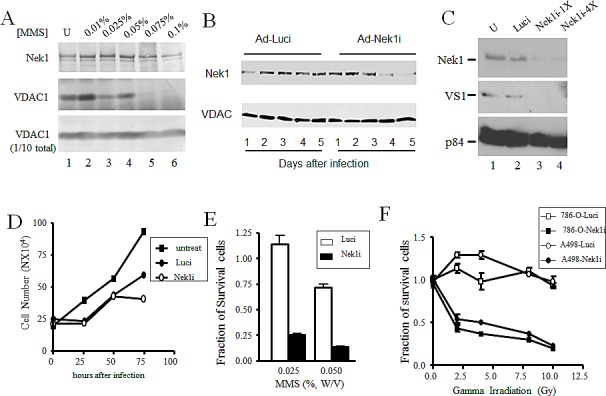
Figure 5. Downregulation of Nek1 expression allows RCC cells more sensitive to genotoxic agents.

A. Dose-response: compared to HK2 cells, a higher MMS dose is required in RCC cells to cause the dissociation of Nek1 from VDAC1. A498 cells were treated with the indicated doses of MMS for 1 hour. After washing three times with PBS, cells were lysed and Nek1-VDAC1 protein complexes were analyzed by Western blotting, as in Fig. 4. B. Downregulation of Nek1 expression in A498 cells by RNAi interference. Short inhibitory RNA, in an adenovirus-based DNA expression system, was used to silence expression of Nek1 in A498 cells. Days after infection of A498 cells with the siRNA constructs, separated proteins from cell lysates were probed for either the Nek1 expression (upper panel) or VDAC1 (lower panel). Nek1 expression was markedly reduced by day 4 (96 hours) after an infection with the Ad-Nek1i. Infection with a control construct to inhibit firefly luciferase (Ad-Luci) had no effect on VDAC1 expression. C. 72 hours after the infection, there was a loss of VDAC1 phosphorylation on S193 in A498 cells expressing Nek1i. Gel-separated proteins from cell lysates were probed for Nek1 expression (upper panel), VDAC1 phosphorylated on S193 (VS1, middle panel), and p84 (bottom panel, to control for loading). U, mock-infected cells; Luci, cells infected with Ad-Luci, used as a negative control; Nek1-1X, cells infected with a construct containing a single copy of the siRNA oligonucleotide specific for Nek1; Nek1-4X, cells infected with an otherwise identical construct containing 4 tandem repeats of the same siRNA oligonucleotide. D. Growth curve of cells after adenovirus infections. A498 cells were plated out at 1X10⁵ cells per 60mm dishes and infected with either Ad-Luci or Ad-Nek1i 24 hours later. Surviving cells (those excluding trypan blue vital dye) were counted every 24 hours and cell number was plotted. The adenovirus infections did not alter the cell growth during the first 60 hours, and Nek1i infected cells started to show no cell number increase after 72 hours (3 experiments). E. Increased sensitivity to the toxicity of MMS in cells expressing Nek1i siRNA. A498 cells were plated out and infected with either Ad-Luci or Ad-Nek1i. Sixty hours after the infection, the cells were either mock-treated or treated with 0.025% or 0.05% (W/V) MMS for 1 hour. After neutralization with sodium thiosulfate, the cells were washed three times with PBS and re-fed with fresh media. Twenty-four hours later, surviving cells were counted and expressed as fractions of identical, mock-treated cells (5 experiments). F. Greater sensitivity to ionizing radiation in cells with Nek1 expression downregulated. A498 and 786-O cells were infected with Ad-Luci or Ad-Nek1i. Sixty hours after the infection, the cells were irradiated with different doses of IR and surviving cells (those excluding the trypan blue vital dye) and were counted 24 hours later (5 experiments).
