

Abstract
INTRODUCTION
Ochronosis is a rare disorder which is defined as the deposition of metabolites of oxidation and polymerization of homogentisic acid, which have high affinity to collogen, in the connective tissues. It is a clinical condition characterized with ochronotic pigmentation of tissues, degenerative arthropathy of especially large joints and black discoloration of urine. In this paper we present a case of ochronosis diagnosed with biopsy and additional tests when a black discoloration of menisci and joint cartilage were detected during arthroscopic intervention for a degenerative meniscus tear.
PRESENTATION OF CASE
A forty two year-old male patient was operated for lateral meniscus tear of his right knee. The arthroscopic examination of right knee revealed black colored synovial hypertrophy and torn lateral meniscus. Partial meniscectomy was performed. The diagnosis of ochronosis was made after histopathologic examination.
DISCUSSION
Ochronotic pigment can accumulate in hyaline cartilage, tendon, skin, teeth, nail, sclera, tympanic membrane, heart valves, renal tubular cells, duramater, pancreas and walls of large arteries. In ochronosis the most frequently involved joints are knee and hip. In ochronotic arthropathy, articular cartilage become more sensitive to mechanical stresses. Our patient had meniscal tear, cartilage damage and black discoloration of synovial tissues and meniscus.
CONCLUSION
Arthroscopy may be helpful in diagnosis of ochronotic arthropathy.
Keywords: Ochronosis, Arthroscopy, Knee
1. Introduction
Ochronosis is defined as the accumulation of metabolites of oxidation and polimerization of homogentisic acid (HGA) in the connective tissues, which have high affinity to collogen.1,2 Because of the defect occurred in the gene which codes homogentisate 1,2 dioxygenase located on the 3q chromosome, homogentisic acid accumulates in tissues and excreted in urine.1 Ochronosis may affect cartilage, intervertebral discs, skin and sclera. Symptoms related to cardiovascular, genitourinary and pulmonary systems may be present.3
Ochronosis is seen mostly in adults and peaks in the forth decade.1,4 With increasing age complaints of hip, knee, shoulder and spine joints may appear. Although ochronotic pigmentation of tissues, black discoloration of urine due to sodium hydroxide (NAOH), degenerative arthropathy of large joints are classical findings, definitive diagnosis is made with quantitative detection of homogentisic acid in the urine and detection of ochronotic pigmentation in the histopathologic examination of tissue biopsies.4,5
In this paper we present a case complaining of knee pain and having arthroscopically detected black discoloration of menisci and cartilage which led us to the biopsy and further laboratory tests and finally diagnosed as ochronosis.
2. Presentation of case
A forty two year-old male admitted to our outpatient clinic with right knee pain and clicking on exertion for two years. There was no history of trauma. Physical examination revealed knee joint effusion. There was complete range of motion despite pain during the last 30° of flexion. There was pain on the lateral joint line by palpation and Mc Murray test was positive. There was no instability. X-ray revealed arthrosis in the lateral tibiofemoral and patellofemoral joint spaces. Magnetic resonance imaging (MRI) of the right knee revealed effusion, partially torn anterior cruciate ligament, thickening of quadriceps tendon. We decided to perform an arthroscopic intervention because of failed conservative treatment.
The arthroscopic examination of right knee revealed black colored synovial hypertrophy. There were 3 cm × 2 cm in diameter Outerbridge stage 4 chondral defect in patellar joint surface and medial femoral condyle. Defective areas were black colored and hardened (Fig. 1). ACL was partially torn but the posterior cruciate ligament (PCL) was intact. Arthroscopic view of the lateral joint space revealed a large, yellow and black colored grade 4 chondral defect in the lateral tibial condyle. Lateral meniscus was thickened, black colored at the inferior surface and torn (Fig. 2). The partial meniscectomy was performed. Biopsy was taken from the black colored areas of femoral condyle (Fig. 3a and b).
Fig. 1.

Chondral lesion on medial femoral condyle.
Fig. 2.
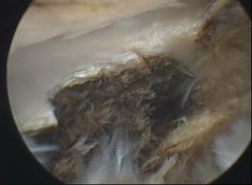
Arthroscopic vision of torn lateral meniscus.
Fig. 3.

Bone biopsy specimen shows the dark discoloration of the bony surface.
We have done further investigation with the suspect of ochronosis because of the black discoloration of tissues seen in the arthroscopic intervention. Hip, knee, vertebra and shoulder radiographs of the patient revealed osteoarthritis. The HGA level was detected high in the urine. Urine was normal in color but when treated with sodium hydroxide (NaOH) color of the urine became darker (Fig. 4). Echocardiography revealed moderate mitral valve insufficiency and left ventricular dysfunction. Dermatologic, urologic and ophthalmologic consultations did not reveal any pathology. Histopathologic examination of arthroscopic biopsy specimens were reported as ochronosis. The patient is under follow up by cardiology for valvular insufficiency and orthopedics for ochronotic arthropathy.
Fig. 4.

The normal colored urine of the patient turned in to black after treatment with sodium hydroxide.
3. Discussion
Ochronosis is a very rare disease with a prevalence of 1/250,000–1,000,000 (1). It was first described by Virchow in a postmortem examination of a 67 year-old patient in 1866. It is characterized by deposition of ochronotic pigment in the tissues due to homogentisic oxidase enzyme deficiency in the tyrosine metabolism. Clemens reported pigmentation of joint cartilage in 1907. Ochronotic pigment can accumulate in hyaline cartilage, tendon, skin, teeth, nail, sclera, tympanic membrane, heart valves, renal tubular cells, duramater, pancreas and walls of great arteries.6,7 Ochronosis is easily recognized with ochronotic pigmentation of tissues, degenerative arthropathy, especially in large joints and dark discoloration of urine because of alcalinisation. There are few cases that are reported after arthroscopic diagnosis.
Our case is the seventh case in the literature. All patients in the literature were examined with knee arthroscopy with different reasons and the diagnosis of ochronosis is made through a further investigation of the intraarticular tissues because of the black discoloration.2,4,8–11 As in the other cases, black discoloration of cartilage and meniscal tissues was detected in our case. Moreover, we detected osteoarthritis and vacuum phenomenon in the intervertebral joints, arthrosis of large joints like hip, knee and glenohumeral joints and increased homogentisic oxidase in urine. Unlike other cases we detected thickening of quadriceps tendon and asymptomatic supraspinatus tear in the MRI and mitral valve insufficiency as an extraarticular finding.
Increasing homogentisic acid levels in the blood and resulting accumulation in tissues become symptomatic especially in the forth decade along with decreasing renal functions.7 Black discoloration of napkins may lead to diagnosis in the neonatal period. History of our case revealed black colored urination during childhood. However the patient did not admit to a doctor.
In ochronosis the most frequently involved joints are knee and hip. In ochronotic arthropathy, articular cartilage becomes more sensitive to mechanic stresses causing fragmentation and resulting in nonspecific tenosynovitis. The 50% of the patients present with knee effusion.7 Our patient admitted to our clinic with knee effusion. High grade glenohumeral arthropathy is very rare but must be kept in mind in ochronotic arthropathy. Ochronotic arthropathy differs from osteoarthritis by presenting with less osteophytes and subchondral cysts.12
Homogentisic acid polyphenol oxidase enzyme, which is located especially in tendons and ligaments, produces free radicals through oxidation and causes tendon and ligament ruptures.1,13 MRI of our patient revealed partial rupture of ACL, a thickened quadriceps tendon and asymptomatic rupture of supraspinatus tendon.
Ochronosis may affect extraarticular tissues. Calcifications in coronary arteries, valvular insufficiencies must be checked by echocardiography. Ultrasonography may show calcifications in prostate and kidneys.7 Pigmentations may be detected in sclera, auricular and nasal cartilages. We detected mitral valve insufficiency in our case.
There is no proven effective treatment of ochronosis. High dose ascorbic acid (100 mg/kg) may reduce HGA excretion in the urine but does not prevent development of arthropathy.5 Because the modalities addressing the cause of the disease are unclear, treatment of ochronotic arthropathy is symptomatic. Nonsteroid antiinflammatory drugs and preparates containing glucosamine and chondroitin sulphate, intraarticular hyaluronic acid and steroid injections, arthroscopic debridement and arthroplasty are the treatment options.2,5,14,15
4. Conclusion
Ochronosis is a rare disease affecting connective tissues. Arthroscopy is helpful in diagnosis of ochronotic arthropathy and may lead to further investigation and treatment of concomitant pathologies.
Conflict of interest
Authors declare that there is no conflict of interest.
Funding
None.
Ethical approval
Written informed consent was obtained from the patient for publication of this case report and case series and accompanying images. A copy of the written consent is available for review by the Editor-in-Chief of this journal on request.
Author contributions
Adnan Kara: study concept or design, data collection, data analysis or interpretation, writing the paper.
Haluk Celik: data collection, writing the paper.
Ali Seker: writing the paper.
Hasan Basri Sezer: writing the paper.Bekir Eray Kilinc: writing the paper.Metin Uzun: writing the paper.
Key learning point.
-
•
Ochronosis is a rare disease affecting connective tissues. Arthroscopy is helpful in diagnosis of ochronotic arthropathy and may lead to further investigation and treatment of concomitant pathologies.
References
- 1.Keller J.M., Macaulay W., Nercessian O.A., Jaffe I.A. New developments in ochronosis: review of the literature. Rheumatol Int. 2005;25:81–85. doi: 10.1007/s00296-004-0498-1. [DOI] [PubMed] [Google Scholar]
- 2.Raaijmaakers M., Steenbrugge F., Dierickx C. Ochronosis arthroscopy of a black knee: a case report and review of the literature. Knee Surg Sports Traumatol Arthrosc. 2008;16:182–184. doi: 10.1007/s00167-007-0413-x. [DOI] [PubMed] [Google Scholar]
- 3.Zhao B.H., Chen B.C., Shao de C, Zhang Q. Osteoarthritis? Ochronotic arthritis! A case study and review of the literature. Knee Surg Sports Traumatol Arthrosc. 2009;17:778–781. doi: 10.1007/s00167-009-0778-0. [DOI] [PubMed] [Google Scholar]
- 4.Delialioglu O.M., Daglar B., Bayrakci K., Ceyhan E., Tezel K., Erekul S., Gunel U. Ochronosis: complicated tear of black meniscus. Knee Surg Sports Traumatol Arthrosc. 2010;18:540–542. doi: 10.1007/s00167-009-0906-x. [DOI] [PubMed] [Google Scholar]
- 5.Cetinus E., Cever I., Kural C., Erturk H., Akyildiz M. Ochronotic arthritis: case reports and review of the literature. Rheumatol Int. 2005;25:465–468. doi: 10.1007/s00296-004-0538-x. [DOI] [PubMed] [Google Scholar]
- 6.Manoj Kumar R.V., Rajasekaran S. Spontaneous tendon ruptures in alkaptonuria. J Bone Joint Surg Br. 2003;85:883–886. [PubMed] [Google Scholar]
- 7.Hamdi N., Cooke T.D., Hassan B. Ochronotic arthropathy: case report and review of the literature. Int Orthop. 1999;23:122–125. doi: 10.1007/s002640050325. [DOI] [PMC free article] [PubMed] [Google Scholar]
- 8.Kural C., Cetinus E.M., Kural A., Uğraş A.A., Kaya I. Knee ochronotic arthropathy and arthroscopic findings. Acta Orthop Traumatol Turc. 2009;43:67–71. doi: 10.3944/AOTT.2009.067. [DOI] [PubMed] [Google Scholar]
- 9.Thacker M., Garude S., Puri A. Ochronotic arthropathy: arthroscopic findings in the shoulder and the knee. Arthroscopy. 2003;19:14–17. doi: 10.1016/s0749-8063(03)00744-8. [DOI] [PubMed] [Google Scholar]
- 10.Tudisco C., Mariani P.P., D’Arrigo C. Knee arthroscopy in a case of ochronotic arthropathy. Ital J Orthop Traumatol. 1992;18:107–110. [PubMed] [Google Scholar]
- 11.Lurie D.P., Musil G. Knee arthropathy in ochronosis: diagnosis by arthroscopy with ultrastructural features. J Rheumatol. 1984;11:101–103. [PubMed] [Google Scholar]
- 12.Sahin G., Milcan A., Bağiş S., Köktürk A., Pata C., Erdoğan C. A case of ochronosis: upper extremity involvement. Rheumatol Int. 2001;21:78–80. doi: 10.1007/s002960100136. [DOI] [PubMed] [Google Scholar]
- 13.Phornphutkul C., Introne W.J., Perry M.B., Bernardini I., Murphey M.D., Fitzpatrick D.L. Natural history of alkaptonuria. N Engl J Med. 2002;347:2111–2121. doi: 10.1056/NEJMoa021736. [DOI] [PubMed] [Google Scholar]
- 14.Spencer J.M., Gibbons C.L., Sharp R.J., Carr A.J., Athanasou N.A. Arthroplasty for ochronotic arthritis: no failure of 11 replacements in 3 patients followed 6–12 years. Acta Orthop Scand. 2004;75:355–358. doi: 10.1080/00016470410001321. [DOI] [PubMed] [Google Scholar]
- 15.Aydoğdu S., Cullu E., Ozsoy M.H., Sur H. Cementless total knee arthroplasty in ochronotic arthropathy: a case report with a 4-year follow-up. J Arthroplasty. 2000;15:539–543. doi: 10.1054/arth.2000.4228. [DOI] [PubMed] [Google Scholar]