

Abstract
The study was undertaken to evaluate the reliability of different facial measurements for determination of vertical dimension of occlusion in edentulous subjects using accepted facial dimensions recorded from dentulous subjects. The hypothesis was that facial measurements can be used to obtain the vertical dimension of occlusion for edentulous patients where no pre-extraction records exist. A total of 180 subjects were selected in the age groups of 50–60 years, consisting of 75 dentate male and 75 dentate female subjects for whom different facial measurements were recorded including vertical dimension of occlusion and rest, and 15 edentulous male and 15 edentulous female subjects for whom all the facial measurements were recorded including the vertical dimension of rest and occlusion following construction of upper and lower complete dentures. The left outer canthus of eye to angle of mouth distance and the right Ear–Eye distance were found to be as valuable adjuncts in the determination of occlusal vertical dimension. The Glabella–Subnasion distance, the Pupil–Stomion distance, the Pupil–Rima Oris distance and the distance between the two Angles of the Mouth did not have a significant role in the determination of the occlusal vertical dimension. The vertical dimension can be determined with reasonable accuracy by utilizing other facial measurements for patients for whom no pre-extraction records exist.
Keywords: Vertical dimension at rest, Vertical dimension of occlusion, Digital vernier caliper
Introduction
Complete denture prosthodontics involves the replacement of the lost natural dentition and the associated structures of the maxilla and mandible for the patients who have lost their remaining natural teeth or are soon to lose them. Owing to the lack of reliable parameters, the most critical and contentious aspect of complete denture construction is the determination of the maxillo-mandibular relations especially the occlusal vertical dimension. The resulting imprecision creates a whole series of problems, both esthetic and functional thus, compromising the success of prosthetic rehabilitation. There are numerous beliefs and theories put forward as to the determination of vertical dimension. Some believe that the vertical dimension restored should be the same as probably what existed prior to the edentulous situation [1]. Although many techniques to determine the correct vertical dimension of occlusion have been proposed like the use of preextraction records, physiologic rest position, closing forces (boos bimeter method), tactile sense, phonetics, esthetic appearance, open rest method, facial measurements, deglutition and the electromyographic method [2]. Finding a reliable method to determine the correct vertical dimension of occlusion has always been a challenge for the clinicians in the field of complete denture prosthodontics.
One of the famous artist’s Leonardo Da Vinci [3] in the fifteenth century, gave simple ratios for drawing the face. This concept was picked up by Ivy [4] who applied it to complete denture construction. In his belief the face could be divided into 4 equal proportions and these ratios were used during complete denture construction. Goodfriend [5] modified the guidelines given by Ivy. He stated that the pupil-rima oris distance would equal the chin- nose distance. Willis [6] in support of Goodfriend popularized these measurements in 1930s and designed a special measuring gauge for this purpose (Willi’s Gauge). Fenn et al. [7] later proposed the use of outer canthus of eye to the angle of mouth distance as a guide to the correct vertical dimension of occlusion in 1953.
Despite the advances in techniques and materials are being made in prosthodontics, still no accurate method of assessing the vertical dimension of occlusion in edentulous patients is available to the dentists [8]. “Clinical judgement” continues to play a major role in the assessment of this important component in the construction of complete dentures. The methods for the successful construction of a set of complete dentures should be as accurate as possible and should be based on scientific principles. Complete dentures are constructed today by incorporating almost the same techniques and concepts as given 30 years ago.
The present study was undertaken to impart objectivity to this critical phase of complete denture construction by determining the reliability of different facial measurements as mentioned in the literature in obtaining the occlusal vertical dimension.
Materials and Methodology
180 subjects, all of North Indian origin, between the age groups of 50–60 years were selected from the dental OPD & Prosthodontic clinic at ITS-CDSR. For the purpose of the study the patients were divided into 4 groups i.e. Group I: Dentate Male Patients (n = 75); Group II: Dentate Female Patients (n = 75); Group III: Edentulous Male Patients (n = 15) & Group IV: Edentulous Female Patients (n = 15).
For all the subjects facial measurements were recorded following history & clinical examination. For edentulous subjects all the facial measurements were recorded before & after complete denture fabrication. For recording the facial measurements the subjects were instructed to sit straight with the head unsupported & the following soft tissue points were palpated & then marked on the face with an indelible pencil:-
Glabella (G): Point of greatest prominence between the two eyebrows.
Subnasion (S): The base of the nose.
Tip of the nose (N).
Menton (M): The lowest most point on the symphysis (i.e. the tip of the chin).
Centre of the pupil (P).
Rima Oris (R): The line between the upper and lower lips.
Stomion (S): The line joining the lips in median line and not the corner of the mouth.
Lateral border of the bony orbit (outer canthus of the eye) (E).
Anterior most point of the external auditory meatus (e).
Three readings were taken for each measurement by two investigators and their average was recorded (ICC value = 0.98). The facial measurements recorded were recorded using a modified digital Vernier Caliper (Fig. 1):
The pupil-rima oris distance [P R] (Fig. 2).
The chin-nose distance [C N] (Fig. 4).
The subnasion-menton distance [S M] (Fig. 5).
The distance between the lateral corner of the mouth and outer canthus of the eye. [M E] (Fig. 6).
Fig. 1.

Modified digital vernier caliper
Fig. 2.

Pupil-Rima Oris & Glabella–Subnasion distance & measurement of Pupil–Rima Oris distance
Fig. 3.
The measurement of Glabella–Subnasion distance
Fig. 4.

Chin–Nose distance & its measurement
Fig. 5.

Subnasion–Menton distance & its measurement
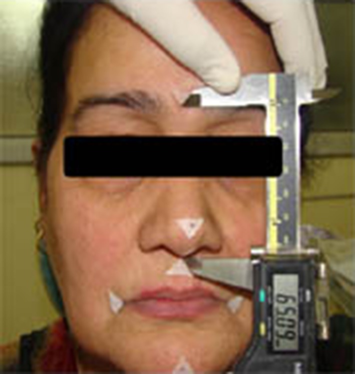
Fig. 6.

Outer canthus Eye–Lateral corner of the mouth distance & its measurement
-
6.
The distance between the centre of the pupil to stomion [P S] (Fig. 7).
-
7.
The distance between the two angles of mouth [A A] (Fig. 8).
-
8.
The Ear–Eye distance [E e] (Fig. 9).
For Edentulous Subjects, after history and clinical examination, upper and lower complete dentures were fabricated & the above mentioned facial dimensions including the occlusal vertical dimension at rest (with & without dentures in mouth) were measured.
Fig. 7.

Centre of pupil to stomion distance & its measurement
Fig. 8.

Distance between the two angles of mouth & its measurement
Fig. 9.

Eye–Ear distance & its measurement
Statistical Analysis
A total of 4680 parameters were recorded from clinical measurements performed on 180 subjects. A master chart was prepared for all the data and the data were fed into a computer using the SPSS (Version 10.0) software package. The data were checked twice for any errors during data-feeding. 95 % Confidence Limit was calculated between the Chin–Nose distance in occlusion & rest with all the observed facial measurements so as to get an interval within which 95 % of differences between measurements were expected to lie. Student’s t test was done to determine the level of significance in the difference of means of the various facial dimensions between groups I–II and III–IV.
Results
The study consisted of 180 subjects, having class I molar relationship (for dentate patients) & Class I Jaw relationship (for edentulous subjects) for whom different facial measurements were recorded so as to find out if at all their existed any relationship between VDO & the observed facial measurements by calculating the 95 % confidence interval between them (Tables 1, 2, 3, 4, 5, 6, 7, 8, 9);. The facial measurements were recorded using a modified digital Vernier Calliper. The study sample was divided into 4 groups depending upon age, sex & dentulousness. 24 measurements were recorded in the dentate subjects and 36 in the edentulous subjects. The following conclusions were arrived at from this study. The Chin–Nose distances revealed a strong association with the Left Outer Canthus of Eye to Angle of Mouth distances in Group I (Dentate Male subjects) and Group III (Edentulous Male Subjects). In Group II (Dentate Female subjects) the Chin–Nose distances revealed a strong association with the Left Outer Canthus of Eye to Angle of Mouth distances and the Right Ear–Eye distances and in Group IV (Edentulous Female subjects) the Chin–Nose distances revealed a strong association with the Left Outer Canthus of Eye to Angle of Mouth distances and the Right Ear–Eye distances. The results of the study thus revealed that, Left Outer Canthus of Eye to Angle of Mouth distance and the Right Ear–Eye distance can be used as a valuable adjunct in the determination of Occlusal Vertical Dimension which is a decisive step of Complete Denture fabrication.
Table 1.
Difference between Chin–Nose distance in occlusion and other observed facial measurements group I (dentate male subjects) [n = 75]
| S. No | Observed facial measurements | Mean difference ± standard deviation | 95 % Confidence Limit | |
|---|---|---|---|---|
| Lower limit | Upper limit | |||
| 1. | Outer canthus of eye to angle of mouth distance (Lt) [R] | −3.4 ± 5.4 | −13.98 | 7.18 |
| 2. | Outer canthus of eye to angle of mouth distance (Lt) [O] | −2.9 ± 5.4 | −13.43 | 7.63 |
The left outer canthus of eye to angle of mouth distances at rest and occlusion showed minimum average differences from the Chin–Nose distance in occlusion and their confidence intervals are (−13.98–7.18); (−13.43–7.63) and respectively
Lt left, Rt right, R at rest O in occlusion
Table 2.
Difference between Chin–Nose distance at rest and other observed facial measurements group I (dentate male subjects) [n = 75]
| S. No | Observed facial measurements | Mean Difference ± standard deviation | 95 % Confidence limit | |
|---|---|---|---|---|
| Lower limit | Upper limit | |||
| 1. | Outer canthus of eye to angle of mouth distance (Lt) [R] | 0.6 ± 5.1 | −9.3 | 10.51 |
| 2. | Outer canthus of eye to angle of mouth distance (Lt) [O] | 0.7 ± 5.2 | −9.49 | 10.87 |
The left outer canthus of eye to angle of mouth distances at rest and occlusion showed minimum average differences from the Chin–Nose distance at Rest and their confidence intervals are (−9.3–10.51); (−9.49–10.87) respectively
Lt left, Rt right, R at rest, O in occlusion
Table 3.
Difference between Chin–Nose distance in occlusion and other observed facial measurements group II (dentate female subjects) [n = 75]
| S. No | Observed facial measurements | Mean difference ± standard deviation | 95 % Confidence limit | |
|---|---|---|---|---|
| Lower limit | Upper limit | |||
| 1. | Ear–Eye distance (Rt) [R] | −6.2 ± 7.0 | −19.92 | 7.52 |
| 2. | Ear–Eye distance (Rt) [O] | −6.1 ± 7.1 | −20.01 | 7.81 |
| 3. | Outer canthus of eye to angle of mouth distance (Lt) [R] | −4.0 ± 5.4 | −14.58 | 6.58 |
| 4. | Outer canthus of eye to angle of mouth distance (Lt) [O] | −4.0 ± 5.4 | −14.58 | 6.5 |
Lt left, Rt right, R at rest, O in occlusion
The left outer canthus of eye to angle of mouth distances at rest and occlusion and the right Ear–Eye distance at rest and occlusion showed minimum average differences from the Chin–Nose distance in occlusion and their confidence intervals are (−9.3–10.51); (−9.49–10.87); (−19.92–7.52) and (−20.01–7.18) respectively
Table 4.
Difference between Chin–Nose distance at rest and other observed facial measurements group II (dentate female subjects) [n = 75]
| S. No | Observed facial measurements | Mean difference ± standard deviation | 95 % Confidence limit | |
|---|---|---|---|---|
| Lower limit | Upper limit | |||
| 2. | Ear–Eye distance (Rt) [R] | −3.6 ± 6.8 | −16.92 | 9.72 |
| 4. | Ear–Eye distance (Rt) [O] | −3.6 ± 6.8 | −16.92 | 9.72 |
| 13. | Outer canthus of eye to angle of mouth distance (Lt) [R] | −1.4 ± 5.1 | −11.39 | 8.59 |
| 15. | Outer canthus of eye to angle of mouth distance (Lt) [O] | −1.4 ± 5.1 | −11.39 | 8.59 |
Lt left, Rt right, R at rest, O in occlusion
The left outer canthus of eye to angle of mouth distances at rest and occlusion and the right Ear–Eye distance at rest and occlusion showed minimum average differences from the Chin–Nose distance at rest and their confidence intervals are narrow (−11.39–8.59), (−11.39–8.59), (−16.92–9.72) and (−16.92–9.72) respectively
Table 5.
Difference Between Chin–Nose Distance in Occlusion and Other Observed Facial Measurements Group III (Edentulous Male Subjects) [n = 15]
| S. No | Observed facial measurements | Mean difference ± standard deviation | 95 % Confidence Limit | |
|---|---|---|---|---|
| Lower Limit | Upper Limit | |||
| 1. | Outer canthus of eye to angle of mouth distance (Lt) [R] | −2.6 ± 3.7 | −9.85 | 4.65 |
| 2. | Outer canthus of eye to angle of mouth distance (Lt) [O] | −2.6 ± 3.7 | −9.85 | 4.65 |
Lt left, Rt right, R at rest, O in occlusion
The left outer canthus of eye to angle of mouth distances at rest and occlusion showed minimum average differences from the Chin–Nose distance in occlusion and their confidence intervals are (−9.58–4.65) and (−9.58–4.65)
Table 6.
Difference between Chin–Nose distance at rest and other observed facial measurements group III (edentulous male subjects) [n = 15]
| S. No | Observed facial measurements | Mean difference ± standard deviation | 95 % Confidence limit | |
|---|---|---|---|---|
| Lower limit | Upper limit | |||
| 1. | Outer canthus of eye to angle of mouth distance (Lt) [R] | 0.003 ± 3.6 | −7.11 | 7.11 |
| 2. | Outer Canthus of Eye to Angle of Mouth Distance (Lt) [O] | −1.5 ± 13.4 | −27.76 | 24.76 |
Lt left, Rt right, R at rest, O in occlusion
The left outer canthus of eye to angle of mouth distances at rest and occlusion showed minimum average differences from the Chin–Nose distance at rest and their confidence intervals are (−7.11–7.11); (−27.76–24.76) respectively
Table 7.
Difference between Chin–Nose distance in occlusion and other observed facial measurements group IV (edentulous female subjects) [n = 15]
| S. No | Observed facial measurements | Mean difference ± standard deviation | 95 % Confidence limit | |
|---|---|---|---|---|
| Lower limit | Upper limit | |||
| 1. | Ear–Eye distance (Rt) [R] | 1.0 ± 7.7 | −14.09 | 16.09 |
| 2. | Ear–Eye distance (Rt) [O] | 1.0 ± 7.7 | −14.09 | 16.09 |
| 3. | Outer canthus of eye to angle of mouth distance (Lt) [R] | −0.9 ± 5.9 | −12.46 | 10.66 |
| 4. | Outer canthus of eye to angle of mouth distance (Lt) [O] | −1.0 ± 2.0 | −4.92 | 2.92 |
Lt left, Rt right, R at rest, O in occlusion
The left outer canthus of eye to angle of mouth distances at rest and occlusion and the right Ear–Eye distance at rest and occlusion showed minimum average differences from the Chin–Nose distance in occlusion and their confidence intervals are (−12.46–10.66), (−4.92–2.92), (−14.09–16.09) and (−14.09–16.09) respectively
Table 8.
Difference between Chin–Nose distance at rest and other observed facial measurements group IV (edentulous female subjects) [n = 15]
| S. No | Observed facial measurements | Mean difference ± standard deviation | 95 % Confidence limit | |
|---|---|---|---|---|
| Lower limit | Upper limit | |||
| 1. | Ear–Eye distance (Rt) [R] | 4.0 ± 7.6 | −10.89 | 18.89 |
| 2. | Ear–Eye distance (Rt) [O] | 4.0 ± 7.6 | −10.89 | 18.89 |
| 3. | Outer canthus of eye to angle of mouth distance (Lt) [R] | 2.0 ± 6.0 | −9.76 | 13.76 |
| 4. | Outer canthus of eye to angle of mouth distance (Lt) [O] | 1.8 ± 1.9 | −1.92 | 5.52 |
Lt left, Rt right, R at rest, O in occlusion
The left outer canthus of eye to angle of mouth distances at rest and occlusion and the right Ear–Eye distance at rest and occlusion showed minimum average differences from the Chin–Nose distance at rest and their confidence intervals are (−9.76–13.76), (−1.92–5.52), (−10.89–18.89) and (−10.89–18.89) respectively
Table 9.
Comparison of mean and standard deviation of the observed facial measurements between dentulous & edentulous subjects (group I–II and III–IV) {student’s t test}
| S.No. | Measurements (in mm) | Dentate (n = 150) Mean ± SD | Edentulous (n = 30) Mean ± S.D. | t Value |
|---|---|---|---|---|
| 1. | Ear–Eye distance (Lt) [R] | 72.04 ± 3.8 | 71.25 ± 3.8 | 1.02 |
| 2. | Ear–Eye distance (Rt) [R] | 73.45 ± 4.5 | 72.47 ± 4.6 | 1.07 |
| 3. | Ear–Eye distance (Lt) [O] | 72.03 ± 3.8 | 71.25 ± 3.8 | 1.00 |
| 4. | Ear–Eye distance (Rt) [O] | 73.37 ± 4.6 | 72.47 ± 4.6 | 0.96 |
| 5. | Pupil–Stomion distance (Lt) [R] | 69.11 ± 5.1 | 71.50 ± 6.2 | −2.22* |
| 6. | Pupil–Stomion distance (Rt) [R] | 67.42 ± 4.8 | 70.70 ± 5.9 | −3.27** |
| 7. | Pupil–Stomion distance (Lt) [O] | 69.07 ± 5.2 | 71.50 ± 6.2 | −2.26** |
| 8. | Pupil–Stomion distance (Rt) O] | 67.22 ± 4.9 | 70.70 ± 5.9 | −3.39** |
| 9. | Pupil–Rima Oris distance (Lt) [R] | 68.76 ± 5.2 | 72.37 ± 4.7 | −3.49*** |
| 10. | Pupil–Rima Oris distance (Rt) [R] | 69.14 ± 4.9 | 72.37 ± 4.7 | −3.30*** |
| 11. | Pupil–Rima Oris distance (Lt) [O] | 68.82 ± 5.2 | 72.37 ± 4.7 | −3.44* |
| 12. | Pupil–Rima Oris distance (Rt) O] | 69.03 ± 4.9 | 72.37 ± 4.7 | −3.41* |
| 13. | Outer canthus of eye to angle of mouth distance (Lt) [R] | 70.80 ± 4.2 | 74.21 ± 8.7 | −2.07** |
| 14. | Outer canthus of eye to angle of mouth distance (Rt) [R] | 70.22 ± 4.1 | 73.25 ± 9.4 | −1.71** |
| 15. | Outer canthus of eye to angle of mouth distance (Lt) [O] | 70.74 ± 4.1 | 74.21 ± 8.7 | −2.16** |
| 16. | Outer canthus of eye to angle of mouth distance (Rt) [O] | 70.38 ± 4.1 | 73.25 ± 9.4 | −1.62** |
| 17. | Angle to angle of mouth distance [R] | 67.78 ± 5.9 | 68.70 ± 6.7 | −0.77 |
| 18. | Angle to angle of mouth distance [O] | 67.39 ± 5.9 | 68.70 ± 6.7 | −1.08 |
| 19. | Glabela–Subnasion distance [R] | 61.27 ± 4.5 | 62.25 ± 5.0 | −1.06 |
| 20. | Glabela–Subnasion distance [O] | 61.35 ± 4.6 | 62.25 ± 5.0 | −0.94 |
| 21. | Subnasion–Menton distance [R] | 58.07 ± 6.2 | 59.23 ± 4.5 | −1.18 |
| 22. | Subnasion–Menton distance [O] | 54.77 ± 6.2 | 56.58 ± 4.3 | −1.90 |
| 23. | Chin–Nose distance [R] | 70.39 ± 6.2 | 75.34 ± 5.4 | −4.41*** |
| 24. | Chin–Nose distance [O] | 67.25 ± 6.5 | 72.54 ± 5.5 | −4.62*** |
Lt left, Rt right, R at rest, O in occlusion, SD standard deviation
* p < 0.05; ** p < 0.01; *** p < 0.001
The results of comparison (Student’s t test) between the facial measurements between the dentate subjects i.e. Groups I and II and the edentulous subjects i.e. Groups III and IV revealed that the Chin–Nose distances, the Pupil–Stomion distances, the Pupil–Rima oris distances and the angle of eye to angle of mouth distances showed a highly statistically significant differences between the two groups (p < 0.05). These distances were greater in the edentulous subjects. All the other facial measurements were statistically similar in the two groups
Discussion
The determination and establishment of vertical dimension has always been a challenge to the prosthodontist in different eras, as it is the most significant and intricate step in the construction of a complete denture for the rehabilitation of an edentulous patient. This has ultimately led to establishing the vertical dimension by employing various means. Methods to establish the occlusal vertical dimension can either be subjective or objective. The subjective methods comprise evaluation of esthetics, phonetics, swallowing and patient comfort. The objective methods comprise electromyographical records, biting power and the utilization of facial measurements.
The traditional methods including the judgement of facial esthetics and patient comfort sounds well subjectively but are too nonspecific scientifically. Aids such as tooth display, lip support, harmonious relationships, and facial pictures do not substantiate for those patients in whom no factual records exist. The objective methods like electromyography and biting forces are impractical as they necessitate the use of complex devices and cannot be routinely used. As there is still no positive method recapturing the original position and pitch of the upper anterior teeth in case of an edentulous patient. Hence, the use of facial dimensions for establishing the Occlusal Vertical Dimension can be considered to be more practical objectively and subjectively.
The establishment of occlusal vertical dimension by utilizing the facial dimensions for complete denture construction has been substantiated in literature and is not a new conception. It is also very rational that to attain facial harmony something as basic yet integral to esthetics like other facial dimensions be used. The use of facial measurements in relation to dentistry was first mentioned by Ivy [4]. He suggested that the face could be simply divided into four equivalent proportions–from top of head to front roots of hair; from hair to root of nose (between the eyes); from thence to bottom of the nose and from bottom of nose to bottom of chin. In the past most of the researchers believed that these correlations could be converted into simple ratios. Chou et al. [9] who studied the relationship of Ear–Eye distance with the occlusal vertical dimension substantiated that these correlations are not simple ratios but rather they are complex equations. The recent past argued regarding the reliability of facial measurements in dentistry. Various researchers have given a variety of measurements that can be used. Out of the different measurements suggested, the work of Willis [6] and Wright [10] in 1930s provided impulsion to delve into this field.
The present study highlights the relationship of the vertical dimension of rest and occlusion with the other facial measurements. These facial measurements were chosen from various studies quoted in literature. These included the glabella-subnasion distance, the pupil-rima oris distance, the chin-nose distance, the subnasion-menton distance, the distance between the lateral corner of the mouth and lateral corner of eye, the distance between the centre of the pupil to stomion, the distance between the two angles of mouth and the Ear–Eye distance [12–14, 16].
For the purpose of measuring vertical dimension 2 points were selected i.e. one on a fixed (tip of the nose) & one on a movable member (menton; the lowest most point on the bony chin) as suggested by Niswonger [11]. The facial measurements were recorded on both the left and the right sides and also at physiologic rest position and in occlusion to ascertain the validity of any association. Keeping in mind the existence of normal asymmetry as evidenced by Brodie [12] i.e. the left and right halves of the face which should ideally be indistinguishable exhibit minor degree of asymmetry.
After recording the facial dimensions including the VDO for all the 150 dentate subjects [males (n = 75) and females (n = 75)] having an Angle’s Class I occlusion, the same measurements were recorded for 30 edentulous subjects [males (n = 15) and females (n = 15)] after fabrication of upper & lower complete dentures following the principles of Dentogenic concept.
Calculation of the 95 % Confidence Limit i.e. the difference between the Chin–Nose distances and the other observed facial measurements was done to find out an alternative facial measurement for the determination of occlusal vertical dimension in case of absence of other pre–extraction records. As anticipated Chin–Nose distances demonstrated a strong positive association with the other facial dimensions. This could simply be stated that if the face is large, there is a probability that the vertical dimension would also be large’. Conversely, if the vertical dimension is identified to be large, it may be safely assumed that the face would also be large. The Chin–Nose distances revealed a strong association with the Left Outer Canthus Eye to Angle of Mouth distances in Group I (Dentate Male subjects) and Group III (Edentulous Male Subjects). In Group II (Dentate Female subjects) the Chin–Nose distances revealed a strong association with the Left Outer Canthus of Eye to Angle of Mouth distances and the Right Ear–Eye distances and in Group IV (Edentulous Female subjects) the Chin–Nose distances revealed a strong association with the Left Outer Canthus of Eye to Angle of Mouth distances and the Right Ear–Eye distances. These were in agreement with the findings of Goodfriend [5], Willis [6], McGee [13], Harvey [14], Schweitzer [15], Fenn et al. [7] and Chou et al. [9]. The other facial measurements used were of little or no significance in the determination of Occlusal Vertical Dimension.
Our results varied from those given by Chou et al. [9] for the left Ear–Eye distance, which might be due to the fact that their study was being carried out on Asian (Mongoloid) and Caucasian individuals while the present study was done on North Indian subjects. They too reported diverse relationships in each of the four groups (divided on the basis of sex and dentulousness).
In the edentulous male and female subjects i.e. Groups III and IV, it was observed that the Left Outer Canthus of Eye to Angle of Mouth distance could be used as a substitute for the Chin–Nose distance at Rest (with dentures) with a high level of accuracy, which was comparable to the work of Fenn et al. [7]. Furthermore, the Left Outer Canthus of Eye to Angle of Mouth distance could also be used as a substitute with a very high level of accuracy for the Chin–Nose distance in Occlusion which is in total agreement with Chou et al. [9]. In addition to this, in Group IV i.e. Edentulous Female patients the Right Ear–Eye distance can also be used precisely as a measurement for the Vertical Dimension at Rest and in Occlusion.
As far as the Dentate Male and Female subjects i.e. Groups I and II are concerned, the Chin–Nose distances were found to be most appreciably equal to the Left Outer Canthus of Eye to Angle of Mouth distances with the addition of the Right Ear–Eye distance which was also convincingly close to the Chin–Nose distances in case of Dentate Female subjects i.e. Group II. Chawla et al. [16] (2000) conducted a study including North Indian population with edentulous subjects in the age group ranging between 40–60 years suggesting that the facial measurements i.e. Left Outer Canthus of Eye to Angle of Mouth distance and the Left Ear–Eye distance could be used with reasonable accuracy to determine the Occlusal Vertical Dimension.
The establishment of a superior relationship of the Occlusal Vertical Dimension with the left facial measurements rather than the right in both Dentate and Edentulous subjects is similar to what has been advocated by Chou et al. [9]. Except the Right Ear–Eye distance which according to the present study can be used for both Dentate and Edentulous female subjects. This could be because of the fact that measurements were taken for young dentate subjects between 20–30 years of age in their study.
Thus, the results of the present study signify that the utilization of facial measurements (Left Outer Canthus of Eye to Angle of Mouth distance and the Right Ear–Eye distance) for determination of the Occlusal Vertical dimension seems to be valid. However the Glabella–Subnasion distance, the distance between the two Angles of the Mouth, the Pupil–Stomion distance and the Pupil–Rima Oris distance were highly variable for the determination of Occlusal Vertical Dimension. Thus, contradicting the findings of McGee [13] and Pound [17]. They also pointed out that as these dimensions are soft tissue measurements they demonstrated a high degree of unpredictability.
Conclusion
We may finally conclude from our study that the facial measurements (i.e. the Left Outer Canthus of Eye to Angle of Mouth distance and the Right Ear–Eye distance) can be used as a valuable adjunct in the determination of Occlusal Vertical Dimension which is an imperative step of Complete Denture fabrication. However further studies using radiographs as an adjunct to evaluate the reliability of the above mentioned facial dimensions can be undertaken with a larger sample size.
References
- 1.Tallgren A. Changes in adult face height due to aging, wear and loss of teeth and prosthetic treament. Acta Odont Scandinav. 1957;15:1–122. doi: 10.3109/00016355709041090. [DOI] [Google Scholar]
- 2.Sears VH. Principles and techniques of complete denture construction. CV Mosby Co: St. Louis; 1949. pp. 105–120. [Google Scholar]
- 3.Photo–data book. General Electric Co., 1945; pp 6
- 4.Ivy RS. Dental and facial types. The American system of dentistry. Operative & prosthetic dentistry. Pentland: Edinburg; 1887. p. 1030. [Google Scholar]
- 5.Goodfriend DJ (1933) Symptomatology and treatment of abnormalities of mandibular articulation. Dent Cosmos 75: 844, 947, 1106
- 6.Willis FM. Features of the face involved in full denture prosthesis. Dent Cosmos. 1935;77:851–854. [Google Scholar]
- 7.Fenn HRB, Liddelow KP, Gimson AP (1953) Clinical dental prosthesis. Ed.1 Staples press, London pp191
- 8.den Haan R, Witter DJ. Occlusal vertical dimension in removable complete dentures. Ned Tijdschr Tandheeldk. 2011;118(2):640–645. doi: 10.5177/ntvt.2011.12.11166. [DOI] [PubMed] [Google Scholar]
- 9.Chou TM, Moore DJ, Young L, Glaros AG. A diagnostic craniometric method for determining occlusal vertical dimension. J Prosthet Dent. 1994;71:568–574. doi: 10.1016/0022-3913(94)90439-1. [DOI] [PubMed] [Google Scholar]
- 10.Wright WH. Use of intra–oral jaw relation wax records in complete denture prosthesis. J Am Dent Assoc. 1939;26:542–557. [Google Scholar]
- 11.Niswonger ME. Obtaining the vertical relation in edentulous cases that existed prior to extraction. J Am Dent Assoc. 1938;25:1842–1847. [Google Scholar]
- 12.Brodie AG. Growth pattern of human head from the third month to eighth year of life. Am J Anat. 1941;68:209–262. doi: 10.1002/aja.1000680204. [DOI] [Google Scholar]
- 13.McGee GF. Use of facial measurements in determining the vertical dimension. J Am Dent Assoc. 1947;35:342–350. doi: 10.14219/jada.archive.1947.0361. [DOI] [PubMed] [Google Scholar]
- 14.Harvey W. Investigation and survey of malocclusion and ear symptoms with particular otic barotrauma. Br Dent J. 1948;85:221–225. [PubMed] [Google Scholar]
- 15.Schweitzer JM. The vertical dimension. J Am Dent Assoc. 1942;29:419. [Google Scholar]
- 16.Chawla C, Parkash H, Duggal R. Facial measurements as a means of determination of vertical dimension. J Indian Prosthodont Soc. 2000;11:33–41. [Google Scholar]
- 17.Pound E. The mandibular movements of speech and their seven related values. J Prosthet Dent. 1966;16:835–843. doi: 10.1016/0022-3913(66)90006-0. [DOI] [PubMed] [Google Scholar]