Abstract
Anaerococcus provenciensis strain 9402080T sp. nov. is the type strain of A. provenciensis sp. nov., a new species within the genus Anaerococcus. This strain was isolated from a cervical abscess sample. A. provenciensis is a Gram-positive anaerobic cocci. Here, we describe the features of this organism, together with the complete genome sequence and annotation. The 2.26 Mbp long genome contains 2099 protein-coding and 57 RNA genes including 8 rRNA genes and exhibits a G+C content of 33.48%.
Keywords: Anaerococcus provenciensis, genome
Introduction
Anaerococcus provenciensis strain 9402080T (= CSUR P121 = DSM 26345) is the type strain of A. provenciensis sp. nov. This bacterium is a Gram-positive, non spore-forming, indole negative, anaerobic and non-motile cocci, that was isolated from a cervical abscess sample, during a study prospecting anaerobic isolates from deep samples [1]. Currently, to classify prokaryotes, a polyphasic approach is preferred, combining phenotypic and genotypic characteristics to describe a new isolate [2]. It was recently proposed to integrate genomic features in the description of new bacterial species, because, as a result of decreasing of genomic sequencing costs, more than 3,000 bacterial genome have been sequenced to date [3] providing much information [4-15].
The genus Anaerococcus belongs to the order Clostridiales, and the family Clostridiales Family XI Incertae Sedis [16]. This is a heterogeneous family, grouping anaerobic cocci and rods, and it is mainly defined on the basis of phylogenetic analyses of 16S rRNA gene sequences. Actually, 11 genera are found in the group Clostridiales Family XI Incertae Sedis, among which are the genera Anaerococcus and Peptoniphilus. The genus Anaerococcus was first described in 2001 [17], and contains 7 species, A. prevotii, A. hydrogenalis, A. lactolyticus, A. murdochii, A. octavius, A. tetradius and A. vaginalis. The type species is A. prevotii (type strain ATCC 9321). It was first described in 1948 by Foubert and Douglas [18]. Members of the genus Anaerococcus are anaerobic Gram-positive non motile cocci, and formerly belonged to the genus Peptostreptococcus sp. bubt were reclassified in 2001 by Ezaki et al., based on phylogenetic and metabolic features [17]. They are mostly found in human vagina, and can also be found in nasal cavity or skin. They have also been implicated in human pathology, and were isolated from several infectious site, such as ovarian, peritoneal, sacral, digital and cervical abscesses, vaginoses, bacteremias, foot ulcers, a sternal wound, and an arthritic knee [17,19-22]. Moreover, uncultured Anaerococcus sp. can be detected in metagenomes from the human skin flora [23].
The two species most closely related to Anaerococcus provenciensis sp. nov, are Anaerococcus prevotii and Anaerococcus tetradius, based on the comparison of their 16S rRNA gene sequence.
Here we present a summary classification and a set of features for A. provenciensis sp. nov. strain 9402080T (= CSUR P121 = DSM 26345), together with a description of the complete genomic sequencing and annotation. These characteristics support the circumscription of the A. provenciensis species.
Classification and features
A cervical abscess sample was collected from a patient during a study designed to prospect for emerging anaerobes using MALDI-TOF and 16S rRNA gene sequencing, in Marseille [1]. The specimen was preserved at -80°C after sampling. Strain 9402080T (Table 1) was isolated in April 2009 by cultivation on 5% sheep blood-enriched Columbia agar (BioMerieux, Marcy l’Etoile, France), under anaerobic conditions.
Table 1. Classification and general features of Anaerococcus provenciensis strain 9402080T according to the MIGS recommendations [24].
| MIGS ID | Property | Term | Evidence codea |
|---|---|---|---|
| Domain Bacteria | TAS [25] | ||
| Phylum Firmicutes | TAS [26-28] | ||
| Class Clostridia | TAS [29,30] | ||
| Current classification | Order Clostridiales | TAS [31,32] | |
| Family XI Incertae Sedis | TAS [16] | ||
| Genus Anaerococcus | TAS [17] | ||
| Species Anaerococcus provenciensis | IDA | ||
| Type strain 9402080T | IDA | ||
| Gram stain | positive | IDA | |
| Cell shape | cocci | IDA | |
| Motility | Non-motile | IDA | |
| Sporulation | Non-sporulating | IDA | |
| Temperature range | mesophile | IDA | |
| Optimum temperature | 37°C | IDA | |
| MIGS-6.3 | Salinity | Weak growth in BHI medium 5% NaCl | IDA |
| MIGS-22 | Oxygen requirement | anaerobic | IDA |
| Carbon source | unknown | ||
| Energy source | unknown | ||
| MIGS-6 | Habitat | human | IDA |
| MIGS-15 | Biotic relationship | free living | IDA |
| MIGS-14 | Pathogenicity Biosafety level Isolation |
unknown 2 Cervical abscess |
|
| MIGS-4 | Geographic location | France | IDA |
| MIGS-5 | Sample collection time | April 2009 | IDA |
| MIGS-4.1 | Latitude Longitude |
43.296482 5.36978 |
IDA IDA |
| MIGS-4.3 | Depth | surface | IDA |
| MIGS-4.4 | Altitude | 0 m above sea level | IDA |
aEvidence codes - IDA: Inferred from Direct Assay; TAS: Traceable Author Statement (i.e., a direct report exists in the literature); NAS: Non-traceable Author Statement (i.e., not directly observed for the living, isolated sample, but based on a generally accepted property for the species, or anecdotal evidence). These evidence codes are from the Gene Ontology project [33]. If the evidence is IDA, then the property was directly observed for a live isolate by one of the authors or an expert mentioned in the acknowledgements.
This strain exhibited the highest 16S rDNA nucleotide sequence similarities with a number of Anaerococcus species, including A. octavius (96%), A. prevotii (95%), A. tetradius (95%), A. lactolyticus (94%), A. vaginalis (93%), and A. hydrogenalis (93%) (Figure 1). These values are lower than the 98.7% 16S rRNA gene sequence threshold recommended by Stackebrandt and Ebers to delineate a new species without carrying out DNA-DNA hybridization [35].
Figure 1.

Phylogenetic tree showing the position of Anaerococcus provenciensis strain 9402080T relative to other type strains within the genus Anaerococcus. GenBank accession numbers are indicated in parentheses. Sequences were aligned using CLUSTALW, and phylogenetic inferences obtained using the maximum-likelihood method within the MEGA 4 software [34]. Numbers at the nodes are bootstrap values obtained from 500 replicates used to generate a majority consensus tree. Clostridium butyricum was used as the outgroup. The scale bar represents a 2% nucleotide sequence divergence.
Seven different growth temperatures (23°C, 25°C, 28°C, 32°C, 35°C, 37°C, 50°C) were tested ; no growth occurred at 50°C; growth occurred in 3 days between 23° and 37°C and optimal growth was observed in 2 days at 35°C and 37°C.
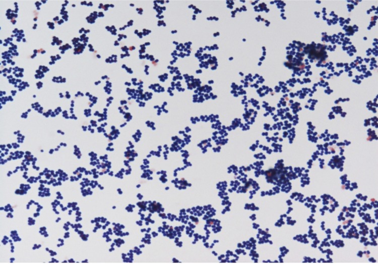
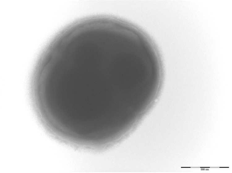
Colonies are small, 1mm in diameter, light grey, smooth and round on blood-enriched Columbia agar under anaerobic conditions using GENbag anaer (BioMérieux). Bacteria were grown on blood-enriched Columbia agar (Biomerieux), on BHI agar medium, on BHI agar medium supplemented with 1% NaCl, in BHI broth medium and in Trypticase-soja TS broth medium. Agar plates were incubated under anaerobic conditions using GENbag anaer (BioMérieux), under microaerophilic conditions using GENbag microaer (BioMérieux) and in the presence of air, with or without 5%CO2. Growth was achieved anaerobically and weakly after 3 days under microaerophilic conditions, on blood-enriched Columbia agar and in TS broth medium. Growth on BHI agar medium, and on BHI agar medium supplemented with 1% NaCl was also weak, and occurred after 72h. Gram staining showed non spore-forming Gram-positive cocci (Figure 2). The motility test was negative. Cells grown anaerobically in TS broth medium have a mean diameter of 1.12 µm (min = 0.98µm; max = 1.33 µm), as determined using electron microscopic observation after negative staining with a 3% ammonium molybdate solution (Figure 3).
Figure 2.
Gram stain of A. provenciensis strain 9402080T
Figure 3.
Transmission electron micrograph of A. provenciensis strain 9402080T, using a Morgani 268D (Philips) at an operating voltage of 60kV. The scale bar represents 1 µm.
Strain 9402080T exhibited catalase activity and no oxidase activity. Using an API 20A strip (BioMerieux, Marcy l’Etoile), positive reactions could be observed for D-Glucose, D-Lactose, D-Saccharose, D-Maltose, Salicin, D-Xylose, Gelatinase, Esculin, D-Mannose, and D-Trehalose. Using an API ZYM strip positive reactions were obtained for alkaline phosphatase (5nmol of hydrolyzed substrate), esterase (5nmol), esterase lipase (5nmol), leucine arylamidase (40nmol), acid phosphatase (5nmol), naphtophosphohydrolase (20nmol), and hyaluronidase (30nmol). Using an Api rapid id 32A, positive reactions could be observed for Arginine Dihydrolase, Beta Galactosidase, Beta Glucosidase, Beta Glucuronidase, N-Acetyl-beta-Glucosaminidase, Alpha-fucosidase, Mannose fermentation, Alkaline phosphatase, Arginine arylamidase, Leucine arylamidase, Pyroglutamate arylamidase, and Histidine arylamidase.
Regarding antibiotic susceptibility, A. provenciensis was susceptible to penicillin G, amoxicillin, cefotetan, imipenem, metronidazole and vancomycin. When compared to the representative species within the genus Anaerococcus, A. provenciensis exhibits the phenotypic characteristics details in Table 2.
Table 2. Differential characteristics of Anaerococcus provenciensis sp. nov., strain 9402080T, A. octavius strain NCTC 9810T, A. prevotii strain NCTC 11806T, and A. tetradius strain DSM 2951T.
| Properties | A. provenciensis | A. octavius | A.prevotii | A. tetradius |
|---|---|---|---|---|
| Cell diameter (µm) | 0.98-1.33 | 0.7-0.9 | 0.6-1.5 | 0.5-1.8 |
| Oxygen requirement | Anaerobic | Anaerobic | Anaerobic | Anaerobic |
| Gram stain | Positive | Positive | Positive | Positive |
| Optimal growth temperature | 35-37°C | na | na | na |
| Habitat | Human | Human | Human | Human |
| Enzyme production | ||||
| Indole | - | - | - | - |
| Alkaline Phosphatase | + | - | - | - |
| Urease | - | - | + | + |
| Catalase | + | - | + | - |
| Gelatinase | +/- | na | na | na |
| Activity of | ||||
| Phosphatase | Acid phosphatase | na | na | na |
| Naphtolphosphohydrolase | ||||
| Saccharolytic enzymes | ß-glucuronidase | - | α-glucosidase | α-glucosidase |
| ß-glucuronidase | ß-glucosidase | |||
| ß-glucuronidase | ||||
| Proteolytic enzymes | Leucine arylamidase | Proline arylamidase | Arginine arylamidase | Arginine arylamidase |
| Pyroglutamyl arylamidase | Pyroglutamyl arylamidase | Histidine arylamidase | ||
| Histidine arylamidase | ||||
| Utilization of | ||||
| Glucose | + | + | - | + |
| Mannose | - | + | + | + |
| Lactose | + | - | - | - |
| Raffinose | - | - | + | + |
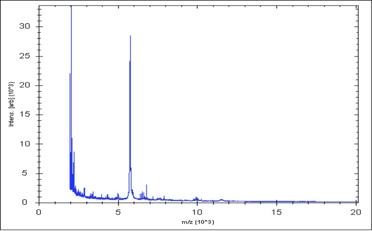
Matrix-assisted laser-desorption/ionization time-of-flight (MALDI-TOF) MS protein analysis was carried out as previously described [36]. Briefly, a pipette tip was used to pick an isolated bacterial colony from a culture agar plate and spread it as a thin film on a MTP 384 MALDI-TOF target plate (Bruker Daltonics, Germany). Ten distinct deposits were done for strain A. provenciensis strain 9402080T, from ten isolated colonies. Each smear was overlaid with 2µL of matrix solution (saturated solution of alpha-cyano-4-hydroxycinnamic acid) in 50% acetonitrile, 2.5% tri-fluoracetic acid, and allowed to dry for five minutes. Measurements were performed with a Microflex spectrometer (Bruker). Spectra were recorded in the positive linear mode for the mass range of 2,000 to 20,000 Da (parameter settings: ion source 1 (ISI), 20kV; IS2, 18.5 kV; lens, 7 kV). A spectrum was obtained after 675 shots at a variable laser power. The time of acquisition was between 30 seconds and 1 minute per spot. The ten 9402080T spectra were imported into the MALDI BioTyper software (version 2.0, Bruker) and analyzed by standard pattern matching (with default parameter settings) against the main spectra of 5,697 bacteria that were used as reference data in the BioTyper database. The method of identification includes the m/z from 3,000 to 15,000 Da. For every spectrum, 100 peaks at most were taken into account and compared with the spectra in database. A score enabled the presumptive identification and discrimination of the tested species from those in a database: a score > 2 with a validated species enabled the identification at the species level; a score > 1.7 but < 2 enabled the identification at the genus level; and a score < 1.7 did not enable any identification. For strain 9402080T, no significance score was obtained, thus suggesting that our isolate was not a member of a known species. We added the spectrum from strain 9402080T (Figure 4) to our database. A dendrogram was constructed with the MALDI Bio Typer software (version 2.0, Bruker), comparing the reference spectrum of strain 9402080T with reference spectra of 24 bacterial species, all belonging to the order of Clostridiales. In this dendrogram, strain 9402080T appears on a separate branch within the genus Anaerococcus (Figure 5).
Figure 4.
Reference mass spectrum from A. provenciensis strain 9402080T. Spectra from 10 individual colonies were compared and a reference spectrum was generated.
Figure 5.
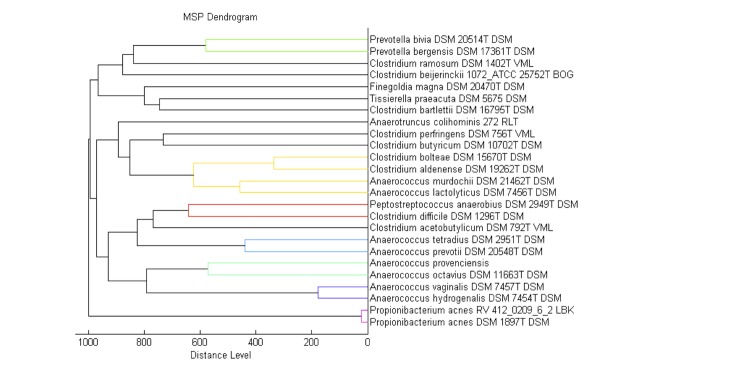
A dendrogram based on the comparison of the A. provenciensis strain 9402080T MALDI-TOF reference spectrum with the spectra from 24 other species of the order of Clostridiales
Genome sequencing information
Genome project history
The organism was selected for sequencing on the basis of its phylogenetic position and 16S rDNA similarity to other members of the Anaerococcus genus, and is part of a study for recovering and analyzing anaerobic bacteria from deep samples. It was the 8th genome of an Anaerococcus species and the first genome of Anaerococcus provenciensis sp. nov. The Genbank accession number is CAJU020000000 (CAJU020000001-CAJU020000026) and consists of 26 contigs. Table 3 shows the project information and its association with MIGS version 2.0 compliance [24].
Table 3. Project information.
| MIGS ID | Property | Term |
|---|---|---|
| MIGS-31 | Finishing quality | High-quality draft |
| MIGS-28 | Libraries used | Two 454 paired end 3-kb library |
| MIGS-29 | Sequencing platforms | 454 GS FLX+ Titanium |
| MIGS-31.2 | Fold coverage | 43.71 |
| MIGS-30 | Assemblers | Newbler version 2.8 |
| MIGS-32 | Gene calling method | Prodigal |
| INSDC ID | PRJEB85 | |
| Genbank ID | CAJU020000000 | |
| Genbank Date of Release | May 28, 2013 | |
| Project relevance | Study of the human gut microbiome |
Growth conditions and DNA isolation
Anaerococcus provenciensis sp. nov. strain 9402080T, CSUR P121 = DSM 26345 was grown anaerobically on blood agar medium at 37°C. 10 petri dishes were spread and resuspended in 3x100µl of G2 buffer. A first mechanical lysis was performed with glass powder on the Fastprep-24 device(Sample Preparation system) from MP Biomedicals, USA using 2x20 seconds pulses. DNA was then incubated with lysozyme (30 minutes at 37°C) and extracted through the BioRobot EZ 1 Advanced XL (Qiagen).The DNA was then concentrated and purified on a Qiamp kit (Qiagen). The yield and the concentration were measured by the Quant-it Picogreenkit (Invitrogen) on the Genios_Tecan fluorometer at 21.1ng/µl.
Genome sequencing and assembly
Two paired end library were pyrosequenced on the 454 Roche Titanium. This project was loaded twice on a 1/4 region for the 3 kb insert libraries on PTP Picotiterplates. 5µg of DNA was mechanically fragmented on the Hydroshear device (Digilab, Holliston, MA,USA) with an enrichment size at 3-4kb. The DNA fragmentation was visualized through the Agilent 2100 BioAnalyzer on a DNA LabChip 7500 with an optimal size of 3.82 kb. The library was constructed according to the 454 Titanium paired end protocol supplied by the manufacturer. Circularization and nebulization were performed and generated a pattern with a maximum at 575 bp. After PCR amplification through 15 cycles followed by double size selection, the single stranded paired end libraries was then quantified on the Agilent 2100 BioAnalyzer on a RNA Pico 6000 LabChip at 135pg/µL. The library concentration equivalence was calculated at 4.31x1008 molecules/µL. The library was stored at -20°C until use.
The 3kb paired end library was clonally amplified with 0.5 and 1 cpb in 4 emPCR reactions per condition with the GS Titanium SV emPCR Kit (Lib-L) v2 .The yield of the emPCR was 5.56 and 9.79% respectively according to the quality expected by the range of 5 to 20% from the Roche procedure.
Two times 790,000 beads were loaded on the GS FLX Titanium PicoTiterPlates PTP Kit 70×75 and sequenced with the GS FLX Titanium Sequencing Kit XLR70.
The 454 sequencing generated 650,718 reads (104,82 Mb) assembled into contigs and scaffolds using Newbler version 2.8 (Roche) and Opera software v1.2 [37] combined with GapFiller V1.10 [38] and some finishing using CLC Genomics Workbench. Finally, the available genome consists of 8 scaffolds and 26 contigs, with a 43.71× coverage.
Genome annotation
Non-coding genes and miscellaneous features were predicted using RNAmmer [39], ARAGORN [40], Rfam [41], and PFAM [42]. Open Reading Frames (ORFs) were predicted using Prodigal [43] with default parameters. The predicted ORFs were excluded if they spanned a sequencing gap region. The functional annotation was achieved using BLASTP [44] against the GenBank database [45] and the Clusters of Orthologous Groups (COG) database [46] [47].
Genome properties
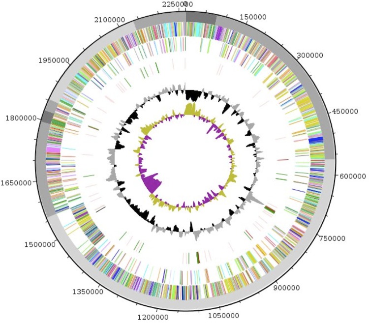
The genome of Anaerococcus provenciensis strain 9402080T is estimated to be 2.26 Mb long with a G+C content of 33.48% (Figure 5 and Table 4). A total of 2,099 protein-coding and 96 RNA genes, including 8 rRNA genes, 48 tRNA, 1 tmRNA and 39 miscellaneous other RNA were found. The majority of the protein-coding genes were assigned a putative function (74.8%); the remainder were annotated as hypothetical proteins. The distribution of genes into COGs functional categories is presented in Table 5 and Figure 6. The properties and the statistics of the genome are summarized in Tables 4 and 5.
Table 4. Nucleotide content and gene count levels of the genome.
| Attribute | Value | % of total a |
|---|---|---|
| Genome size (bp) | 2,265,283 | 100 |
| DNA coding region (bp) | 2,024,670 | 89.37 |
| DNA G+C content (bp) | 677.859 | 33.48 |
| Total genes | 2195 | 100 |
| rRNA | 8 | 0.36 |
| tRNA | 48 | 2.19 |
| tmRNA | 1 | 0.04 |
| miscRNA | 39 | 1.78 |
| Protein-coding genes | 2099 | 95.62 |
| Genes with function prediction | 1570 | 74.79 |
| Genes assigned to COGs | 2077 | 98.95 |
a The total is based on either the size of the genome in base pairs or the total number of protein coding genes in the annotated genome
Table 5. Number of genes associated with the 25 general COG functional categories.
| Code | Value | %agea | Description |
|---|---|---|---|
| J | 152 | 6.76 | Translation |
| A | 4 | 0.18 | RNA processing and modification |
| K | 174 | 7.74 | Transcription |
| L | 178 | 7.92 | Replication, recombination and repair |
| B | 4 | 0.18 | Chromatin structure and dynamics |
| D | 40 | 1.78 | Cell cycle control, mitosis and meiosis |
| Y | 0 | 0 | Nuclear structure |
| V | 92 | 4.09 | Defense mechanisms |
| T | 82 | 3.65 | Signal transduction mechanisms |
| M | 99 | 4.41 | Cell wall/membrane biogenesis |
| N | 14 | 0.62 | Cell motility |
| Z | 5 | 0.22 | Cytoskeleton |
| W | 0 | 0 | Extracellular structures |
| U | 51 | 2.27 | Intracellular trafficking and secretion |
| O | 78 | 3.48 | Posttranslational modification, protein turnover, chaperones |
| C | 130 | 5.78 | Energy production and conversion |
| G | 221 | 9.83 | Carbohydrate transport and metabolism |
| E | 125 | 5.56 | Amino acid transport and metabolism |
| F | 64 | 2.85 | Nucleotide transport and metabolism |
| H | 59 | 2.62 | Coenzyme transport and metabolism |
| I | 51 | 2.27 | Lipid transport and metabolism |
| P | 127 | 5.65 | Inorganic ion transport and metabolism |
| Q | 17 | 0.75 | Secondary metabolites biosynthesis, transport and catabolism |
| R | 238 | 10.6 | General function prediction only |
| S | 220 | 9.79 | Function unknown |
| - | 22 | 0.98 | Not in COGs |
a The percentage is based on the total number of protein coding genes in the annotated genome.
Figure 6.
Graphical circular map of the genome. From outside to the center: scaffolds are in grey (unordered), genes on forward strand (colored by COG categories), genes on reverse strand (colored by COG categories), RNA genes (tRNAs green, rRNAs red, tm RNAs black, misc_RNA pink), GC content (black/grey), and GC skew (purple/olive).
Insights into the genome sequence
We made some brief comparisons of Anaerococcus provenciensis against Anaerococcus prevotii DSM 20548 (NC_013171) which is currently the closest available genome. This genome contains 1 chromosome (accession number: NC_013171) and 1 plasmid (accession number: NC_013164).
The draft genome sequence of Anaerococcus provenciensis is bigger than that of Anaerococcus prevotii (2.26 Mbp and 1.99 Mbp, respectively). The G+C content (33.48%) is slightly lower than that of Anaerococcus prevotii (35.7%). Anaerococcus provenciensis has more coding-genes (2,099 predicted genes against 1,916 genes), but the ratios of the number of genes per Mbp genome size are relatively close (1079.22 – 962.81).
Table 6. Comparison of the percentage of genes associated with the 25 general COG functional categories for Anaerococcus provenciensis and Anaerococcus prevotii DSM 20548.
| Code | A. provenciensis % of total | A. prevotii % of total | Difference (in %) |
COG description |
|---|---|---|---|---|
| J | 6.76 | 7.53 | -0.77 | Translation |
| A | 0.18 | 0.10 | 0.08 | RNA processing and modification |
| K | 7.74 | 6.91 | 0.83 | Transcription |
| L | 7.92 | 6.13 | 1.79 | Replication, recombination and repair |
| B | 0.18 | 0.16 | 0.02 | Chromatin structure and dynamics |
| D | 1.78 | 1.56 | 0.22 | Cell cycle control, mitosis and meiosis |
| Y | 0 | 0.05 | -0.05 | Nuclear structure |
| V | 4.09 | 3.43 | 0.66 | Defense mechanisms |
| T | 3.65 | 3.17 | 0.48 | Signal transduction mechanisms |
| M | 4.41 | 5.24 | -0.83 | Cell wall/membrane biogenesis |
| N | 0.62 | 0.36 | 0.26 | Cell motility |
| Z | 0.22 | 0.16 | 0.06 | Cytoskeleton |
| W | 0 | 0 | 0 | Extracellular structures |
| U | 2.27 | 1.92 | 0.35 | Intracellular trafficking and secretion |
| O | 3.48 | 3.63 | -0.15 | Posttranslational modification, protein turnover, chaperones |
| C | 5.78 | 6.59 | -0.81 | Energy production and conversion |
| G | 9.83 | 8.41 | 1.42 | Carbohydrate transport and metabolism |
| E | 5.56 | 6.65 | -1.09 | Amino acid transport and metabolism |
| F | 2.85 | 3.69 | -0.84 | Nucleotide transport and metabolism |
| H | 2.62 | 3.58 | -0.96 | Coenzyme transport and metabolism |
| I | 2.27 | 2.34 | -0.07 | Lipid transport and metabolism |
| P | 5.65 | 6.80 | -1.15 | Inorganic ion transport and metabolism |
| Q | 0.75 | 0.78 | -0.03 | Secondary metabolites biosynthesis, transport and catabolism |
| R | 10.6 | 11.21 | -0.61 | General function prediction only |
| S | 9.79 | 9.61 | 0.18 | Function unknown |
| - | 0.98 | 0.99 | -0.01 | Not in COGs |
Table 6 presents the difference in gene numbers (in percentage) from each COG category between Anaerococcus provenciensis and Anaerococcus prevotii DSM 20548. The totals are highly similar in the two species. The biggest difference is in the COG "Carbohydrate Metabolism and transportation" category, which does not exceed 1.42%.
Conclusion
On the basis of phenotypic, phylogenetic and genomic analysis, we formally propose the creation of Anaerococcus provenciensis sp. nov. that contains the strain 9402080T. This bacterium has been found in Marseille, France.
Description of Anaerococcus provenciensis sp. nov.
Anaerococcus provenciensis (pro.ven.ci.en’cis; L. gen. masc. n. provenciensis, pertaining to Provence, the name of the aeae, south-east of France, where the type strain was isolated). Isolated from a cerebral abscess sample from a patient from Marseille. A. provenciensis is a Gram-positive cocci, obligately anaerobic, non-spore-forming bacterium. Grows at 37°C in anaerobic atmosphere. Negative for indole. Non-motile. The G+C content of the genome is 33.48%. The type strain is 9402080T(= CSUR P121 = DSM 26345).
References
- 1.La Scola B, Fournier PE, Raoult D. Burden of emerging anaerobes in the MALDI-TOF and 16S rRNA gene sequencing era. Anaerobe 2011; 17:106-112 10.1016/j.anaerobe.2011.05.010 [DOI] [PubMed] [Google Scholar]
- 2.Genome Online Database http://www.genomesonline.org/cgi-bin/GOLD/index.cgi
- 3.Tindall BJ, Rossello-Mora R, Busse HJ, Ludwig W, Kampfer P. Notes on the characterization of prokaryote strains for taxonomic purposes. Int J Syst Evol Microbiol 2010; 60:249-266 10.1099/ijs.0.016949-0 [DOI] [PubMed] [Google Scholar]
- 4.Kokcha S, Mishra AK, Lagier JC, Million M, Leroy Q, Raoult D, Fournier PE. Non contiguous-finished genome sequence and description of Bacillus timonensis sp. nov. Stand Genomic Sci 2012; 6:346-355 10.4056/sigs.2776064 [DOI] [PMC free article] [PubMed] [Google Scholar]
- 5.Lagier JC, El Karkouri K, Nguyen TT, Armougom F, Raoult D, Fournier PE. Non-contiguous finished genome sequence and description of Anaerococcus senegalensis sp. nov. Stand Genomic Sci 2012; 6:116-125 10.4056/sigs.2415480 [DOI] [PMC free article] [PubMed] [Google Scholar]
- 6.Mishra AK, Gimenez G, Lagier JC, Robert C, Raoult D, Fournier PE. Non-contiguous finished genome sequence and description of Alistipes senegalensis sp. nov. Stand Genomic Sci 2012; 6:304-314 10.4056/sigs.2625821 [DOI] [PMC free article] [PubMed] [Google Scholar]
- 7.Lagier JC, Armougom F, Mishra AK, Ngyuen TT, Raoult D, Fournier PE. Non-contiguous finished genome sequence and description of Alistipes timonensis sp. nov. Stand Genomic Sci 2012; 6:315-324 10.4056/sigs.2685971 [DOI] [PMC free article] [PubMed] [Google Scholar]
- 8.Mishra AK, Lagier JC, Robert C, Raoult D, Fournier PE. Non-contiguous finished genome sequence and description of Clostridium senegalense sp. nov. Stand Genomic Sci 2012; 6:386-395 [DOI] [PMC free article] [PubMed] [Google Scholar]
- 9.Mishra AK, Lagier JC, Robert C, Raoult D, Fournier PE. Non-contiguous finished genome sequence and description of Peptoniphilus timonensis sp. nov. Stand Genomic Sci 2012; 7:1-11 10.4056/sigs.2956294 [DOI] [PMC free article] [PubMed] [Google Scholar]
- 10.Mishra AK, Lagier JC, Rivet R, Raoult D, Fournier PE. Non-contiguous finished genome sequence and description of Paenibacillus senegalensis sp. nov. Stand Genomic Sci 2012; 7:70-81 10.4056/sigs.3056450 [DOI] [PMC free article] [PubMed] [Google Scholar]
- 11.Lagier JC, Gimenez G, Robert C, Raoult D, Fournier PE. Non-contiguous finished genome sequence and description of Herbaspirillum massiliense sp. nov. Stand Genomic Sci 2012; 7:200-209 [DOI] [PMC free article] [PubMed] [Google Scholar]
- 12.Roux V, El Karkouri K, Lagier JC, Robert C, Raoult D. Non-contiguous finished genome sequence and description of Kurthia massiliensis sp. nov. Stand Genomic Sci 2012; 7:221-232 10.4056/sigs.3206554 [DOI] [PMC free article] [PubMed] [Google Scholar]
- 13.Kokcha S, Ramasamy D, Lagier JC, Robert C, Raoult D, Fournier PE. Non-contiguous finished genome sequence and description of Brevibacterium senegalense sp. nov. Stand Genomic Sci 2012; 7:233-245 10.4056/sigs.3256677 [DOI] [PMC free article] [PubMed] [Google Scholar]
- 14.Ramasamy D, Kokcha S, Lagier JC, N’Guyen TT, Raoult D, Fournier PE. Non-contiguous finished genome sequence and description of Aeromicrobium massilense sp. nov. Stand Genomic Sci 2012; 7:246-257 10.4056/sigs.3306717 [DOI] [PMC free article] [PubMed] [Google Scholar]
- 15.Lagier JC, Ramasamy D, Rivet R, Raoult D, Fournier PE. Non-contiguous finished genome sequence and description of Cellulomonas massiliensis sp. nov. Stand Genomic Sci 2012; 7:258-270 10.4056/sigs.3316719 [DOI] [PMC free article] [PubMed] [Google Scholar]
- 16.Ludwig W, Schleifer KH, Whitman WB. Revised road map to the phylum Firmicutes. In: Bergey's Manual of Systematic Bacteriology, 2nd ed., vol. 3 (The Firmicutes) (P. De Vos, G. Garrity, D. Jones, N.R. Krieg, W. Ludwig, F.A. Rainey, K.-H. Schleifer, and W.B. Whitman, eds.), Springer-Verlag, New York. (2009) pp. 1-13. [Google Scholar]
- 17.Ezaki T, Kawamura Y, Li N, Li ZY, Zhao L, Shu S. Proposal of the genera Anaerococcus gen. nov., Peptoniphilus gen. nov. and Gallicola gen. nov. for members of the genus Peptostreptococcus. Int J Syst Evol Microbiol 2001; 51:1521-1528 [DOI] [PubMed] [Google Scholar]
- 18.Foubert EL, Douglas HC. Studies on the Anaerobic Micrococci: I. Taxonomic Considerations. J Bacteriol 1948; 56:25-34 [DOI] [PubMed] [Google Scholar]
- 19.Song Y, Liu C, Finegold SM. Peptoniphilus gorbachii sp. nov., Peptoniphilus olsenii sp. nov., and Anaerococcus murdochii sp. nov. isolated from clinical specimens of human origin. J Clin Microbiol 2007; 45:1746-1752 10.1128/JCM.00213-07 [DOI] [PMC free article] [PubMed] [Google Scholar]
- 20.Jain S, Bui V, Spencer C, Yee L. Septic arthritis in a native joint due to Anaerococcus prevotii. J Clin Pathol 2008; 61:775-776 10.1136/jcp.2007.053421 [DOI] [PubMed] [Google Scholar]
- 21.La Scola B, Fournier PE, Raoult D. Burden of emerging anaerobes in the MALDI-TOF and 16S rRNA gene sequencing era. Anaerobe 2011; 17:106-112 10.1016/j.anaerobe.2011.05.010 [DOI] [PubMed] [Google Scholar]
- 22.Pépin J, Deslandes S, Giroux G, Sobela F, Khonde N, Diakite S, Demeule D, Labbé AC, Carrier N, Frost E. The complex vaginal flora of west african women with bacterial vaginosis. PLoS ONE 2011; 6:e25082 10.1371/journal.pone.0025082 [DOI] [PMC free article] [PubMed] [Google Scholar]
- 23.Grice EA, Kong HH, Conlan S, Deming CB, Davis J, Young AC, NISC Comparative Sequencing Program, Bouffard GG, Blakesley RW, Murray PR, et al. Topographical and temporal diversity of the human skin microbiome. Science 2009; 324:1190-1192 10.1126/science.1171700 [DOI] [PMC free article] [PubMed] [Google Scholar]
- 24.Field D, Garrity G, Gray T, Morrison N, Selengut J, Sterk P, Tatusova T, Thomson N, Allen MJ, Angiuoli SV, et al. The minimum information about a genome sequence (MIGS) specification. Nat Biotechnol 2008; 26:541-547 10.1038/nbt1360 [DOI] [PMC free article] [PubMed] [Google Scholar]
- 25.Woese CR, Kandler O, Wheelis ML. Towards a natural system of organisms: proposal for the domains Archae, Bacteria, and Eukarya. Proc Natl Acad Sci USA 1990; 87:4576-4579 10.1073/pnas.87.12.4576 [DOI] [PMC free article] [PubMed] [Google Scholar]
- 26.Garrity GM, Holt JG. The Road Map to the Manual. In: Garrity GM, Boone DR, Castenholz RW (eds), Bergey's Manual of Systematic Bacteriology, Second Edition, Volume 1, Springer, New York, 2001, p. 119-169 [Google Scholar]
- 27.Gibbons NE, Murray RGE. Proposals Concerning the Higher Taxa of Bacteria. Int J Syst Bacteriol 1978; 28:1-6 10.1099/00207713-28-1-1 [DOI] [Google Scholar]
- 28.Murray RGE. The Higher Taxa, or, a Place for Everything...? In: Holt JG (ed), Bergey's Manual of Systematic Bacteriology, First Edition, Volume 1, The Williams and Wilkins Co., Baltimore, 1984, p. 31-34. [Google Scholar]
- 29.List of new names and new combinations previously effectively, but not validly, published. List no. 132. Int J Syst Evol Microbiol 2010; 60:469-472 10.1099/ijs.0.022855-0 [DOI] [PubMed] [Google Scholar]
- 30.Rainey FA. Class II. Clostridia class nov. In: De Vos P, Garrity G, Jones D, Krieg NR, Ludwig W, Rainey FA, Schleifer KH, Whitman WB (eds), Bergey's Manual of Systematic Bacteriology, Second Edition, Volume 3, Springer-Verlag, New York, 2009, p. 736. [Google Scholar]
- 31.Skerman VBD, Sneath PHA. Approved list of bacterial names. Int J Syst Bacteriol 1980; 30:225-420 10.1099/00207713-30-1-225 [DOI] [Google Scholar]
- 32.Prevot AR. Dictionnaire des bactéries pathogens. In: Hauduroy P, Ehringer G, Guillot G, Magrou J, Prevot AR, Rosset, Urbain A (eds). Paris, Masson, 1953, p.1-692. [Google Scholar]
- 33.Ashburner M, Ball CA, Blake JA, Botstein D, Butler H, Cherry JM, Davis AP, Dolinski K, Dwight SS, Eppig JT, et al. Gene ontology: tool for the unification of biology. The Gene Ontology Consortium. Nat Genet 2000; 25:25-29 10.1038/75556 [DOI] [PMC free article] [PubMed] [Google Scholar]
- 34.Tamura K, Dudley J, Nei M, Kumar S. MEGA4: Molecular Evolutionary Genetics Analysis (MEGA) software version 4.0. Mol Biol Evol 2007; 24:1596-1599 10.1093/molbev/msm092 [DOI] [PubMed] [Google Scholar]
- 35.Stackebrandt E, Ebers J. Taxonomic parameters revisited: tarnished gold standards. Microbiol Today 2006; 33:152-155 [Google Scholar]
- 36.Seng P, Drancourt M, Gouriet F, La Scola B, Fournier PE, Rolain JM, Raoult D. Ongoing revolution in bacteriology: routine identification of bacteria by matrix-assisted laser desorption ionization time-of-flight mass spectrometry. Clin Infect Dis 2009; 49:543-551 10.1086/600885 [DOI] [PubMed] [Google Scholar]
- 37.Gao S, Sung W, Nagarajan N. Opera: reconstructing optimal genomic scaffolds with high-throughput paired-end sequences. J Comput Biol 2011; 18:1681-1691 10.1089/cmb.2011.0170 [DOI] [PMC free article] [PubMed] [Google Scholar]
- 38.Boetzer M, Pirovano W. Toward almost closed genomes with GapFiller. Genome Biol 2012; 13:R56 10.1186/gb-2012-13-6-r56 [DOI] [PMC free article] [PubMed] [Google Scholar]
- 39.Lagesen K, Hallin P, Rødland EA, Staerfeldt H, Rognes T, Ussery DW. RNAmmer: consistent and rapid annotation of ribosomal RNA genes. Nucleic Acids Res 2007; 35:3100-3108 10.1093/nar/gkm160 [DOI] [PMC free article] [PubMed] [Google Scholar]
- 40.Laslett D, Canback B. ARAGORN, a program to detect tRNA genes and tmRNA genes in nucleotide sequences. Nucleic Acids Res 2004; 32:11-16 10.1093/nar/gkh152 [DOI] [PMC free article] [PubMed] [Google Scholar]
- 41.Griffiths-Jones S, Moxon S, Marshall M, Khanna A, Eddy SR, Bateman A. Rfam: annotating non-coding RNAs in complete genomes. Nucleic Acids Res 2005; 33:D121-D124 10.1093/nar/gki081 [DOI] [PMC free article] [PubMed] [Google Scholar]
- 42.Punta M, Coggill PC, Eberhardt RY, Mistry J, Tate J, Boursnell C, Pang N, Forslund K, Ceric G, Clements J, et al. The Pfam protein families database. Nucleic Acids Res 2012; 40:D290-D301 10.1093/nar/gkr1065 [DOI] [PMC free article] [PubMed] [Google Scholar]
- 43.Hyatt D, Chen G, Locascio PF, Land ML, Larimer FW, Hauser LJ. Prodigal: prokaryotic gene recognition and translation initiation site identification. BMC Bioinformatics 2010; 11:119 10.1186/1471-2105-11-119 [DOI] [PMC free article] [PubMed] [Google Scholar]
- 44.Camacho C, Coulouris G, Avagyan V, Ma N, Papadopoulos J, Bealer K, Madden TL. BLAST+: architecture and applications. BMC Bioinformatics 2009; 10:421 10.1186/1471-2105-10-421 [DOI] [PMC free article] [PubMed] [Google Scholar]
- 45.Benson DA, Cavanaugh M, Clark K, Karsch-Mizrachi I, Lipman DJ, Ostell J, Sayers EW. GenBank. Nucleic Acids Res 2013; 41:D36-D42 10.1093/nar/gks1195 [DOI] [PMC free article] [PubMed] [Google Scholar]
- 46.Tatusov RL, Koonin EV, Lipman DJ. A genomic perspective on protein families. Science 1997; 278:631-637 10.1126/science.278.5338.631 [DOI] [PubMed] [Google Scholar]
- 47.Tatusov RL, Galperin MY, Natale DA, Koonin EV. The COG database: a tool for genome-scale analysis of protein functions and evolution. Nucleic Acids Res 2000; 28:33-36 10.1093/nar/28.1.33 [DOI] [PMC free article] [PubMed] [Google Scholar]


