
Figure 1.
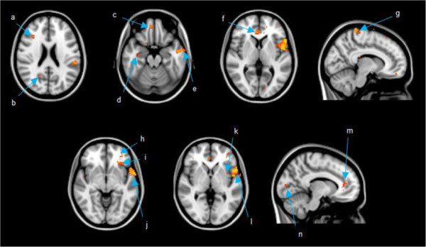
Images are presented in radiological view where right is the patient’s left side and left is the patient’s right side. Coordinates presented in MNI space. a) middle frontal gyrus (36 28 22); b) precuneus (20 -58 25); c) gyrus rectus (6 48 -20); d) hippocampus (34 -14 -20) e) middle temporal gyrus (-58 -4 -20); f) anterior cingulate (4 38 5); g) paracentral lobule (10 -36 75); h) medial orbitofrontal gyrus (-32 44 -5); i) inferior orbitofrontal gyrus (-32 28 -5); j) superior temporal gyrus (-65 2 -5); k) insular cortex (-36 6 0); l) inferior frontal operculum (-54 10 0); m) anterior cingulate (-10 38 10); n) calcarine cortex (-12 -86 5).