. 2014 Aug 13;3:e02949. doi: 10.7554/eLife.02949

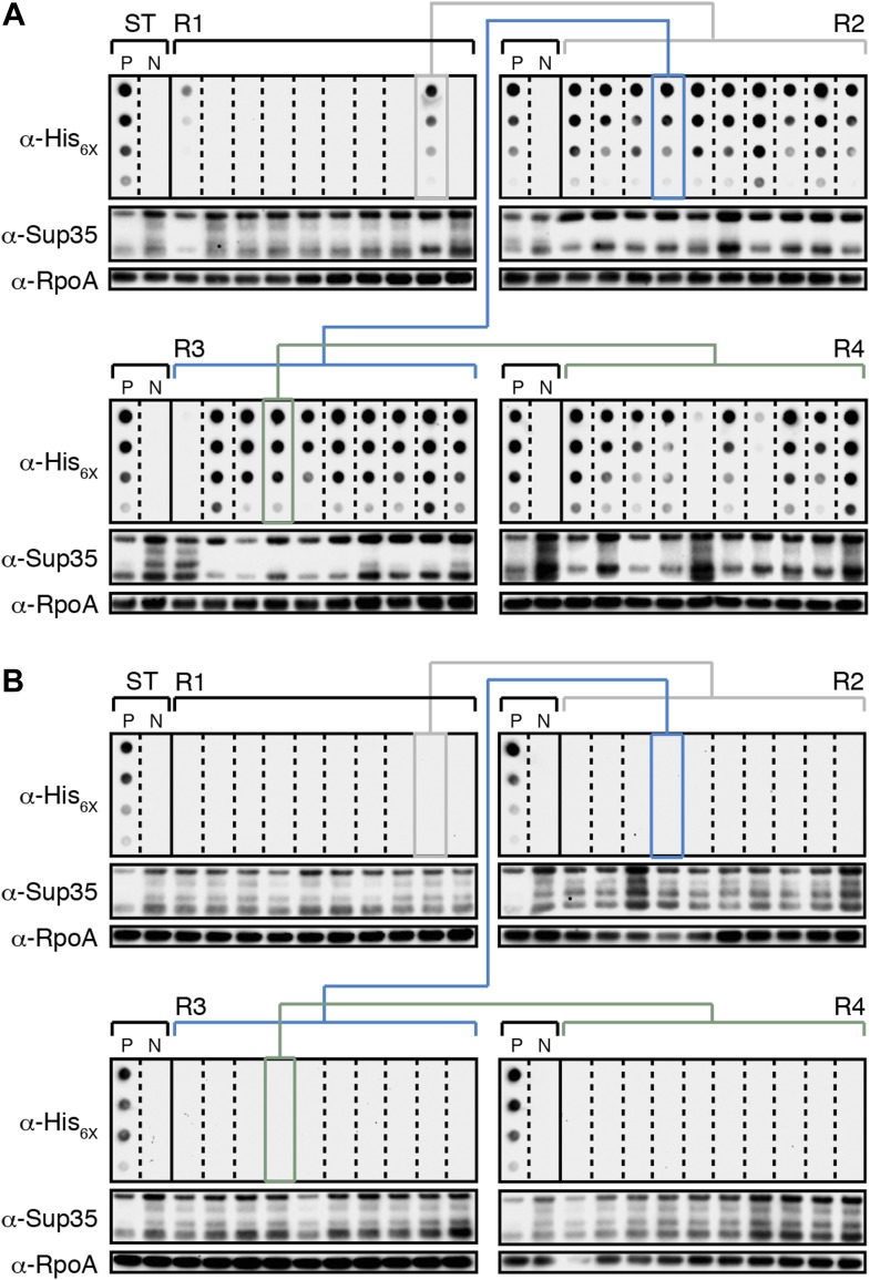
Figure 4. E. coli can propagate the Sup35 NM prion over ∼100 generations.

(A) Experimental Lineage 1 (L1E). An aggregate-positive Round 1 (R1) experimental clone (gray box) derived from a starter culture (ST) of cells containing Sup35 NM and New1 is identified and restreaked to yield progeny Round 2 (R2) clones (gray bracket). All 10 R2 clones analyzed contain detectable SDS-stable Sup35 NM aggregates. An aggregate-positive R2 clone (blue box) is identified and restreaked to yield progeny Round 3 (R3) clones (blue bracket). Again, all 10 R3 clones analyzed contain SDS-stable Sup35 NM aggregates. An aggregate-positive R3 clone (green box) is identified and restreaked to yield progeny Round 4 (R4) clones (green bracket). 9 of 10 R4 clones analyzed contain SDS-stable Sup35 NM aggregates. The filter retention assay is used to detect SDS-stable Sup35 NM aggregates. Intracellular Sup35 NM fusion protein levels are comparable in all 40 samples as assessed by Western blot analysis. Starter cultures of cells containing Sup35 NM and New1 and cells containing Sup35 NM alone serve as positive (P) and negative (N) controls, respectively. The α-His6X and α-Sup35 antibodies recognize the Sup35 NM-mCherry-His6X fusion protein, and the α-RpoA antibody recognizes the α subunit of E. coli RNA polymerase. (B) Control Lineage 1 (L1C). An aggregate-negative R1 control clone (gray box) derived from a starter culture of cells containing Sup35 NM alone is identified and restreaked to yield progeny R2 clones (gray bracket), an aggregate-negative R2 clone (blue box) is identified and restreaked to yield progeny R3 clones (blue bracket), and an aggregate-negative R3 clone (green box) is identified and restreaked to yield progeny R4 clones (green bracket). No SDS-stable Sup35 NM aggregates are detectable in any sample. Intracellular Sup35 NM fusion protein levels are comparable in all 40 samples as assessed by Western blot analysis.

DOI: http://dx.doi.org/10.7554/eLife.02949.007

Figure 4.

Figure 4—figure supplement 1. The fate of Sup35 NM in experimental Lineages 2-4.

Figure 4—figure supplement 1.

(A) Experimental Lineage 2 (L2E). An aggregate-positive Round 1 (R1) clone (gray box) derived from a starter culture (ST) of cells containing Sup35 NM and New1 is identified and restreaked to yield progeny Round 2 (R2) clones (gray bracket). 8 of 10 R2 clones analyzed contain detectable SDS-stable Sup35 NM aggregates as assessed by filter retention analysis. An aggregate-positive R2 clone (blue box) is restreaked to yield progeny Round 3 (R3) clones (blue bracket). 0 of 10 R3 clones analyzed contain detectable SDS-stable Sup35 NM aggregates. The apparent loss of aggregates coincides with the loss of detectable Sup35 NM fusion protein as assessed by Western blot analysis. Assaying the 10 Round 4 (R4) progeny clones derived from an aggregate-negative R3 clone (green box) reveals that fusion protein levels can be restored without the recovery of SDS-stable Sup35 NM aggregates (green bracket). Starter cultures of cells containing Sup35 NM and New1 and cells containing Sup35 NM alone serve as positive (P) and negative (N) controls, respectively. The α-His6X and α-Sup35 antibodies recognize the Sup35 NM-mCherry-His6X fusion protein, and the α-RpoA antibody recognizes the α subunit of E. coli RNA polymerase. (B) Experimental Lineage 3 (L3E). 9 of 10 R2 clones analyzed contain detectable SDS-stable Sup35 NM aggregates, which are retained in 8 of 10 R3 progeny clones and 7 of 10 R4 progeny clones. (C) Experimental Lineage 4 (L4E). 8 of 10 R2 clones analyzed contain detectable SDS-stable Sup35 NM aggregates, which are retained in 9 of 10 R3 progeny clones. 0 of 10 R4 clones analyzed contain detectable SDS-stable Sup35 NM aggregates. As in R3 of L2E (A), the apparent loss of aggregates coincides with a dramatic drop in Sup35 NM fusion protein levels.
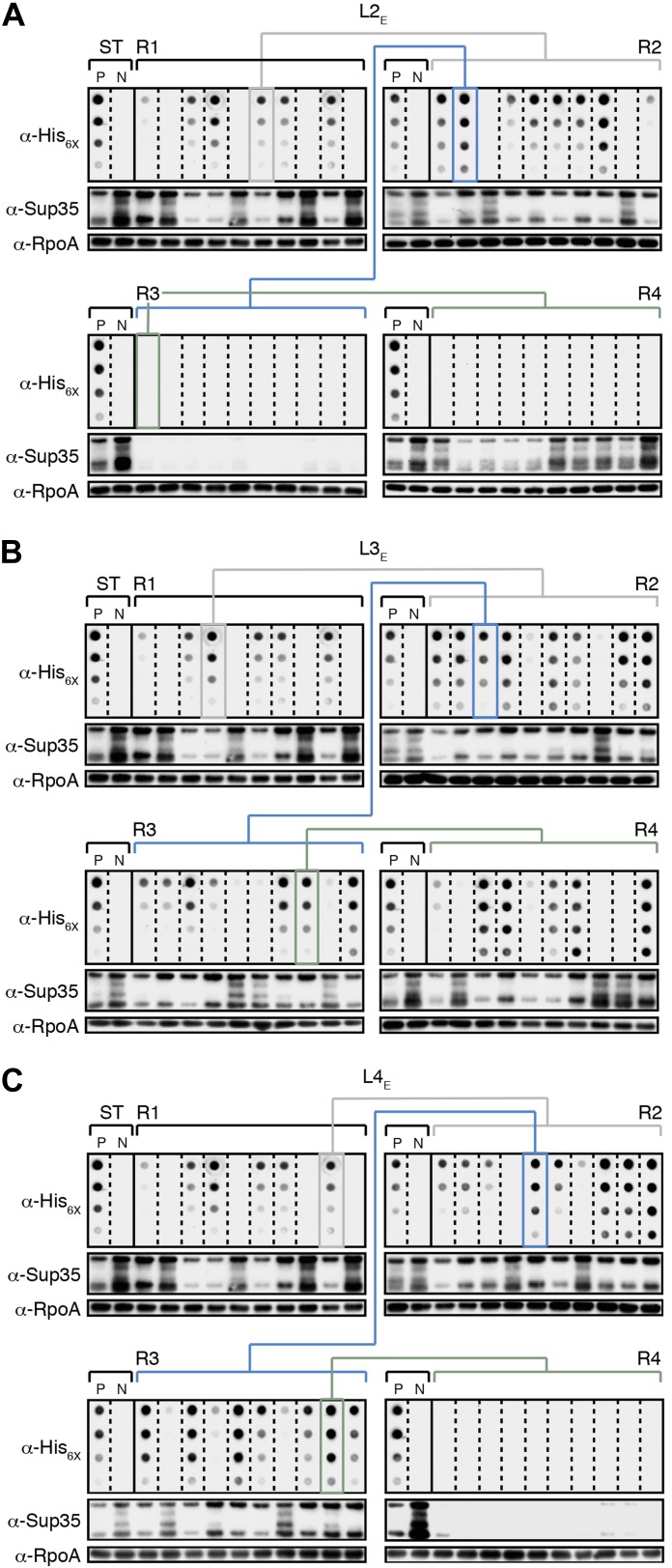
Figure 4—figure supplement 2. The fate of Sup35 NM in control Lineages 2–4.

Figure 4—figure supplement 2.

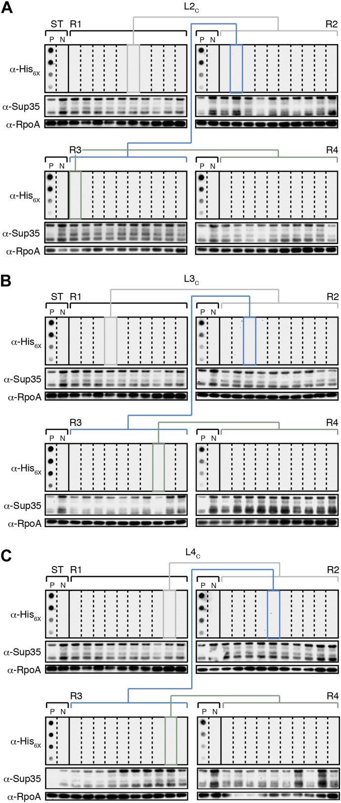
(A) Control Lineage 2 (L2C). An aggregate-negative Round 1 (R1) clone (gray box) derived from a starter culture (ST) of cells containing Sup35 NM alone is identified and restreaked to yield progeny Round 2 (R2) clones (gray bracket). 0 of 10 R2 clones analyzed contain detectable SDS-stable Sup35 NM aggregates as assessed by filter retention analysis. An aggregate-negative R2 clone (blue box) is restreaked to yield progeny R3 clones (blue bracket). 0 of 10 R3 clones analyzed contain detectable SDS-stable Sup35 NM aggregates. An aggregate-negative R3 clone (green box) is restreaked to yield progeny R4 clones (green bracket). 0 of 10 R4 clones analyzed contain detectable SDS-stable Sup35 NM aggregates. Starter cultures of cells containing Sup35 NM and New1 and cells containing Sup35 NM alone serve as positive (P) and negative (N) controls, respectively. The α-His6X and α-Sup35 antibodies recognize the Sup35 NM-mCherry-His6X fusion protein, and the α-RpoA antibody recognizes the α subunit of E. coli RNA polymerase. (B) Control Lineage 3 (L3C). As in L2C (A), no SDS-stable Sup35 NM aggregates are detectable in progeny R2, R3, or R4 clones. (C) Control Lineage 4 (L4C). As in L2C (A) and L3C (B), no SDS-stable Sup35 NM aggregates are detectable in progeny R2, R3, or R4 clones.