Abstract
Early life stress (ELS) is associated with increased vulnerability for diseases in later life, including psychiatric disorders. Animal models and human studies suggest that this effect is mediated by epigenetic mechanisms. In humans, epigenetic studies to investigate the influence of ELS on psychiatric phenotypes are limited by the inaccessibility of living brain tissue. Due to the tissue-specific nature of epigenetic signatures, it is impossible to determine whether ELS induced epigenetic changes in accessible peripheral cells, for example, blood lymphocytes, reflect epigenetic changes in the brain. To overcome these limitations, we applied a cross-species approach involving: (i) the analysis of CD34+ cells from human cord blood; (ii) the examination of blood-derived CD3+ T cells of newborn and adolescent nonhuman primates (Macaca mulatta); and (iii) the investigation of the prefrontal cortex of adult rats. Several regions in MORC1 (MORC family CW-type zinc finger 1; previously known as: microrchidia (mouse) homolog) were differentially methylated in response to ELS in CD34+ cells and CD3+ T cells derived from the blood of human and monkey neonates, as well as in CD3+ T cells derived from the blood of adolescent monkeys and in the prefrontal cortex of adult rats. MORC1 is thus the first identified epigenetic marker of ELS to be present in blood cell progenitors at birth and in the brain in adulthood. Interestingly, a gene-set-based analysis of data from a genome-wide association study of major depressive disorder (MDD) revealed an association of MORC1 with MDD.
Introduction
Early life stress (ELS) is associated with increased vulnerability for diseases in later life, including psychiatric disorders.1, 2, 3, 4, 5, 6, 7, 8 Previous studies suggest that the effect of ELS on lifelong phenotypes is mediated by epigenetic mechanisms.9, 10, 11 Weaver et al.,12 reported lifelong epigenetic modifications of the glucocorticoid receptor gene (NR3C1) in the rat hippocampus in response to stress induced by low levels of maternal care. More recent studies in rodents have reported ELS effects on DNA methylation for additional genes, including AVP, BDNF, NR4A1, SLC6A4, HSD11B2, RELN, Esr1 and CRH.13, 14, 15, 16, 17, 18, 19, 20, 21 Similarly, in humans ELS has been shown to be associated with altered methylation of NR3C1, BDNF and SLC6A4.22, 23, 24, 25, 26, 27, 28, 29, 30, 31 Recently, Klengel et al.32 reported that demethylation of a glucocorticoid response element in the stress response regulator FKBP5 predicted risk for posttraumatic stress disorder, and that this association was dependent upon risk allele and childhood trauma status. Although the above studies focused on candidate genes, two recent studies investigating ELS effects on an epigenome-wide level identified a variety of epigenetic alterations due to ELS.33,34
Studies of ELS in humans are limited. First, since brains of living humans are not accessible for epigenetic studies, it is impossible to ascertain if alterations in DNA methylation of peripheral cells after exposure to ELS reflect brain DNA methylation changes triggered by ELS. Second, excluding underlying causes of changes in DNA methylation other than ELS is problematic, as genetic background and/or predisposing environmental factors cannot be randomized and controlled in human studies. And third, to address the issue of a temporal relationship between ELS, DNA methylation changes and adult phenotypes, a longitudinal investigation of a human cohort from birth until adulthood would be required.
To partially address these limitations, we investigated the effects of ELS on the methylome using a convergent approach. This involved: (i) a case–control analysis of human CD34+ cells from cord blood; (ii) the examination of CD3+ T cells from the peripheral blood of nonhuman primates (Macaca mulatta) at ages 14–30 days and 2 years; and (iii) the analysis of the prefrontal cortex (PFC) of adult rats.
To limit the influence of later environmental factors in human studies that might affect DNA methylation independently of ELS, an investigation as early in life as possible must be performed. We analyzed cord blood, as this is the earliest accessible biomaterial in humans although we recognize that the DNA methylation pattern might be impacted intrauterine by multiple exposures. We focused on CD34+ cells, progenitors of white blood cells, as white blood cells have an important role in the immune response of an individual, and they are therefore likely to bear environmental-exposure-related DNA methylation signatures.35 Any DNA methylation state that is triggered by prenatal stress and persists later in life in white blood cells must have originated in progenitor CD34+ cells as they contribute throughout life to the renewing pool of mature peripheral white blood cells. However, additional cell-specific DNA methylation changes, which are not present in CD34+ cells at birth, are likely to occur during cell differentiation.
We identified five single-copy genes, whose promoter DNA methylation state of individual transcripts is affected the same way (hypo- or hypermethylation, respectively) after exposure to ELS in all the three species, at all the investigated time points and, in the peripheral tissues, as well as in the rodent brain. A gene-set-based analysis of available genome-wide association study (GWAS) data on major depressive disorder (MDD)36 revealed an association between one of those genes, MORC1 (MORC family CW-type zinc finger 1) and human depression.
Materials and Methods
For detailed methodological descriptions, see Supplementary Information.
Human cohort
Data were obtained from a cohort of mothers and their infants (n=180) recruited during the third trimester of pregnancy in the Rhine-Neckar Region of Germany. For inclusion and exclusion criteria see Supplementary Information. The study protocol was approved by the Ethics Committee of the Medical Faculty Mannheim of the University of Heidelberg. The study was conducted in accordance with the Declaration of Helsinki. All mothers provided written informed consent before participation.
A structured interview and questionnaires were used for risk factor assessment (Supplementary Table S1a). A composite score was calculated to take three different dimensions of stress into account: (a) maternal psychopathology (primarily depressive and anxiety symptoms); (b) perceived stress; and (c) socioeconomic and psychosocial stress (Supplementary Table S1c). Stressful prenatal adverse conditions were also considered to define 10 infants with extremely high and 10 infants with extremely low levels of prenatal ELS, respectively. The sociodemographic and medical characteristics of the extreme groups are shown in Supplementary Table S1b. For the comparison of the extreme groups, two-tailed t-tests for independent samples were used. All results are expressed as means±standard deviation or as a percentage, as appropriate. The epigenome-wide data sets of two infants in the low ELS group did not pass our quality control filters, therefore, the group size decreased to n=8.
Animals
Rhesus monkeys were reared as previously described.35 Venous blood samples were obtained from 14–30 days old and 2-year-old monkeys.
Pregnant rats were randomly assigned to control (Ctrl; n=7) or prenatal stressed (PS; n=9) conditions. The stress paradigm was carried out as previously described.37 Male offspring PFCs were dissected on postnatal day 62.
In the case of rhesus macaques, protocols for the use of experimental animals were approved by the Institutional Animal Care and Use Committee of the NICHD. Rat handling and experimental procedures were performed in accordance with the EC guidelines (EC Council Directive 86/609 1987) and with the Italian legislation on animal experimentation (Decreto L.vo 116/92). All animal experiments were carried out in accordance with the National Institute of Health Guide for the Care and Use of Laboratory Animals. All efforts were made to minimize animal suffering and to reduce the number of animals used.
Separation of CD34+ cells from human cord blood
Cord blood was collected immediately after birth and drawn into ethylenediaminetetraacetic acid (EDTA)-coated tubes. Peripheral blood mononuclear cells (PBMCs) were isolated through centrifugation with Ficoll-Paque (GE Healthcare, Munich, Germany), and CD34+ cells were extracted from PBMCs using the Dynal CD34 Progenitor Cell Selection System (Life Technologies, Darmstadt, Germany).
Separation of CD3+ T cells from monkey peripheral blood
This was performed as previously described.35 In brief, peripheral blood was drawn into EDTA-coated tubes. PBMCs were isolated through centrifugation with Ficoll-Paque (GE Healthcare, Burnaby, BC, Canada), and T cells were isolated from the PBMCs using CD3+ Dynabeads (Life Technologies, Burlington, ON, Canada).
Extraction of DNA
Genomic DNA was extracted using Qiagen (Hilden, Germany) or Promega (Madison, WI, USA) systems, sheared by sonication and quantified using the Qubit system (Life Technologies, Burlington, ON, Canada).
Analysis of genome-wide promoter DNA methylation
The procedure used for MeDIP analysis was adapted from previously published protocols.35,38 Briefly, 2 μg of DNA were sonicated, and methylated DNA was immunoprecipitated using anti-5-methyl-cytosine (Eurogentec, Fremont, CA, USA). The DNA-antibody complex was immunoprecipitated with protein G, and the methylated DNA was resuspended in digestion buffer (50 mM TRisHCl pH8; 10 mM EDTA; 0.5% SDS) and treated with proteinase K overnight at 55 °C. The input and bound fraction were purified, amplified using the Whole Genome Amplification Kit (Sigma-Aldrich, St. Louis, MO, USA), and labeled for microarray hybridization with Cy3-dUTP and Cy5-dUTP, respectively, using the CGH Enzymatic Labeling Kit (Agilent Technologies, Mississauga, ON, Canada) in accordance with the manufactureŕs instructions. Custom designed tiling arrays were used (Agilent Technologies). All steps of the hybridization, washing, scanning and feature extraction procedures were performed in accordance with the Agilent Technologies protocol for chip-on-chip analysis. Extracted microarray intensities were processed and analyzed using the R software environment for statistical computing (http://www.r-project.org/).
Validation
Gene-specific validation of MeDIP data was performed applying quantitative-real time PCR (QPCR) using the 2−ΔΔCt method. Data are expressed as group means±s.e.m. The Graphpad 5 software was used to perform one-tailed Mann–Whitney U-tests.
RNA extraction from human cord blood
Cord blood for RNA extraction was collected immediately after birth and drawn into PAXgene Blood RNA tubes (PreAnalytiX, Hombrechtikon, Switzerland). RNA was extracted using the PAXgene Blood RNA Kit (Qiagen). The quality and quantity of RNA samples were analyzed using an Agilent 2100 Bioanalyzer (Agilent Technologies, Böblingen, Germany). For one of the infants from the high ELS group, no RNA was available for analysis.
Analysis of gene expression in human cord blood
Reverse transcription was performed using the high-capacity cDNA Reverse Transcription Kit (Life Technologies, Darmstadt, Germany). Gene expression levels were analyzed using QPCR and the 2−ΔΔCt method. Data are expressed as group means±s.e.m. Statistical significance was tested using one-tailed Mann–Whitney U-tests.
Pyrosequencing
The promoter region of MORC1 (NC_000003.11, assembly: CRCh37/hg19, position: 108.838.104–108.838.644) was analyzed by pyrosequencing. In brief, three fragments of bisulfite-treated DNA (EpiTect Bisulfite Kit, Qiagen) were amplified by PCR (HotStar Taq DNA Polymerase, Qiagen; primer information see Supplementary Table S2) using an unmodified forward primer and a biotin-labeled reverse primer (Eurofins, Ebersberg, Germany). Pyrosequencing was performed using a PyroMark Q24 Advanced system (Qiagen; primer information see Supplementary Table S2) in accordance with the manufacturer's protocol. Methylated and unmethylated EpiTect control DNA samples (Qiagen) were used as controls for bisulfite conversion, amplification and pyrosequencing. The percentage of methylation at each CpG site was quantified using the PyroMark Q24 Advanced software version 3.0.0 (Qiagen) Sequencing was performed in triplicate. Quality control filtering and statistical analyses of the pyrosequencing results were conducted using R Version 2.15.3 (http://www.r-project.org). Measurements marked as unreliable by the Pyromark software were removed from the data set. Triplicate measurements were averaged after the removal of outliers (values deviating more than 3%). A Mann–Whitney U-test was used to compare the mean percentage of methylation of CpG sites for the ELS versus control groups. Data are presented as the mean±s.e.m.
Genetic analysis
Genotypes were investigated using GWAS data of a previous study of MDD.36 dbSNP (http://www.ncbi.nlm.nih.gov/projects/SNP) was searched for SNPs (single-nucleotide polymorphisms) across the genes. The set-based test, as implemented in PLINK (v1.0.7)39 was performed (with default options and 105 permutations), to test for association between MDD and the whole set of genetic variants in the data set.
Results
Effects of ELS on genome-wide promoter methylation in human newborns
To shortlist candidate genes that: (i) are affected by ELS in humans, rats and monkeys; (ii) persist into adulthood; and (iii) are differentially methylated in both peripheral tissue and the brain, a series of MeDIP genome-wide methylation analyses were performed. For the human sample, CD34+ hematopoietic stem cells derived from the umbilical cord blood of extreme ELS groups were analyzed. A total of 3405 distinct genes were significantly associated with prenatal stress. Of these genes, 1786 were hypomethylated and 1750 were hypermethylated after exposure to ELS. For 131 genes, a mixed methylation pattern was observed. Supplementary Table S3a provides a list of genes associated with the 25 most significant probes. Interestingly, this list includes B3GAT2 for which an association with schizophrenia has been previously reported.40
Effects of ELS on genome-wide promoter methylation in peripheral tissues of nonhuman primates
Persistent changes in gene methylation secondary to ELS exposure that can be identified in venous blood cells were of particular interest, as these genes are of potentially high value in follow-up studies in humans. Venous blood of human infants cannot be obtained for ethical reasons. Therefore, the ELS signature of CD3+ T cells derived from venous blood of newborn (14–30 days old) and adolescent (2 years old) rhesus monkeys exposed to different rearing conditions (maternally reared versus surrogate-peer reared) have been analyzed. CD3+ T cells have been chosen, as CD34+ stem cells, which are progenitors of CD3+ T cells, are not sufficiently abundant in venous blood.
In the CD3+ T cells of 14–30-day-old monkeys, a total of 4924 distinct genes were significantly associated with postnatal stress. Of these genes, 2803 were hypomethylated and 2424 were hypermethylated in surrogate-peer reared monkeys and 303 genes displayed a mixed methylation pattern. Supplementary Table S3b provides a list of genes associated with the 25 most significant probes.
In the CD3+ T cells of adolescent monkeys (2 years old), a total of 2547 distinct genes associated with postnatal stress have been identified. Of these genes, 1744 were hypomethylated and 873 were hypermethylated in surrogate-peer reared monkeys and 70 genes displayed a mixed methylation pattern. Supplementary Table S3c provides a list of genes associated with the 25 most significant probes.
A total of 1180 genes were differentially methylated in both the newborn and adolescent monkeys (P<2.6E−10, hypergeometric test), supporting the hypothesis that ELS is associated with a pervasive signature in the methylome of CD3+ T cells, which arise early after exposure, and that a number of methylation changes persist later in life.
A comparison of genes differentially methylated in human CD34+ with genes differentially methylated in peripheral CD3+ T cells of newborn and adolescent monkeys revealed an overlap of 176 genes (Supplementary Table S4). Gene set analysis of these 176 genes showed enrichments for biological functions such as cancer (P=4.45E−6) or gene expression (P=7.44E−5; data not shown) suggesting that the overlap may not be random, although it is not significant.
Identification of conserved genes responding to ELS in peripheral tissues and in the brain
To identify which of these overlapping genes are differentially methylated in the adult brain, the human and monkey data were compared with genome-wide methylation data derived from PFC of adult rats. A total of 3385 distinct genes were significantly associated with prenatal stress. Of these genes, 1554 were hypomethylated, 1973 were hypermethylated after exposure to ELS and 142 genes presented a mixed methylation pattern. Supplementary Table S3d provides a list of genes associated with the 25 most significant probes.
By comparing the data derived from human and monkey peripheral tissue with the rodent PFC data, we identified 30 genes (overlap not significant) whose methylation status was associated with ELS in all the tissues and species analyzed (Supplementary Table S5). Interestingly, this list includes CACNA1C one of the best-supported and replicated risk genes for affective disorders.41, 42, 43, 44 By restricting the analyses to promoter regions (−2000 to +500 from the transcription start site) of individual transcripts, we identified seven gene promoters whose DNA methylation status is affected the same way (hypo- or hypermethylation, respectively) in all species and tissues. U6, PDE4DIP, ADARB2 and MORC1 were hypomethylated in the ELS groups. 7SK, PRMT5 and CSRNP3 were hypermethylated in the ELS groups.
Association with depression
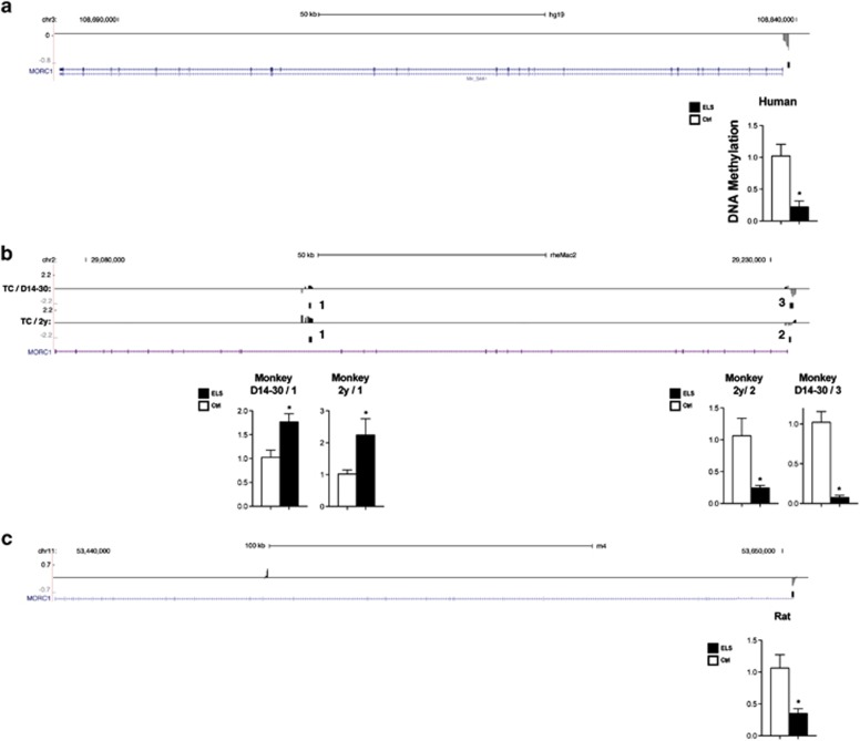
MDD is one of the well-established ELS-associated phenotypes.1, 2, 3, 4 We therefore took advantage of a previous GWAS study on MDD36 and performed a gene-based case–control analysis to test for an association between genetic variants in those overlapping genes and MDD. We excluded U6 and 7SK from our analysis, as multiple copies of those genes exist throughout the genome. In addition, we had to exclude PRMT5 and PDE4DIP as no genetic variants in PDE4DIP and only one genetic variant in PRMT5 were represented in the quality-controlled GWAS data set. Of the remaining three genes, MORC1 and CSRNP3 showed a nominally significant association with MDD (P=0.00483 and 0.03139, respectively). Only the association of MORC1 with MDD withstood Bonferroni correction for the number of genes tested (P=0.01449), thus providing evidence that MORC1 is involved in MDD, a stress-associated disorder. QPCR was used to validate MORC1 methylation changes observed in human CD34+ cells, monkey CD3+ T cells and rat PFC (Figure 1).
Figure 1.
Expanded views from the UCSC genome browser of the MORC1 gene are depicted. (a) Human CD34+ cord blood. (b) 14–30-day-old monkey CD3+ T cells (TC/D14–30), 2-year-old monkey CD3+ T cells (TC/2y). Numbers 1, 2, 3 indicate the locations of DNA amplification for QPCR validations. (c) Rat PFC. For each graph, average methylation probe fold differences (Log2) between control and ELS groups, as well as regions of significant differential methylation are shown. The last track shows the MORC1 gene, as taken from the NCBI reference sequences collection (RefSeq). QPCR analysis of DNA methylation differences in the MORC1 gene between ELS and control groups are displayed. Relative bound fraction concentrations are shown. Error bars represent s.e.m. The symbol ‘*' denotes P-values <0.05, as calculated with the Mann–Whitney U-test. Ctrl, control group; ELS, early life stress; MORC1, MORC family CW-type zinc finger 1; QPCR, quantitative PCR.
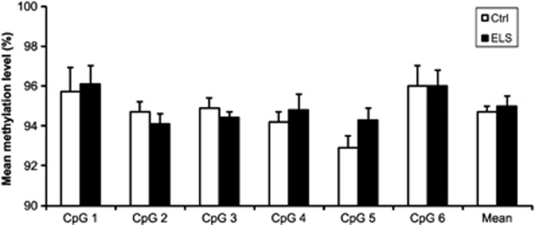
In the human cohort, we attempted to validate the MeDIP results using a pyrosequencing approach. Pyrosequencing revealed very high methylation levels and the significant difference between the ELS and controls was not replicated (Figure 2).
Figure 2.
The bar graph shows the mean methylation levels in the ELS and control group for each of the six CpG sites in MORC1, as determined by pyrosequencing. In addition, the average methylation level of all sites is displayed. Error bars represent s.e.m. No significant difference between the ELS and Ctrl group was detected. Statistical significance was calculated using the Mann–Whitney U-test. Ctrl, control group; ELS, early life stress; MORC1, MORC family CW-type zinc finger 1.
MORC1 gene expression
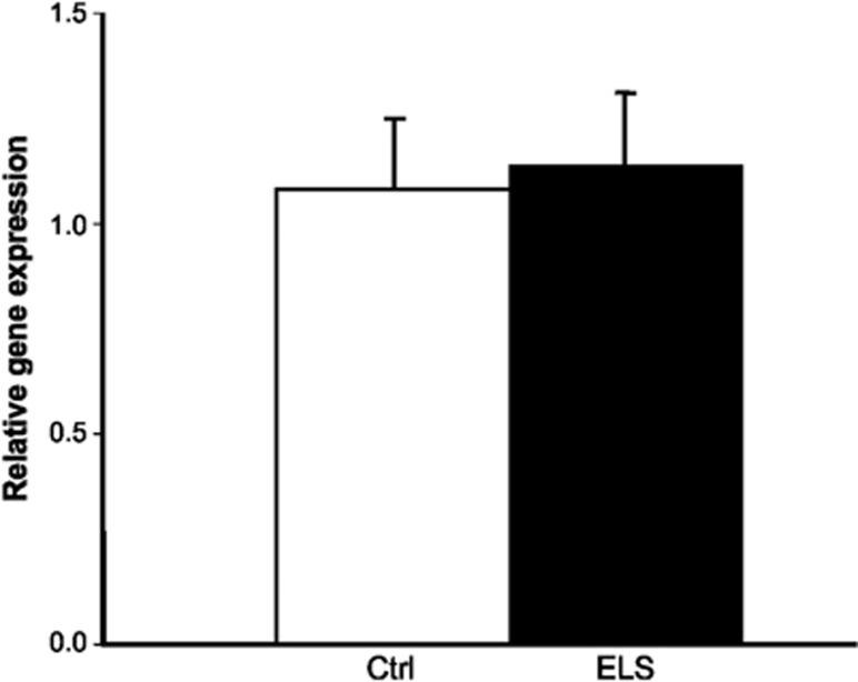
To investigate whether the observed alterations in MORC1 methylation levels secondary to ELS resulted in differential gene expression, MORC1 expression levels were analyzed in cord blood samples derived from the human extreme groups using a QPCR approach. No differences in MORC1 expression were detected between the high and low ELS groups (Figure 3).
Figure 3.
Relative expression of MORC1 in human cord blood (n=9 ELS and n=8 Ctrl). Values are given as mean quantities normalized by β-actin. Error bars represent s.e.m. No significant difference between the ELS and Ctrl group was detected. Statistical significance was calculated using the Mann–Whitney U-test. Ctrl, control group; ELS, early life stress; MORC1, MORC family CW-type zinc finger 1.
Discussion
The present study is the first to apply a cross-species and cross-tissue approach to the identification of ELS effects on the epigenome. As has been postulated before,45 our data provide evidence for genome-wide and system-wide changes in DNA methylation in response to ELS. This information might be useful in predicting lifelong behavioral and physical phenotypes, especially as we could identify such changes very early in life. In this study, differential methylation of 30 genes was associated with ELS immediately after birth in human CD34+ cells, and additionally in peripheral CD3+ T cells of newborn and adolescent monkeys, as well as in the brain of adult rats. A gene-based case–control analysis to test for an association between genetic variants and MDD identified one of those genes—MORC1—as being additionally associated with MDD on a genetic level in a different cohort. Our results provide evidence for a link between the MORC1 gene and depression. MORC1 is an excellent candidate as an immediate ELS responsive gene as it is differentially methylated in the brain and peripheral cells at different ages and could be followed longitudinally in living humans.
Although previous studies reported associations between ELS and DNA methylation in the adult brain tissue or peripheral blood cells, critical questions remained unanswered. First, are ELS-associated DNA methylation changes a consequence of early life experiences, or a cause of the later psychiatric phenotypes resulting from ELS? To address this question, we examined changes in DNA immediately after exposure to ELS. Our data show that in both, monkeys and humans, DNA methylation differences emerged soon after ELS exposure supporting the hypothesis that DNA methylation changes follow ELS, and precede the appearance of the clinical phenotypes later in life.
In humans, disentangling DNA methylation caused by ELS from DNA methylation caused by other preexisting confounding factors such as genetic variation and other environmental factors remains impossible. We bypassed these limitations by applying a convergent, translational approach comparing human data with data derived from animal models as they can be randomized to high and low ELS and their environment can be controlled.
A wide variety of prenatal stressors—including maternal depression during pregnancy, as well as a multitude of socioeconomic and psychosocial stressors—may result in epigenetic alterations and long-lasting effects on the health and behavior of the child. Accordingly, the present study applied a broad definition of prenatal stress in humans. However, all subjects in the high stress group were chronically exposed to multiple socioeconomic and psychosocial stressors as well as to high levels of subjectively perceived stress of the mother. The Macaca mulatta analyses focused on the effects of very early postnatal stress on the epigenome. This experimental condition was specific, standardized and adequately controlled. However, the comparison of a postnatal stress paradigm in the monkey to a gestational stress paradigm in humans could be considered a limitation of our study. Nonetheless, the maternal separation model for ELS applied in the nonhuman primates leads to neurobiological, physiological and behavioral consequences similar to those identified in humans after exposure to early adversity.46, 47, 48, 49 Our study provides additional evidence that ELS causes evolutionary conserved differential methylation of responsive genes as has been postulated previously.50
Although we are aware of sex-specific epigenetic effects, we decided to study the human offspring of the most stressed mothers, independent of sex of the infants. As the gender distribution did not differ significantly between the groups, we would consider this a minor issue. Maternal smoking and alcohol consumption during pregnancy have an impact on the global DNA methylation pattern of the infant.51, 52, 53, 54, 55, 56, 57 Both were significantly higher in the present high ELS group, and this is an obvious confounding factor. However, this effect can be excluded if overlapping differentially methylated genes are found in animal models of ELS, since these animals were exposed to neither cigarette smoke nor alcohol during gestation.
We reasoned that there exist robust and fundamental changes in DNA methylation, which will be conserved not just among species but also among tissues. The overlapping changes identified in both CD34+ and CD3+ cells are consistent with the hypothesis that changes in methylation in response to ELS appear early in progenitor cells which are then passed on to their different daughter lineages. Identification of genes that respond similarly to ELS in brain and blood cells is critical for studying behavioral epigenetics in humans and for potential diagnostics and therapeutic interventions. Future studies will be necessary to identify which specific cerebral cell subtypes are affected by ELS, as we analyzed PFC material without isolating specific cells.
ELS is a condition associated with several health and psychiatric conditions including depression in later life.1, 2, 3, 4 Taking advantage of an available GWAS in MDD, we were able to demonstrate that genetic variants in one of those persistently methylated genes, MORC1, are significantly associated with MDD. This supports the hypothesis that MORC1 is involved in at least one known consequence of ELS, the risk for depression in later life. The genome-wide finding for MORC1 was confirmed in all the three organisms using QPCR, which is the standard method for validation of MeDIP-chip data. Unexpectedly, the additional attempt to replicate the differential methylation of MORC1 using a pyrosequencing approach failed. One possible reason could be that the MeDIP-chip does not provide single base resolution, but instead suggests a region between 800 and 1200 bp harboring CpG sites of interest. The identification of differentially methylated CpGs in this area via pyrosequencing is hampered by the relative short (⩽300 nucleotides) sequencing reads, which complicates selection of the target region for replication. It is therefore possible that we missed the differentially methylated CpGs. Furthermore, results obtained by MeDIP and bisulfite-based sequencing are not necessarily comparable. For example, in a recent study by Jenke et al.,58 methylation levels obtained by MeDIP differed substantially from methylation levels obtained by pyrosequencing. Furthermore, bisulfite conversion of DNA does not distinguish between 5-methylcytosine and 5-hydroxymethylcytosine, whereas MeDIP using an anti-5-methyl-cytosine is selective for DNA methylation. We are therefore unable to exclude the possibility that the region identified by MeDIP as being differentially methylated after ELS is additionally differentially hydroxymethylated after ELS. This could furthermore explain the very high methylation levels obtained by pyrosequencing.
No significant differences in MORC1 expression were detected in cord blood samples from the human extreme groups. Unfortunately, no RNA from CD34+ cells was available for our cohort. Hence gene expression analysis was performed in whole blood samples, whereas DNA methylation was measured in CD34+ cells. The cell-type-specific nature of DNA methylation patterns could therefore explain this unexpected finding. A recent study showed that hypermethylated promoters can be transiently activated through chromatin remodeling without promoter demethylation, thus contradicting the common hypothesis that correlates DNA hypermethylation with silencing of gene expression.59 This finding suggests that promoter DNA methylation might be a programming factor providing an enduring memory for gene silencing rather than a marker of active gene expression. Further research is needed to ascertain if this phenomenon also applies to MORC1 and to investigate the functional consequences of differential MORC1 methylation in more detail.
The role of MORC1 is largely unknown. In mammals, MORC1 is mainly expressed in male germ cells. In the present study, MORC1 expression levels in human cord blood were very low, and this may have hampered the detection of significant differences between the groups. Germ cell MORC1 expression commences in early embryonic development and is important for the completion of prophase of meiosis I during spermatogenesis.60,61
Loss of function mutation in this gene leads to male infertility in mice. For other members of the gene family, expression is not restricted to the testis, which suggests a more general biological function for the MORC gene family. Recent research suggests that MORC1 itself has a more general biological role, as it codes for an evolutionary conserved nuclear protein, which may influence gene silencing and chromatin structure, possibly through the detection of epigenetic marks.62, 63, 64 The identification of MORC1 as a gene whose promoter is differentially methylated after exposure to different forms of ELS in different organisms, different tissues and different time points also suggests that this gene has a more general regulatory role in response to stress. However, as only limited knowledge concerning the role of MORC1 in epigenetic processes is available, the role of MORC1 within the context of ELS remains a matter of speculation. Systematic investigation of this question is warranted.
In addition to the identification of MORC1 as epigenetically modified after exposure to ELS and associated with MDD, differential methylation in the human sample was observed for CACNA1C, ANK3 and PCLO, genes that were identified in previous GWAS and replication studies of affective disorders.41, 42, 43,65
In conclusion, our novel systematic, genome-wide and cross-tissues–cross-species investigation of the effects of ELS on the epigenome identified MORC1 as differentially methylated in all the three investigated organisms, in peripheral tissues as well as in the brain and at different time points in the life-span. Furthermore, an association was demonstrated between MORC1 and MDD, an ELS-associated disorder. We therefore propose MORC1 as a new candidate gene for stress-related disorders whose DNA methylation status: (i) reflects ELS; (ii) is amenable to longitudinal follow-up in peripheral cells; and (iii) may predict the emergence of ELS-related disorders, such as depression, in later life. This has important research, diagnostic and therapeutic implications.
Acknowledgments
This work was supported by an Era-Net Neuron grant to MD, MR, MAR, ML, FC, PG and MS, fonds de recherché Santé Québec (FRSQ) 24419 to MS, by grants 01GS08144 (MMN) and 01GS08147 (MR) from the National Genome Research Network (NGFN-plus) of the German Federal Ministry of Education and Research (BMBF) and from Fondazione CARIPLO - grant no. 2012-0503 to MAR and FC. MD, MR and ML received support from the Dietmar-Hopp Foundation. VN received support from the Olympia-Morata-Programme of the University of Heidelberg and the Boehringer-Ingelheim Fonds. SJS was supported by funds from the Division of Intramural Research, NICHD, HIH. We thank Christine Schmähl for critical reading of the manuscript and gratefully acknowledge Alessandra Berry, Veronica Bellisario and Sara Capoccia for their precious help with the rat prenatal stress model. We are very grateful for the essential contributions of the midwives, and would like to thank all families for their participation.
The authors declare no conflict of interest.
Footnotes
Supplementary Information accompanies the paper on the Translational Psychiatry website (http://www.nature.com/tp)
Supplementary Material
References
- McCrory E, De Brito SA, Viding E. The link between child abuse and psychopathology: a review of neurobiological and genetic research. J Royal Soc Med. 2012;105:151–156. doi: 10.1258/jrsm.2011.110222. [DOI] [PMC free article] [PubMed] [Google Scholar]
- Agid O, Shapira B, Zislin J, Ritsner M, Hanin B, Murad H, et al. Environment and vulnerability to major psychiatric illness: a case control study of early parental loss in major depression, bipolar disorder and schizophrenia. Mol Psychiatry. 1999;4:163–172. doi: 10.1038/sj.mp.4000473. [DOI] [PubMed] [Google Scholar]
- McLaughlin KA, Green JG, Gruber MJ, Sampson NA, Zaslavsky AM, Kessler RC. Childhood adversities and adult psychiatric disorders in the national comorbidity survey replication II: associations with persistence of DSM-IV disorders. Arch Gen Psychiatry. 2010;67:124–132. doi: 10.1001/archgenpsychiatry.2009.187. [DOI] [PMC free article] [PubMed] [Google Scholar]
- Widom CS, DuMont K, Czaja SJ. A prospective investigation of major depressive disorder and comorbidity in abused and neglected children grown up. Arch Gen Psychiatry. 2007;64:49–56. doi: 10.1001/archpsyc.64.1.49. [DOI] [PubMed] [Google Scholar]
- Roseboom TJ, Painter RC, van Abeelen AF, Veenendaal MV, de Rooij SR. Hungry in the womb: what are the consequences? Lessons from the Dutch famine. Maturitas. 2011;70:141–145. doi: 10.1016/j.maturitas.2011.06.017. [DOI] [PubMed] [Google Scholar]
- Afifi TO, Enns MW, Cox BJ, Asmundson GJ, Stein MB, Sareen J. Population attributable fractions of psychiatric disorders and suicide ideation and attempts associated with adverse childhood experiences. Am J Public Health. 2008;98:946–952. doi: 10.2105/AJPH.2007.120253. [DOI] [PMC free article] [PubMed] [Google Scholar]
- Danese A, Moffitt TE, Harrington H, Milne BJ, Polanczyk G, Pariante CM, et al. Adverse childhood experiences and adult risk factors for age-related disease: depression, inflammation, and clustering of metabolic risk markers. Arch Pediatr Adolesc Med. 2009;163:1135–1143. doi: 10.1001/archpediatrics.2009.214. [DOI] [PMC free article] [PubMed] [Google Scholar]
- Heim C, Nemeroff CB. The role of childhood trauma in the neurobiology of mood and anxiety disorders: preclinical and clinical studies. Biol Psychiatry. 2001;49:1023–1039. doi: 10.1016/s0006-3223(01)01157-x. [DOI] [PubMed] [Google Scholar]
- McGowan PO, Szyf M. Environmental epigenomics: understanding the effects of parental care on the epigenome. Essays Biochem. 2010;48:275–287. doi: 10.1042/bse0480275. [DOI] [PubMed] [Google Scholar]
- Weaver IC. Epigenetic programming by maternal behavior and pharmacological intervention. Nature versus nurture: let's call the whole thing off. Epigenetics. 2007;2:22–28. doi: 10.4161/epi.2.1.3881. [DOI] [PubMed] [Google Scholar]
- Gudsnuk KM, Champagne FA. Epigenetic effects of early developmental experiences. Clin Perinatol. 2011;38:703–717. doi: 10.1016/j.clp.2011.08.005. [DOI] [PubMed] [Google Scholar]
- Weaver IC, Cervoni N, Champagne FA, D'Alessio AC, Sharma S, Seckl JR, et al. Epigenetic programming by maternal behavior. Nat Neurosci. 2004;7:847–854. doi: 10.1038/nn1276. [DOI] [PubMed] [Google Scholar]
- Chen J, Evans AN, Liu Y, Honda M, Saavedra JM, Aguilera G. Maternal deprivation in rats is associated with corticotrophin-releasing hormone (CRH) promoter hypomethylation and enhances CRH transcriptional responses to stress in adulthood. J Neuroendocrinol. 2012;24:1055–1064. doi: 10.1111/j.1365-2826.2012.02306.x. [DOI] [PMC free article] [PubMed] [Google Scholar]
- Roth TL, Lubin FD, Funk AJ, Sweatt JD. Lasting epigenetic influence of early-life adversity on the BDNF gene. Biol Psychiatry. 2009;65:760–769. doi: 10.1016/j.biopsych.2008.11.028. [DOI] [PMC free article] [PubMed] [Google Scholar]
- Kinnally EL, Feinberg C, Kim D, Ferguson K, Leibel R, Coplan JD, et al. DNA methylation as a risk factor in the effects of early life stress. Brain Behav Immun. 2011;25:1548–1553. doi: 10.1016/j.bbi.2011.05.001. [DOI] [PMC free article] [PubMed] [Google Scholar]
- Kinnally EL, Capitanio JP, Leibel R, Deng L, LeDuc C, Haghighi F, et al. Epigenetic regulation of serotonin transporter expression and behavior in infant rhesus macaques. Genes Brain Behav. 2010;9:575–582. doi: 10.1111/j.1601-183X.2010.00588.x. [DOI] [PMC free article] [PubMed] [Google Scholar]
- Murgatroyd C, Patchev AV, Wu Y, Micale V, Bockmuhl Y, Fischer D, et al. Dynamic DNA methylation programs persistent adverse effects of early-life stress. Nat Neurosci. 2009;12:1559–1566. doi: 10.1038/nn.2436. [DOI] [PubMed] [Google Scholar]
- Jensen Pena C, Monk C, Champagne FA. Epigenetic effects of prenatal stress on 11beta-hydroxysteroid dehydrogenase-2 in the placenta and fetal brain. PLoS One. 2012;7:e39791. doi: 10.1371/journal.pone.0039791. [DOI] [PMC free article] [PubMed] [Google Scholar]
- Champagne FA, Weaver IC, Diorio J, Dymov S, Szyf M, Meaney MJ. Maternal care associated with methylation of the estrogen receptor-alpha1b promoter and estrogen receptor-alpha expression in the medial preoptic area of female offspring. Endocrinology. 2006;147:2909–2915. doi: 10.1210/en.2005-1119. [DOI] [PubMed] [Google Scholar]
- Kember RL, Dempster EL, Lee TH, Schalkwyk LC, Mill J, Fernandes C. Maternal separation is associated with strain-specific responses to stress and epigenetic alterations to Nr3c1, Avp, and Nr4a1 in mouse. Brain Behav. 2012;2:455–467. doi: 10.1002/brb3.69. [DOI] [PMC free article] [PubMed] [Google Scholar]
- Kundakovic M, Lim S, Gudsnuk K, Champagne FA. Sex-specific and strain-dependent effects of early life adversity on behavioral and epigenetic outcomes. Front Psychiatry. 2013;4:78. doi: 10.3389/fpsyt.2013.00078. [DOI] [PMC free article] [PubMed] [Google Scholar]
- Radtke KM, Ruf M, Gunter HM, Dohrmann K, Schauer M, Meyer A, et al. Transgenerational impact of intimate partner violence on methylation in the promoter of the glucocorticoid receptor. Transl Psychiatry. 2011;1:e21. doi: 10.1038/tp.2011.21. [DOI] [PMC free article] [PubMed] [Google Scholar]
- Perroud N, Paoloni-Giacobino A, Prada P, Olie E, Salzmann A, Nicastro R, et al. Increased methylation of glucocorticoid receptor gene (NR3C1) in adults with a history of childhood maltreatment: a link with the severity and type of trauma. Transl Psychiatry. 2011;1:e59. doi: 10.1038/tp.2011.60. [DOI] [PMC free article] [PubMed] [Google Scholar]
- McGowan PO, Sasaki A, D'Alessio AC, Dymov S, Labonte B, Szyf M, et al. Epigenetic regulation of the glucocorticoid receptor in human brain associates with childhood abuse. Nat Neurosci. 2009;12:342–348. doi: 10.1038/nn.2270. [DOI] [PMC free article] [PubMed] [Google Scholar]
- Mulligan CJ, D'Errico NC, Stees J, Hughes DA. Methylation changes at NR3C1 in newborns associate with maternal prenatal stress exposure and newborn birth weight. Epigenetics. 2012;7:853–857. doi: 10.4161/epi.21180. [DOI] [PMC free article] [PubMed] [Google Scholar]
- Tyrka AR, Price LH, Marsit C, Walters OC, Carpenter LL. Childhood adversity and epigenetic modulation of the leukocyte glucocorticoid receptor: preliminary findings in healthy adults. PLoS One. 2012;7:e30148. doi: 10.1371/journal.pone.0030148. [DOI] [PMC free article] [PubMed] [Google Scholar]
- Labonte B, Yerko V, Gross J, Mechawar N, Meaney MJ, Szyf M, et al. Differential glucocorticoid receptor exon 1(B), 1(C), and 1(H) expression and methylation in suicide completers with a history of childhood abuse. Biol Psychiatry. 2012;72:41–48. doi: 10.1016/j.biopsych.2012.01.034. [DOI] [PubMed] [Google Scholar]
- Toledo-Rodriguez M, Lotfipour S, Leonard G, Perron M, Richer L, Veillette S, et al. Maternal smoking during pregnancy is associated with epigenetic modifications of the brain-derived neurotrophic factor-6 exon in adolescent offspring. Am J Med Genet B Neuropsychiatr Genet. 2010;153B:1350–1354. doi: 10.1002/ajmg.b.31109. [DOI] [PubMed] [Google Scholar]
- Beach SR, Brody GH, Todorov AA, Gunter TD, Philibert RA. Methylation at 5HTT mediates the impact of child sex abuse on women's antisocial behavior: an examination of the Iowa adoptee sample. Psychosomat Med. 2011;73:83–87. doi: 10.1097/PSY.0b013e3181fdd074. [DOI] [PMC free article] [PubMed] [Google Scholar]
- Vijayendran M, Beach SR, Plume JM, Brody GH, Philibert RA. Effects of genotype and child abuse on DNA methylation and gene expression at the serotonin transporter. Front Psychiatry. 2012;3:55. doi: 10.3389/fpsyt.2012.00055. [DOI] [PMC free article] [PubMed] [Google Scholar]
- Kang HJ, Kim JM, Stewart R, Kim SY, Bae KY, Kim SW, et al. Association of SLC6A4 methylation with early adversity, characteristics and outcomes in depression. Prog Neuropsychopharmacol Biol Psychiatry. 2013;44:23–28. doi: 10.1016/j.pnpbp.2013.01.006. [DOI] [PubMed] [Google Scholar]
- Klengel T, Mehta D, Anacker C, Rex-Haffner M, Pruessner JC, Pariante CM, et al. Allele-specific FKBP5 DNA demethylation mediates gene-childhood trauma interactions. Nat Neurosci. 2013;16:33–41. doi: 10.1038/nn.3275. [DOI] [PMC free article] [PubMed] [Google Scholar]
- Labonte B, Suderman M, Maussion G, Navaro L, Yerko V, Mahar I, et al. Genome-wide epigenetic regulation by early-life trauma. Arch Gen Psychiatry. 2012;69:722–731. doi: 10.1001/archgenpsychiatry.2011.2287. [DOI] [PMC free article] [PubMed] [Google Scholar]
- Yang BZ, Zhang H, Ge W, Weder N, Douglas-Palumberi H, Perepletchikova F, et al. Child abuse and epigenetic mechanisms of disease risk. Am J Prev Med. 2013;44:101–107. doi: 10.1016/j.amepre.2012.10.012. [DOI] [PMC free article] [PubMed] [Google Scholar]
- Provencal N, Suderman MJ, Guillemin C, Massart R, Ruggiero A, Wang D, et al. The signature of maternal rearing in the methylome in rhesus macaque prefrontal cortex and T cells. J Neurosci. 2012;32:15626–15642. doi: 10.1523/JNEUROSCI.1470-12.2012. [DOI] [PMC free article] [PubMed] [Google Scholar]
- Rietschel M, Mattheisen M, Frank J, Treutlein J, Degenhardt F, Breuer R, et al. Genome-wide association-, replication-, and neuroimaging study implicates HOMER1 in the etiology of major depression. Biol Psychiatry. 2010;68:578–585. doi: 10.1016/j.biopsych.2010.05.038. [DOI] [PubMed] [Google Scholar]
- Fumagalli F, Bedogni F, Perez J, Racagni G, Riva MA. Corticostriatal brain-derived neurotrophic factor dysregulation in adult rats following prenatal stress. Eur J Neurosci. 2004;20:1348–1354. doi: 10.1111/j.1460-9568.2004.03592.x. [DOI] [PubMed] [Google Scholar]
- Keshet I, Schlesinger Y, Farkash S, Rand E, Hecht M, Segal E, et al. Evidence for an instructive mechanism of de novo methylation in cancer cells. Nat Genet. 2006;38:149–153. doi: 10.1038/ng1719. [DOI] [PubMed] [Google Scholar]
- Purcell S, Neale B, Todd-Brown K, Thomas L, Ferreira MA, Bender D, et al. PLINK: a tool set for whole-genome association and population-based linkage analyses. Am J Hum Genet. 2007;81:559–575. doi: 10.1086/519795. [DOI] [PMC free article] [PubMed] [Google Scholar]
- Kahler AK, Djurovic S, Rimol LM, Brown AA, Athanasiu L, Jonsson EG, et al. Candidate gene analysis of the human natural killer-1 carbohydrate pathway and perineuronal nets in schizophrenia: B3GAT2 is associated with disease risk and cortical surface area. Biol Psychiatry. 2011;69:90–96. doi: 10.1016/j.biopsych.2010.07.035. [DOI] [PubMed] [Google Scholar]
- Ferreira MA, O'Donovan MC, Meng YA, Jones IR, Ruderfer DM, Jones L, et al. Collaborative genome-wide association analysis supports a role for ANK3 and CACNA1C in bipolar disorder. Nat Genet. 2008;40:1056–1058. doi: 10.1038/ng.209. [DOI] [PMC free article] [PubMed] [Google Scholar]
- Liu Y, Blackwood DH, Caesar S, de Geus EJ, Farmer A, Ferreira MA, et al. Meta-analysis of genome-wide association data of bipolar disorder and major depressive disorder. Mol Psychiatry. 2011;16:2–4. doi: 10.1038/mp.2009.107. [DOI] [PMC free article] [PubMed] [Google Scholar]
- Wray NR, Pergadia ML, Blackwood DH, Penninx BW, Gordon SD, Nyholt DR, et al. Genome-wide association study of major depressive disorder: new results, meta- analysis, and lessons learned. Mol Psychiatry. 2012;17:36–48. doi: 10.1038/mp.2010.109. [DOI] [PMC free article] [PubMed] [Google Scholar]
- Green EK, Grozeva D, Jones I, Jones L, Kirov G, Caesar S, et al. The bipolar disorder risk allele at CACNA1C also confers risk of recurrent major depression and of schizophrenia. Mol Psychiatry. 2010;15:1016–1022. doi: 10.1038/mp.2009.49. [DOI] [PMC free article] [PubMed] [Google Scholar]
- Szyf M. The early-life social environment and DNA methylation. Clin Genet. 2012;81:341–349. doi: 10.1111/j.1399-0004.2012.01843.x. [DOI] [PubMed] [Google Scholar]
- Suomi SJ. Early determinants of behaviour: evidence from primate studies. Brit Med Bull. 1997;53:170–184. doi: 10.1093/oxfordjournals.bmb.a011598. [DOI] [PubMed] [Google Scholar]
- Machado CJ, Bachevalier J. Non-human primate models of childhood psychopathology: the promise and the limitations. J Child Psychol Psychiatry. 2003;44:64–87. doi: 10.1111/1469-7610.00103. [DOI] [PubMed] [Google Scholar]
- Harlow HF, Harlow MK. The Effect of Rearing Conditions on Behavior. Int J Psychiatry. 1965;1:43–51. [PubMed] [Google Scholar]
- Conti G, Hansman C, Heckman JJ, Novak MF, Ruggiero A, Suomi SJ. Primate evidence on the late health effects of early-life adversity. Proc Natl Acad Sci USA. 2012;109:8866–8871. doi: 10.1073/pnas.1205340109. [DOI] [PMC free article] [PubMed] [Google Scholar]
- Suderman M, McGowan PO, Sasaki A, Huang TC, Hallett MT, Meaney MJ, et al. Conserved epigenetic sensitivity to early life experience in the rat and human hippocampus. Proc Natl Acad Sci USA. 2012;109:17266–17272. doi: 10.1073/pnas.1121260109. [DOI] [PMC free article] [PubMed] [Google Scholar]
- Breton CV, Byun HM, Wenten M, Pan F, Yang A, Gilliland FD. Prenatal tobacco smoke exposure affects global and gene-specific DNA methylation. Am J Resp Crit Care Med. 2009;180:462–467. doi: 10.1164/rccm.200901-0135OC. [DOI] [PMC free article] [PubMed] [Google Scholar]
- Suter M, Ma J, Harris A, Patterson L, Brown KA, Shope C, et al. Maternal tobacco use modestly alters correlated epigenome-wide placental DNA methylation and gene expression. Epigenetics. 2011;6:1284–1294. doi: 10.4161/epi.6.11.17819. [DOI] [PMC free article] [PubMed] [Google Scholar]
- Flom JD, Ferris JS, Liao Y, Tehranifar P, Richards CB, Cho YH, et al. Prenatal smoke exposure and genomic DNA methylation in a multiethnic birth cohort. Cancer Epidemiol Biomarkers Prev. 2011;20:2518–2523. doi: 10.1158/1055-9965.EPI-11-0553. [DOI] [PMC free article] [PubMed] [Google Scholar]
- Joubert BR, Haberg SE, Nilsen RM, Wang X, Vollset SE, Murphy SK, et al. 450K epigenome-wide scan identifies differential DNA methylation in newborns related to maternal smoking during pregnancy. Environ Health Perspect. 2012;120:1425–1431. doi: 10.1289/ehp.1205412. [DOI] [PMC free article] [PubMed] [Google Scholar]
- Liu Y, Balaraman Y, Wang G, Nephew KP, Zhou FC. Alcohol exposure alters DNA methylation profiles in mouse embryos at early neurulation. Epigenetics. 2009;4:500–511. doi: 10.4161/epi.4.7.9925. [DOI] [PMC free article] [PubMed] [Google Scholar]
- Zhou FC, Chen Y, Love A. Cellular DNA methylation program during neurulation and its alteration by alcohol exposure. Birth Defects Res A Clin Mol Teratol. 2011;91:703–715. doi: 10.1002/bdra.20820. [DOI] [PubMed] [Google Scholar]
- Wilhelm-Benartzi CS, Houseman EA, Maccani MA, Poage GM, Koestler DC, Langevin SM, et al. In utero exposures, infant growth, and DNA methylation of repetitive elements and developmentally related genes in human placenta. Environ Health Perspect. 2012;120:296–302. doi: 10.1289/ehp.1103927. [DOI] [PMC free article] [PubMed] [Google Scholar]
- Jenke AC, Postberg J, Raine T, Nayak KM, Molitor M, Wirth S, et al. DNA methylation analysis in the intestinal epithelium-effect of cell separation on gene expression and methylation profile. PLoS One. 2013;8:e55636. doi: 10.1371/journal.pone.0055636. [DOI] [PMC free article] [PubMed] [Google Scholar]
- Raynal NJ, Si J, Taby RF, Gharibyan V, Ahmed S, Jelinek J, et al. DNA methylation does not stably lock gene expression but instead serves as a molecular mark for gene silencing memory. Cancer Res. 2012;72:1170–1181. doi: 10.1158/0008-5472.CAN-11-3248. [DOI] [PMC free article] [PubMed] [Google Scholar]
- Inoue N, Hess KD, Moreadith RW, Richardson LL, Handel MA, Watson ML, et al. New gene family defined by MORC, a nuclear protein required for mouse spermatogenesis. Hum Mol Genet. 1999;8:1201–1207. doi: 10.1093/hmg/8.7.1201. [DOI] [PubMed] [Google Scholar]
- Watson ML, Zinn AR, Inoue N, Hess KD, Cobb J, Handel MA, et al. Identification of morc (microrchidia), a mutation that results in arrest of spermatogenesis at an early meiotic stage in the mouse. Proc Natl Acad Sci USA. 1998;95:14361–14366. doi: 10.1073/pnas.95.24.14361. [DOI] [PMC free article] [PubMed] [Google Scholar]
- Iyer LM, Anantharaman V, Wolf MY, Aravind L. Comparative genomics of transcription factors and chromatin proteins in parasitic protists and other eukaryotes. Int J Parasitol. 2008;38:1–31. doi: 10.1016/j.ijpara.2007.07.018. [DOI] [PubMed] [Google Scholar]
- Moissiard G, Cokus SJ, Cary J, Feng S, Billi AC, Stroud H, et al. MORC family ATPases required for heterochromatin condensation and gene silencing. Science. 2012;336:1448–1451. doi: 10.1126/science.1221472. [DOI] [PMC free article] [PubMed] [Google Scholar]
- Perry J, Zhao Y. The CW domain, a structural module shared amongst vertebrates, vertebrate-infecting parasites and higher plants. Trends Biochem Sci. 2003;28:576–580. doi: 10.1016/j.tibs.2003.09.007. [DOI] [PubMed] [Google Scholar]
- Sullivan PF, de Geus EJ, Willemsen G, James MR, Smit JH, Zandbelt T, et al. Genome-wide association for major depressive disorder: a possible role for the presynaptic protein piccolo. Mol Psychiatry. 2009;14:359–375. doi: 10.1038/mp.2008.125. [DOI] [PMC free article] [PubMed] [Google Scholar]
Associated Data
This section collects any data citations, data availability statements, or supplementary materials included in this article.


