Abstract
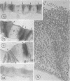
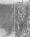
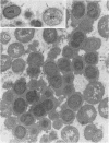
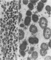
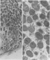
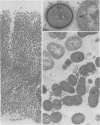
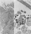
Streptococcus sanguis has been localized ultrastructurally within intact dental plaque by means of an indirect technique which utilizes horseradish peroxidase-labeled antibody. The technique allows for complete diffusion of the reagents to all portions of the plaque specimens. Control procedures can be carried out on serial sections of plaque with a bacterial composition similar to that of the experimental specimen. The 30-mum-thick sections can be examined in the light microscope to localize areas specifically labeled with peroxidase prior to cutting ultra-thin sections for electron microscopy. This study demonstrated that specific bacteria can be localized within intact dental plaque. The results also indicated that S. sanguis grows in dental plaque as columnar shaped microcolonies perpendicular to the tooth surfaces. Growth appears to be by cell division rather than deposition of new cells at the surfaces. Despite their relatively good structural preservation, the cells in the deeper (older) layers of plaque appear to have lost some of their antigenic activity in comparison to the cells near the surface.
Full text
PDF
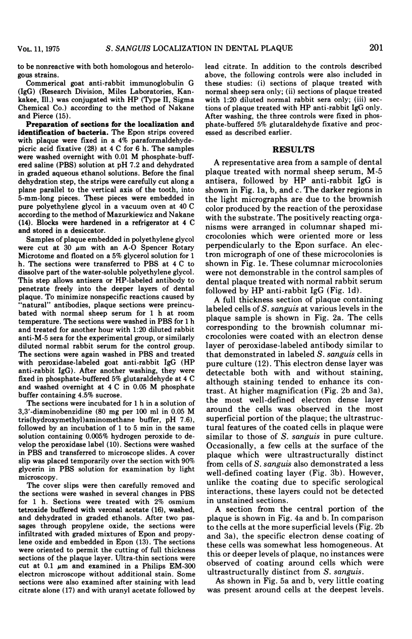
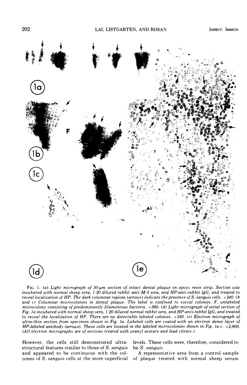
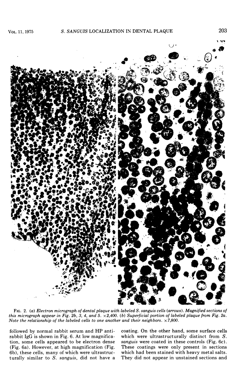
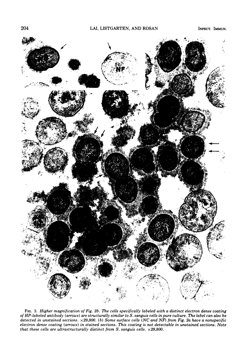
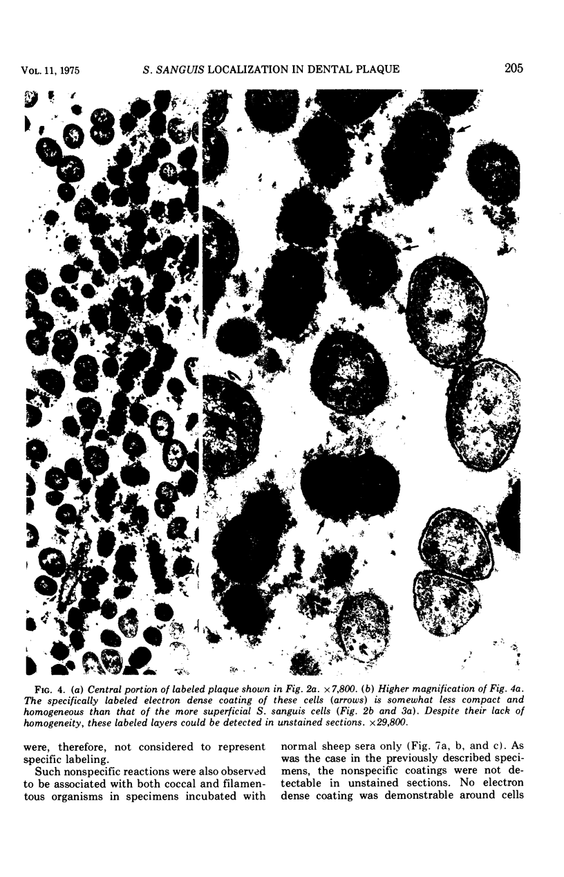
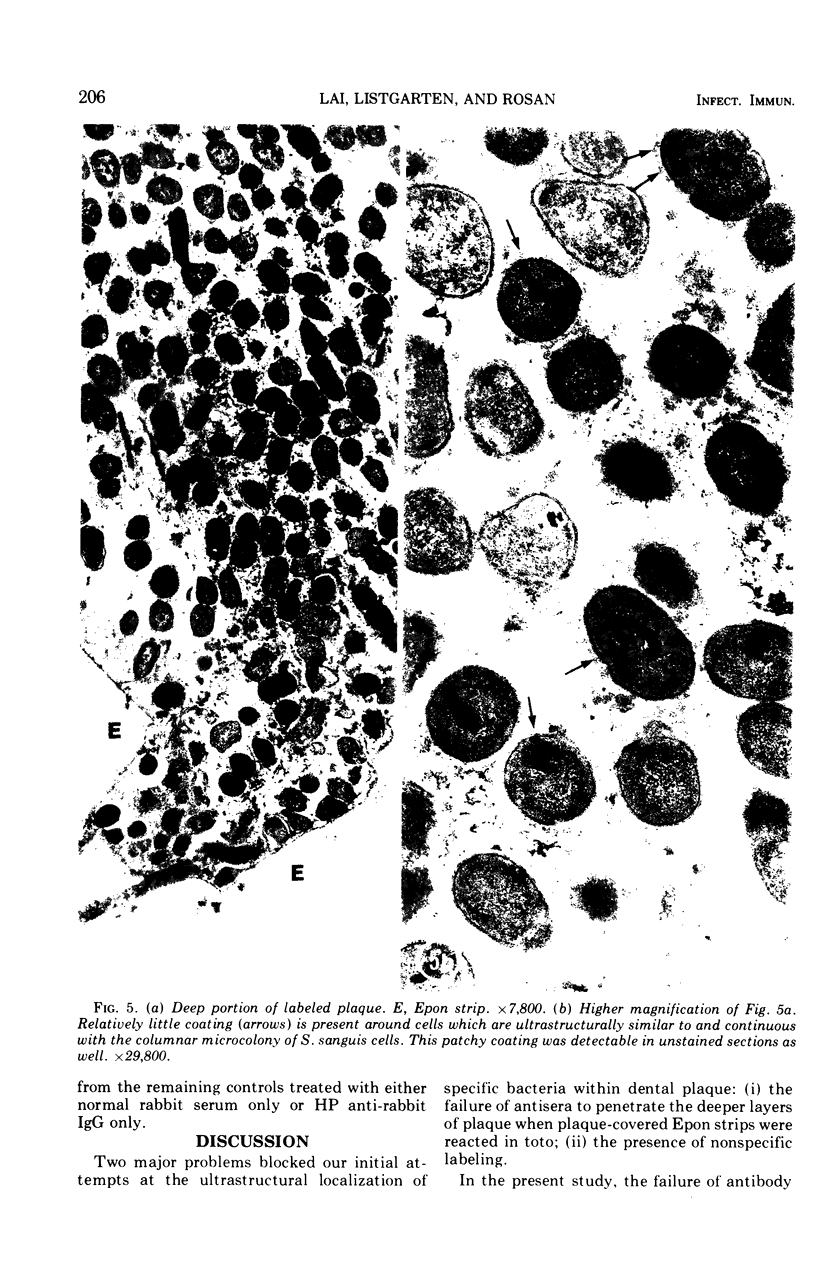
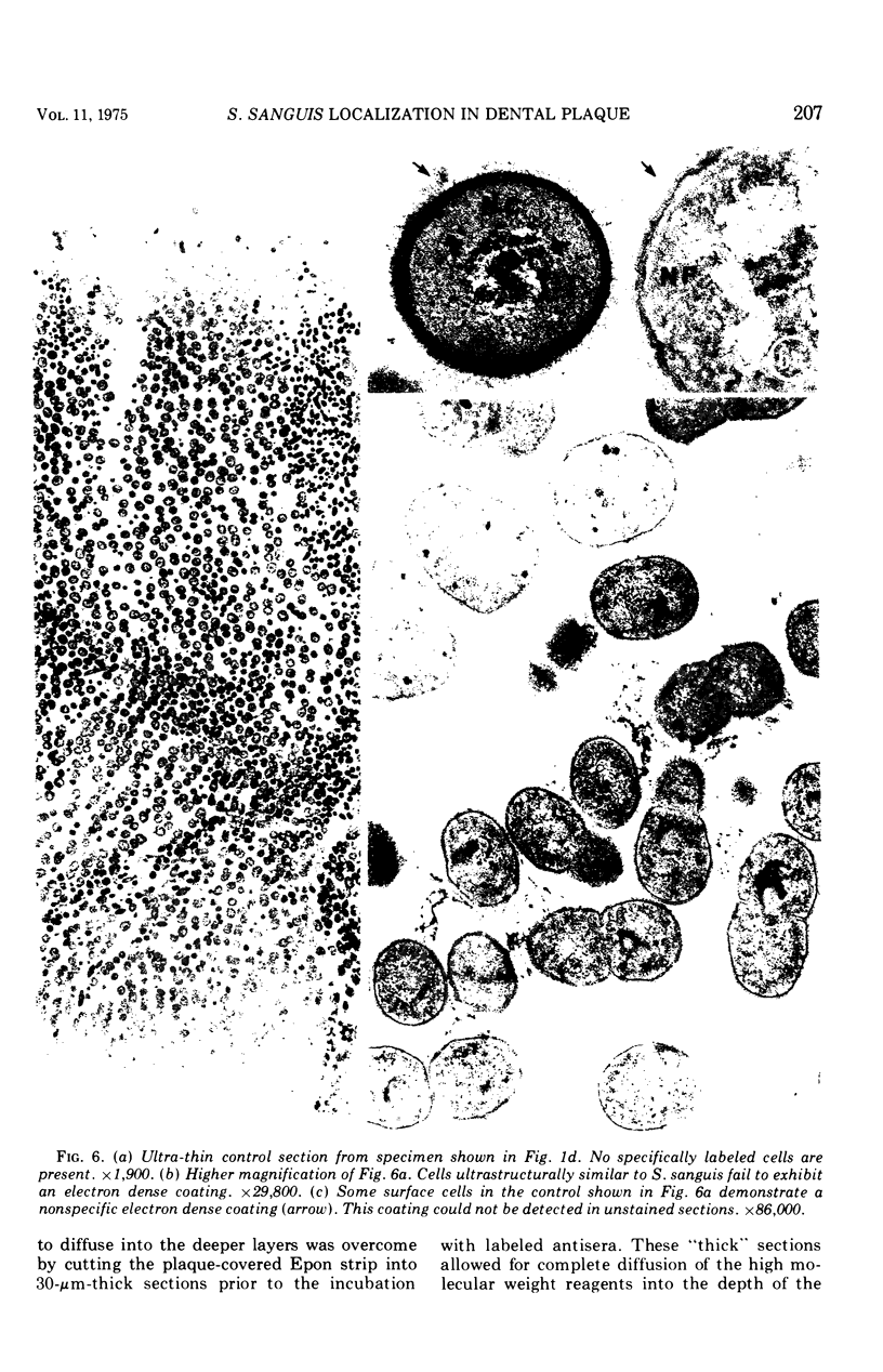
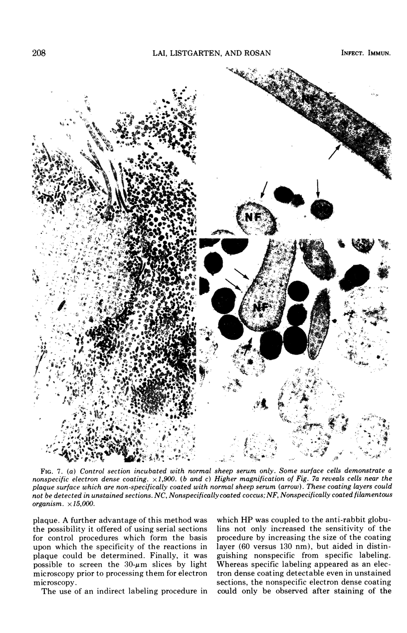
Images in this article
Selected References
These references are in PubMed. This may not be the complete list of references from this article.
- BROWN A. D. ASPECTS OF BACTERIAL RESPONSE TO THE IONIC ENVIRONMENT. Bacteriol Rev. 1964 Sep;28:296–329. doi: 10.1128/br.28.3.296-329.1964. [DOI] [PMC free article] [PubMed] [Google Scholar]
- Barkin M. E. Ultrastructural cytochemical localization of polysaccharides in dental plaque. J Dent Res. 1970 Sep-Oct;49(Suppl):979+–979+. doi: 10.1177/00220345700490054301. [DOI] [PubMed] [Google Scholar]
- Berthold C. H., Berthold P., Söder P. O. The growth of dental plaque on different materials. Sven Tandlak Tidskr. 1971 Dec;64(12):863–877. [PubMed] [Google Scholar]
- Frank R. M., Brendel A. Ultrastructure of the approximal dental plaque and the underlying normal and carious enamel. Arch Oral Biol. 1966 Sep;11(9):883–912. doi: 10.1016/0003-9969(66)90080-x. [DOI] [PubMed] [Google Scholar]
- Genco R. J., Evans R. T., Ellison S. A. Dental research in microbiology with emphasis on periodontal disease. J Am Dent Assoc. 1969 May;78(5):1016–1036. doi: 10.14219/jada.archive.1969.0162. [DOI] [PubMed] [Google Scholar]
- Graham R. C., Jr, Karnovsky M. J. The early stages of absorption of injected horseradish peroxidase in the proximal tubules of mouse kidney: ultrastructural cytochemistry by a new technique. J Histochem Cytochem. 1966 Apr;14(4):291–302. doi: 10.1177/14.4.291. [DOI] [PubMed] [Google Scholar]
- LUFT J. H. Improvements in epoxy resin embedding methods. J Biophys Biochem Cytol. 1961 Feb;9:409–414. doi: 10.1083/jcb.9.2.409. [DOI] [PMC free article] [PubMed] [Google Scholar]
- Lai C. H., Listgarten M. A., Rosan B. Immunoelectron microscopic identification and localization of Streptococcus sanguis with peroxidase-labeled antibody: localization of surface antigens in pure cultures. Infect Immun. 1975 Jan;11(1):193–199. doi: 10.1128/iai.11.1.193-199.1975. [DOI] [PMC free article] [PubMed] [Google Scholar]
- Lai C., Listgarten M., Rosan B. Serology of Streptococcus sanguis: localization of antigens with unlabeled antisera. Infect Immun. 1973 Sep;8(3):475–481. doi: 10.1128/iai.8.3.475-481.1973. [DOI] [PMC free article] [PubMed] [Google Scholar]
- Mazurkiewicz J. E., Nakane P. K. Light and electron microscopic localization of antigens in tissues embedded in polyethylene glycol with a peroxidase labeled antibody method. J Histochem Cytochem. 1972 Dec;20(12):969–974. doi: 10.1177/20.12.969. [DOI] [PubMed] [Google Scholar]
- Nakane P. K., Pierce G. B., Jr Enzyme-labeled antibodies for the light and electron microscopic localization of tissue antigens. J Cell Biol. 1967 May;33(2):307–318. doi: 10.1083/jcb.33.2.307. [DOI] [PMC free article] [PubMed] [Google Scholar]
- PALADE G. E. A study of fixation for electron microscopy. J Exp Med. 1952 Mar;95(3):285–298. doi: 10.1084/jem.95.3.285. [DOI] [PMC free article] [PubMed] [Google Scholar]
- REYNOLDS E. S. The use of lead citrate at high pH as an electron-opaque stain in electron microscopy. J Cell Biol. 1963 Apr;17:208–212. doi: 10.1083/jcb.17.1.208. [DOI] [PMC free article] [PubMed] [Google Scholar]
- RITZ H. L. LOCALIZATION OF NOCARDIA IN DENTAL PLAQUE BY IMMUNOFLUORESCENCE. Proc Soc Exp Biol Med. 1963 Aug-Sep;113:925–929. doi: 10.3181/00379727-113-28533. [DOI] [PubMed] [Google Scholar]
- Ritz H. L. Fluorescent antibody staining of Neisseria, Streptococcus and Veillonella in frozen sections of human dental plaque. Arch Oral Biol. 1969 Sep;14(9):1073–1083. doi: 10.1016/0003-9969(69)90077-6. [DOI] [PubMed] [Google Scholar]
- Ritz H. L. Microbial population shifts in developing human dental plaque. Arch Oral Biol. 1967 Dec;12(12):1561–1568. doi: 10.1016/0003-9969(67)90190-2. [DOI] [PubMed] [Google Scholar]
- Rosan B. Antigens of Streptococcus sanguis. Infect Immun. 1973 Feb;7(2):205–211. doi: 10.1128/iai.7.2.205-211.1973. [DOI] [PMC free article] [PubMed] [Google Scholar]
- Rosan B., Eisenberg R. J. Morphological changes in Streptococcus sanguis associated with growth in the presence of oxygen. Arch Oral Biol. 1973 Nov;18(11):1441–1444. doi: 10.1016/0003-9969(73)90118-0. [DOI] [PubMed] [Google Scholar]
- Schroeder H. E., Hirzel H. C. A method of studying dental plaque morphology. Helv Odontol Acta. 1969 Apr;13(1):22–27. [PubMed] [Google Scholar]
- Schroeder H. E. The structure and relationship of plaque to the hard and soft tissues: electron microscopic interpretation. Int Dent J. 1970 Sep;20(3):353–381. [PubMed] [Google Scholar]
- Snyder M. L., Bullock W. W., Parker R. B. Morphology of gram-positive filamentous bacteria identified in dental plaque by fluorescent antibody technique. Arch Oral Biol. 1967 Nov;12(11):1269–1273. doi: 10.1016/0003-9969(67)90128-8. [DOI] [PubMed] [Google Scholar]
- Socransky S. S. Relationship of bacteria to the etiology of periodontal disease. J Dent Res. 1970 Mar-Apr;49(2):203–222. doi: 10.1177/00220345700490020401. [DOI] [PubMed] [Google Scholar]
- Stefanini M., De Martino C., Zamboni L. Fixation of ejaculated spermatozoa for electron microscopy. Nature. 1967 Oct 14;216(5111):173–174. doi: 10.1038/216173a0. [DOI] [PubMed] [Google Scholar]


