Abstract
A new strategy for encoding polypeptide libraries with photolabile tags is developed. The photoassisted assay, based on conditional release of encoding tags only from bound pairs, can differentiate between peptides which have minor differences in a form of post-translational modifications with epigenetic marks. The encoding strategy is fully compatible with automated peptide synthesis. The encoding pendants are compact and do not perturb potential binding interactions.
Keywords: encoded combinatorial library, spatial proximity test, conditional photorelease of tags, epigenetic marks
1. INTRODUCTION
High throughput combinatorial chemistry is critical for modern drug discovery, with parallel synthesis and screening techniques currently being the industry standard. More than two decades after the introduction of massively high-throughput one-bead-one-compound split-and-pool combinatorial methods,1,2 parallel synthesis and screening still dominate the landscape of big pharma. Hybrid methodologies, such as synthesis in NanoKan containers have been adopted by some companies to take advantage of polymeric bead-supported synthesis while keeping track of the identity of individual compounds being synthesized in the bar-coded NanoKans.
When used, one-bead-one-compound libraries are normally assayed for binding of biological targets through fluorescence-guided mechanical segregation of the winning beads. The required mechanical manipulations impose a lower limit on the size of the particle used as a solid support. Currently there is no simple way for assaying solution phase mixtures. While various iterative deconvolution methods3 have been developed to synthesize and screen soluble sub-libraries, all of them require multiple redundant synthetic steps, are time consuming, and expensive. Solution phase combinatorial chemistry holds great promise, as it is compatible with both divergent and convergent multi-step synthetic schemes, and not constrained to linear synthesis.4 Its immense potential, however, has not yet been fully realized, partly due to the complexity of assaying solution mixtures for binding.
We note that there are currently problems for which (i) on-bead screening is not possible at all, and (ii) parallel approaches are, if not impossible, then prohibitively time-consuming and labor-intensive. One of these problems is high throughput screening of combinatorial libraries for binding between individual members of the library. As an example, understanding homo- or hetero-dimerization, -trimerization and formation of higher order oligomeric assemblies of peptides is of utmost importance in the field of structural biochemistry for a variety of reasons, ranging from fundamental insights into protein folding, protein-protein interactions including aggregation, to the applied considerations of building artificial self-assembled molecular architectures and de novo protein design. Huge combinatorial libraries of peptides are readily available via parallel and especially split-and-pool synthesis on polymeric beads. However, there are no high throughput methods to rapidly screen such libraries for oligomers possessing the tightest di-, tri-, etc. hetero-oligomerization binding constants.
Potentially, one can use parallel libraries to test for such interactions, but the number of possible combinations quickly becomes prohibitively high – for a library of N compounds one would have to conduct N2 binding experiments for the dimers, N3 - for the trimers and so on. For example, in a very modest library of only 625 peptides the number of all possible dimers exceeds 390,000, the number of trimers exceeds 244 million and the number of tetramers exceeds 150 billion.
Various approaches are used to test for interactions between biomolecular entities in solutions: differential scanning calorimetry, titration calorimetry, analytical centrifugation, spectroscopy including NMR, plasmon resonance, and mass spectrometry, of which only the latter has enough "bandwidth" to deal with modest combinatorial collections of biomolecules. Detection of non-covalent interactions between biological molecules by ESI mass-spectrometry was pioneered in early 90's, most prominently by Ganem5a,b(for a review see5c). Later MALDI-TOF methods followed.6 It is still not clear whether or not gas phase affinities, measured by these methods, reflect the actual binding in solution.
Yet, while current analysis is not ideal, the comprehensive understanding of the fundamentals of peptide-peptide interactions is at the core of a number of critical problems in biomedical sciences. One specific problem concerns diseases involving aggregation of peptides or small proteins, especially such neurodegenerative disorders as Alzheimer's disease, Huntington's disease etc.7 It is not surprising then that the studies directed toward better understanding of protein folding (for example in the DeGrado8 or Gellman9 labs) are always intertwined with studies of peptide-peptide interaction and oligomerization. Unfortunately, the progress in the rational design of short peptides capable of forming dimers or trimers is at best spotty. There are few prominent examples of this in the literature, including one from the Imperiali lab.10 Combinatorial approaches were designed to augment rational design, especially for cases of great complexity where theories are lacking or deficient. If there were a high throughput method to test for peptide-peptide interactions one would obtain invaluable information about the very basic motifs of such intermolecular binding. Regrettably, there are no methods currently available to test for such interactions in a truly high-throughput manner.
We have recently developed a method for chemical encoding of combinatorial libraries with photolabile externally sensitized tags, which will allow us to pre-screen solution phase libraries for bound dimers or higher oligomers or, in more complex cases, to dramatically narrow the possibilities to a manageable subset of potential candidates. Our tagging methodology makes use of dithiane-based photolabile molecular systems that are capable of photo induced fragmentation only when in the presence of an external sensitizer. The mechanism of such fragmentation has been shown to involve oxidative single electron transfer (ET) from the dithiane moiety to the excited (most commonly, triplet) ET-sensitizer followed by a mesolytic fragmentation in the generated cation radical.11 In these systems, photo-induced fragmentation is contingent upon the occurrence of a molecular recognition event. In other words, a molecular recognition event is needed to arm the system, after which it becomes photolabile.
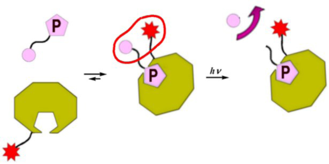
Scheme 1 outlines the general concept of such a system: one component of the molecular recognition pair (green octagon), is outfitted with a compact ET-sensitizer, e.g. xanthone, whereas the dithiane adduct is tethered to the second component (pink pentagon labeled P). Binding brings the sensitizer into the vicinity of the tag adduct, at which point irradiation commences. Xanthone sensitizes fragmentation in the adduct, releasing the dithiane tag into solution. The tags are then detected in a standardized analytical protocol revealing the identity of the bound pairs. A diverse set of substituted dithiane tags is readily available for encoding. Additionally, to further diversify the available variety of tags, we have identified non-dithiane encoding tags, for example derivatives of trithiabicyclo[2.2.2]octanes, which can be used in combinatorial encoding.12
Scheme 1.
Tagged combinatorial libraries are well precedented. In the classical bead-tagging approach, for example the one developed by Clark Still,13 each bead is encoded with the full set of tags needed for subsequent identification of the ligand displayed on the surface of the bead. Still's strategy is not suitable for encoding individual molecules, which simply do not have enough "real estate" to accommodate all the encoding tags without running into a risk of multiple tags interfering with molecular recognition events. Our strategy is to encode individual molecules one tag at a time. Every library member can be encoded with a set of tags so that a certain fraction of its molecules are encoded with the first tag, another fraction – with second, etc., with the net result of each ligand being present in the solution as several sub-populations cumulatively encoded with all the tags necessary for its subsequent identification. Irradiation in this case yields the desired result because all the tags encoding individual molecules in the bound pairs are collectively released into the solution, where they are analyzed revealing the identity of the bound compound.
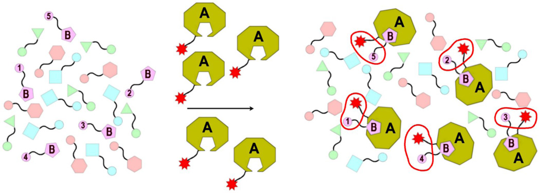
Initially we proved this general concept of tagging the individual (unsupported) library members and screening for molecular recognition using the known binding pair, avidin-biotin.14 In these experiments a mini library of compounds, which included biotin, was encoded by tagging each compound with different dithianes. Scheme 2 gives a general outline for screening of such library of ligands (biotin is depicted by a pink "B" pentagon), encoded with the tethered tags (pink circles 1–5). Avidin (the green octagon "A") is outfitted with a xanthone-based ET-sensitizer, and incubated with the library. The binding event brings the sensitizer into the vicinity of the tag/adducts (encircled with red), which, upon irradiation triggers the release of tags 1–5, i.e. only the tags which encode biotin.
Scheme 2.
So far our dithiane tags-encoding methodology was tested with a model barbiturate-binding artificial receptor15 and validated for the avidin-biotin binding pair,14 known to have a very tight KD. In this study we extend this approach onto biological systems exhibiting low micro molar affinities and demonstrate that it can be used to differentiate between very similar epitopes modified via post-translational modifications (PTMs), such as unmodified and methylated lysine. Our choice of the protein substrate is ING2 PHD (plant homeodomain) finger, which is known to recognize a hexapeptide fragment of the histone H3 tail Ala-Arg-Thr-Lys-Gln-Thr, but only when the lysine residue is trimethylated, i.e. Ala-Arg-Thr-Lys(Me3)-Gln-Thr. The trimethylated epitope is referred to as H3K4me3.
Nuanced aspects of molecular recognition play a pivotal role in biological processes. This is especially important for the emerging field of epigenetics, where PTMs regulate complex signaling cascades, including gene transcription, DNA repair, recombination, and replication and chromatin remodeling.16,17 Epigenetic misregulations have been linked to various human diseases including cancer, premature aging and neurodegenerative disorders, and thus development of experimental approaches to characterize PTM recognitionis essential in understanding the epigenetic mechanisms, PTM-driven functional outcomes, and disease-associated alterations. In the context of this study, massive high throughput screening of encoded split-and-pool libraries of small peptides containing epigenetic marks, in one non-compartmentalized volume is particularly appealing. We will show that (i) compact dithiane tags are readily installed via the same robotic peptide synthesis protocol and that they are not perturbing molecular recognition events; (ii) the photo induced tag release is contingent on a binding event taking place; and (iii) the fidelity of this binding assay is very high as exemplified by a large, easily discernible change (10–300 fold) in the ratio of the released encoding dithianes, detected in the experiments with indiscriminate diffusion controlled release as compared to the experiments where strong bias is introduced by a binding event.
2. MATHERIALS AND METHODS
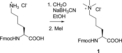
N-Trimethyl lysine1
Fmoc-NH-Lys(Me3)OH, was synthesized as shown using a modified procedure:18 to a stirred solution of 9.0g of Fmoc-lysine hydrochloride, 5mmol, in 600ml of ethanol in ice bath 4.2ml of formalin was added. After stirring for 15 min 4.5g of NaBH3CN was added at 0–5°C. After another 15 min the formalin and NaBH3CN treatment was repeated. Then 10ml of MeI was added, the ice cooling bath was removed and the reaction mixture was stirred overnight. The reaction was quenched with 1.3M HCl solution which was added under stirring for approximately 30min until the evolution of hydrogen gas was completed. The solvent was removed with rotary evaporator. The residue was dissolved in THF and a precipitate was removed by filtering. The filtrate was purified by gel-filtration using a sintered glass funnel with ~6 cm (60mL) silica gel layer, eluting first with THF, THF/water (10:1),then MeOH-water (5:1);2.3g of trimethyl lysine (yield 23%, pure by NMR19) was collected and used without further purification.
Synthesis of 2-alkyl dithianes14 and dithianyl benzylamine-based tagging pendants420 was previously described.
Automated Peptide Synthesis
Two decapeptides containing the Ala-Arg-Thr-Lys-Gln-Thr motif were synthesized using AAPPTEC's Titan-357 robotic synthesizer: one containing the epigenetic mark, trimethylated lysine, and the other – δ-N-unsubstituted lysine. Photoactive tagging glutamines outfitted with two unique 2-alkyl dithianes, 2-propyldithiane and 2-butyldithiane were used to “encode” these peptides respectively. Standard AAPPTEC protocols designed for Rink Amide resin were used for the attachment of the first glycine residue, Fmoc peptide synthesis, and cleavage of the peptide from the resin.21 The only deviation from the standard coupling protocols was the incorporation of the trimethylated lysine, which initially proved challenging. The following optimized procedure was developed: 200mg of the TentaGel RAM resin with Fmoc-protected glycine was subjected to FMOCRE sequence for Fmoc removal. Then 0.5mL of 0.4M LiCl DMF solution, 0.5mL of 0.5M trimethyl Fmoc-lysine solution (114mg in 0.5mL of DMF, 4eq), 1ml of DMSO, 35mg of HOBT (4 eq) and 0.05mL of DIC (4 eq) were added and mixed for 5h, the reactor was drained (K-EM2MIN sequence) and the same coupling procedure was repeated second time. This procedure ensured >95% incorporation of the trimethylated lysine as monitored by LC-MS(ESI).
Sensitizer with tether
N-(10-Xanthonyldecanoyl)-10-aminodecanoic acid2 and its N-hydroxysuccinimide (NHS) derivative3 were synthesized via Suzuki coupling as described previously.22 The length of the tether was previously shown to be adequate for the photo induced dithiane release in the avidin-biotin binding pair.

Expression and Purification of the ING2 PHD finger
The uniformly 15N-labeled ING2 PHD finger was expressed in E. coli BL21(DE3) pLysS (Stratagene) grown in zinc-enriched 15NH4Cl-supplemented (Isotec) M9-minimal media. Bacteria were harvested by centrifugation after IPTG induction and lysed using a French press. The GST-fusion protein was purified on a glutathione Sepharose 4B column (Amersham), cleaved with PreScission protease (Amersham) and concentrated in Millipore concentrators (Millipore) into 20 mM d11-Tris buffer containing 150 mM NaCl, 10 mM d10-dithiothreitol and 1 mM NaN3 in 7% 2H2O/H2O, pH 6.5.
NMR spectroscopy
NMR spectra were collected at 25°C on a Varian INOVA 500 MHz spectrometer. 1H,15N-heteronuclear single quantum coherence (HSQC) spectra of the ING2 PHD finger (0.2 mM) were recorded in the presence of increasing concentrations of BuDTA-H3K4me3 and H3K4me3 peptides. The normalized chemical shift change was calculated using the equation [(ΔδH)2 + (ΔδN/5)2]0.5, where is δ the chemical shift in parts per million (ppm).
Xanthone-tethered ING2 PHD finger
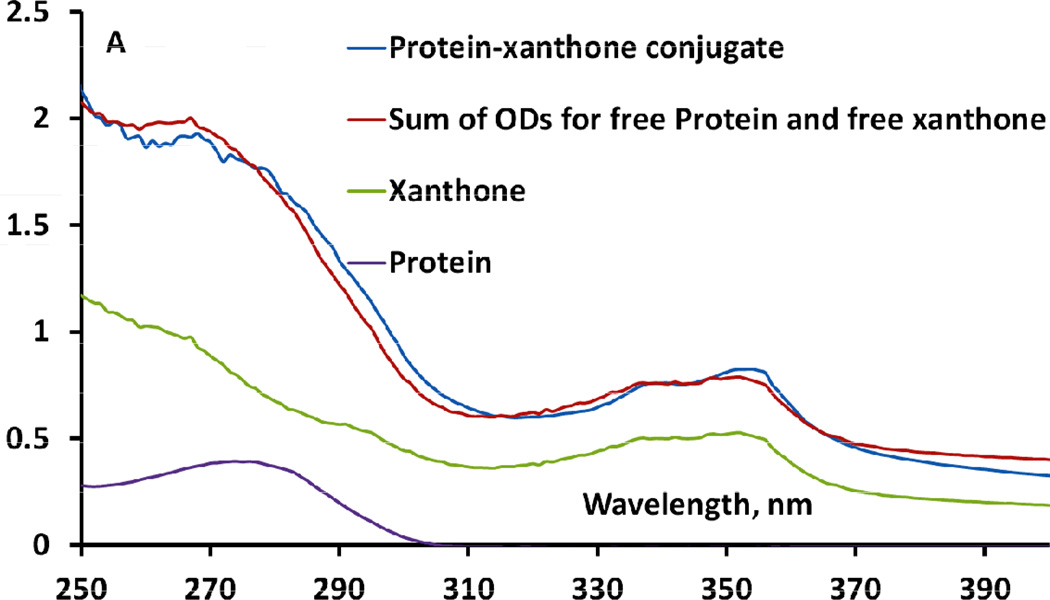
ING2 PHD finger was outfitted with xanthone as an electron transfer photosensitizer as follows. 200µL of 1.07mM of the protein was mixed with 500µL of 0.01M PBS at pH=8. 20µL of Xanthone-tether-NHS3 in DMSO (0.02mg/mL) was added and the reaction was gently shaken for 1 hour at room temperature. Two more 20µL additions were made each 1 hour apart while shaking. After the final addition, the reaction was shaken overnight at room temperature. The conjugate was purified using Sephadex G20 with 0.01M PBS as the eluent. Fractions containing xanthone without protein were discarded. Fractions containing ING2–xanthone conjugates were quantified using UV/Vis spectrophotometry. The extent of xanthone conjugation was determined by fitting the UV spectrum of a conjugate to the sum of ODs for free protein and free xanthone-tether of known concentration between 250 and 400 nm. This fitting gives the average ratio and, at the same time, the conjugate concentration. An example of a fraction with 0.32mM concentration of a 2:3 protein-xanthone conjugate is shown in Figure 1. Thus, on average 1.5 sensitizer pendants were immobilized to one molecule of the protein.
Figure 1.
ING2–xanthone conjugation quantified by OD fitting (green trace "xanthone" refers to free acid 2)
Next, control experiments for indiscriminant release of dithianes from the encoded peptides using free N-(10-xanthonyldecanoyl)-10-aminodecanoic acid were designed to demonstrate that in the absence of ING2 PHD finger, the free bulk sensitizer can trigger release of the dithiane tags indiscriminately from both tagged peptides.
Two peptides, Ala-Arg-Thr-Lys-Gln-Thr-Gly-Gln(PrDTA)-Gly-Gly and Ala-Arg-Thr-Lys(Me3)-Gln- Thr-Gly-Gln(BuDTA)-Gly-Gly, were suspended in 200µL of 20mM micellar DPC with free N-(10-xanthonyldecanoyl)-10-aminodecanoic acid, to achieve equal (20µM) concentration of both peptides and the sensitizer. The micellar solution was irradiated in a Rayonet photoreactor, using RPR-3500 UV lamps (broad emission 300–400nm with a maximum at 350nm), extracted with hexane 2 × 0.5 mL, and 2µL of the hexane extract was injected into a HP6890N GC/MS.
Alkyl dithianes detection was performed with a HP6890N GC/MS instrument in the single ion monitoring mode; start temperature 120°C ramping at 25°C/min to a final temperature of 220°C with a helium flow of 0.8mL/min. Column: 30m × 250µm ID, 5% phenyl methyl siloxane fused silica bonded capillary. Calibration was carried out with a 10µM2-alkyldithianes solution;0.3 min difference in retention times ensured confident assignment of the two dithiane species.
Binding assay
2.5mL 3.70 × 10⁻⁵ M aqueousxanthone-tethered protein with 2.05mL of 0.01M PBS, 92µL of 1mM tagged peptides, Ala-Arg-Thr-Lys-Gln-Thr-Gly-Gln(PrDTA)-Gly-Gly and Ala-Arg-Thr-Lys(Me3)-Gln-Thr-Gly-Gln(BuDTA)-Gly-Gly, in 0.01M PBS, and 12mg of n-dodecylphosphocholine (Avanti) was added to a 10mL vial. Final concentrations: 50µM protein, 20µM peptide, 20mM DPC. The solution was shaken for 1 minute and allowed to incubate at room temperature for 1 hour, followed by 3hrs irradiation using 4 × 250mW 365nm Nichia UV LEDs. After irradiation, the solution was extracted with 0.5mL of distilled hexanes, the hexane layer was pre-concentrated at ambient temperature using rotary evaporator to a volume of 20µL and analyzed by GC-MS. Experiment was run in triplicate, see Figure 5.
Figure 5.
ING2-H3K4me3 binding detected byphototriggered release of BuDTA tag, encoding trimethylated H3K4me3.PrDTA encoding the unmethylated counterpart is barely detectable.
2. RESULTS AND DISCUSSION
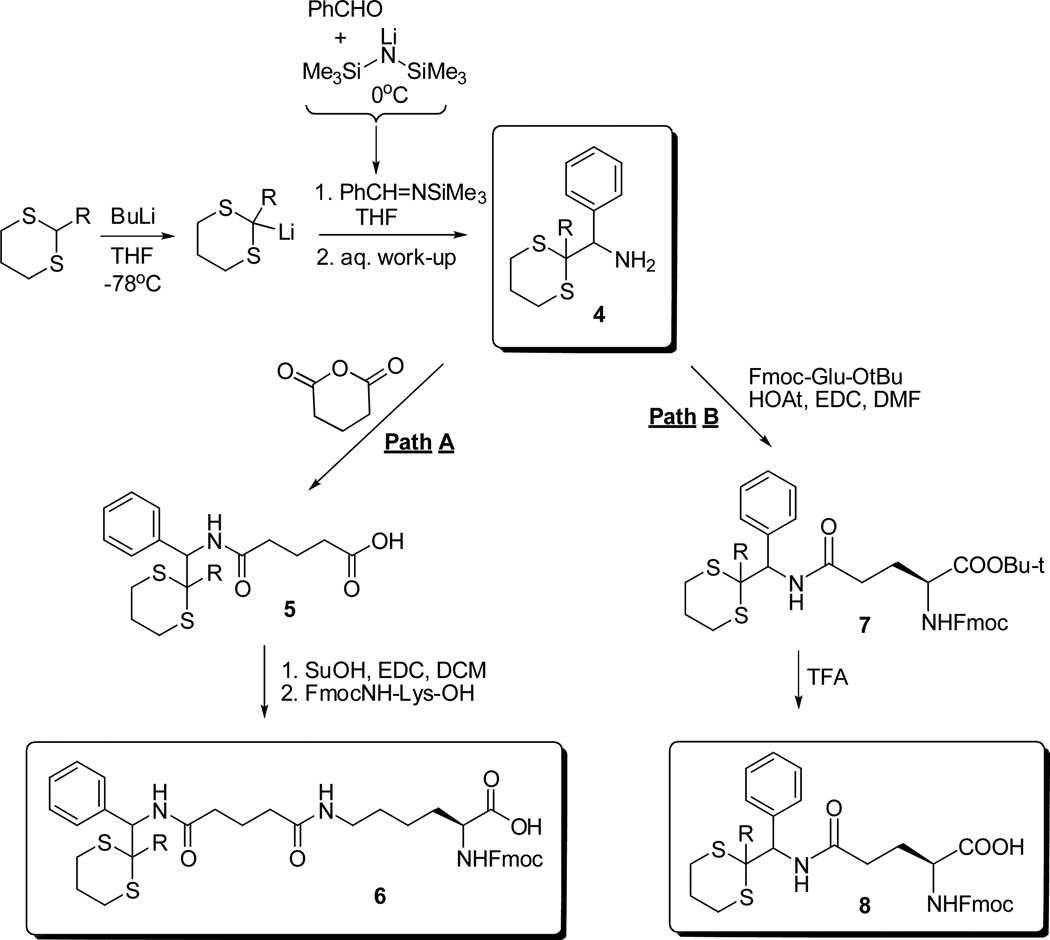
For tagging of the peptides we used the dithiane adducts with benzaldimine, as they offer the most straightforward tethering protocol. They were also found to undergo efficient photo induced electron transfer fragmentation, just like their oxa-counterparts. Scheme 3 shows two types of tagged amino acids, lysine and glutaric acid, with compact dithiane adducts of benzaldimine. Lithiated dithianes were reacted with PhCH=NOSiMe3, pre-formed in situ via the reaction of benzaldehyde and lithium hexamethylsilazide23 to yield dithianyl-benzylamines4 after aqueous work-up as described previously.20 Path A included tethering the tagging pendants using glutaric anhydride with subsequent coupling with Fmoc NH-Lys. This gave a longer-tethered tagging module6. Path B involved coupling the dithianyl-benzylamines4 with Fmoc-Glu-OtBu and deprotecting the resulting tert-butyl ester7 by treatment with TFA. This produced short-tethered compact tagging module8.
Scheme 3.
Synthesis of tagged Fmoc-lysine and Fmoc-glutamine
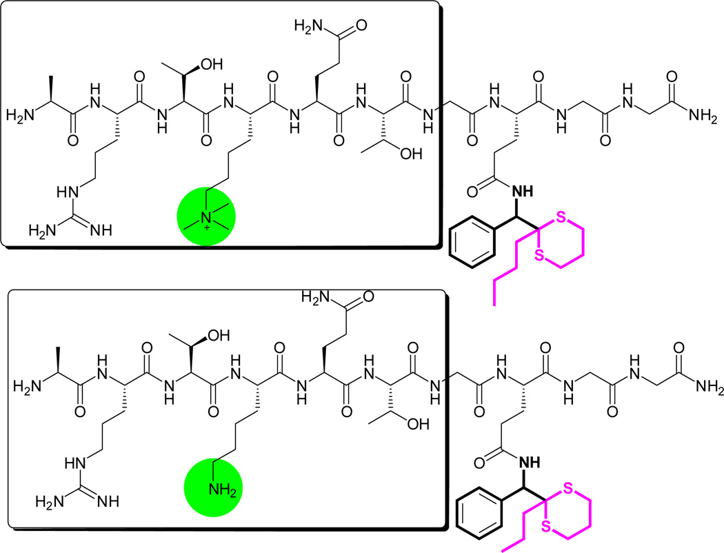
The photoactive Fmoc-protected amino acid derivatives 6 and 8 were introduced into the automated peptide synthesis, as it was critical to demonstrate that the tagged amino acid derivatives are compatible with the automated peptide synthesizer protocols. In this work we used AAPPTEC's Titan-357 36-well robotic synthesizer. In order to facilitate the tag incorporation and to spatially separate the tag from the native hexapeptide epitope we elected to use glycine spacers before and after the dithiane-tagged amino acid. TentaGel® RAM (Rink amide) beads were used. Even with glycine spacers, designed to reduce steric crowding, we experienced difficulty with the lysine-based extended tagging pendants 6, which were eventually abandoned. On the contrary, the shorter glutamine-based pendants were readily incorporated under the standard conditions of automated peptide synthesis as described in Section 2. Figure 2 shows the two encoded peptides Ala-Arg-Thr-Lys*-Gln-Thr-Gly-Gln(Tag)-Gly-Gly in which the tag is flanked by glycine residues.
Figure 2.
H3K4me3 peptide tagged with Gly-Gln(BuDTA)-Gly-Gly (top); and its unmethylated counterpart, tagged with Gly-Gln(PrDTA)-Gly-Gly (bottom). The encoding butyl- and propyldithianes are shown in maroon.
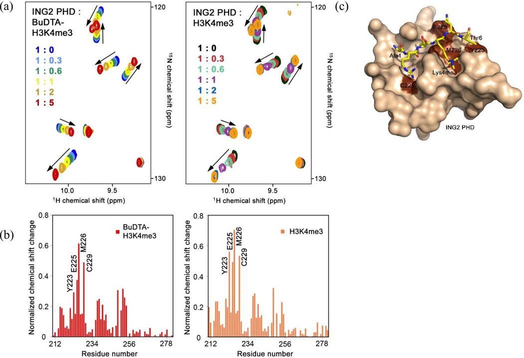
We tested binding of the BuDTA-tagged H3K4me3 peptide to the uniformly 15N-labeled ING2 PHD finger using 1H,15NHSQC NMR titration experiments. Gradual addition of the BuDTA-tagged H3K4me3 peptide caused significant chemical shift changes in the PHD finger, Figure 3.
Figure 3.
BuDTA-H3K4me3 binds to the ING2 PHD finger. (a) Superimposed 1H,15N heteronuclear single quantum coherence (HSQC) spectra of the ING2 PHD finger (0.2 mM) collected while BuDTA-H3K4me3 peptide (left) or H3K4me3 peptide24 (right) was titrated in. Spectra are color coded according to the protein:peptide molar ratio (inset). (b) The normalized chemical shift changes observed in 1H,15N HSQC spectra of the ING2 PHD finger upon binding of BuDTA-H3K4me3 (left) or H3K4me3 (right) as a function of residue. (c) The PHD finger residues that exhibit significant chemical shift changes due to binding of BuDTA-H3K4me3 are colored brown on the surface of the ING2 PHD-H3K4me3 complex24 and labeled. The bound H3K4me3 peptide is shown as a stick model and colored yellow. Thr6, at the periphery of the binding interface, serves as the attachment point for the tagging pendant in BuDTA-H3K4me3.
Particularly, residues comprising the H3K4me3-binding site, including Y223, E225, M226 and C229, were perturbed to the highest degree. Similar changes in the PHD finger were observed upon titration of the untagged H3K4me3 peptide, confirming that BuDTA-H3K4me3 can be used without compromising binding activity of the histone tail.
As described in Section 2, photochemical experiments were carried out in 20mM aqueous micellar solution of dodecylphosphocholine. The DPC concentration was experimentally determined to ensure that the micellar solution of ING2 and the two proteins is transparent and clear. The DPC micelle aggregation number of 53–56 molecules per micelle25 was also taken into the consideration to prevent statistical crowding of non-bound decapeptides with ING2 PHD finger in the same micelle. Under our experimental conditions statistically less than one quarter of micelles were populated, decreasing the probability of random double occupation by non-binding molecules.
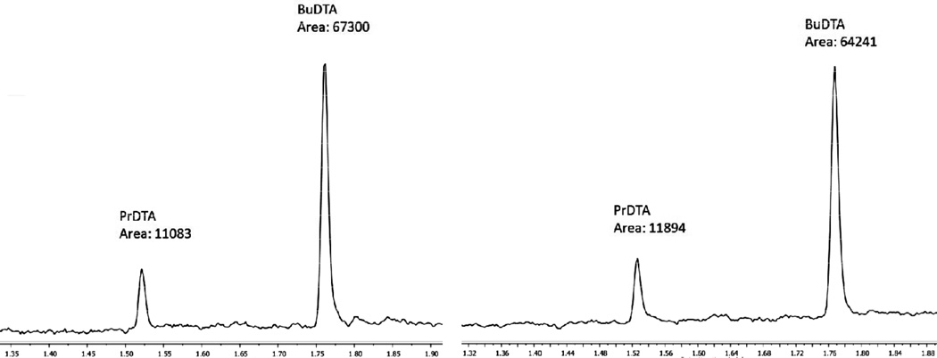
An important control experiment was performed to ensure that in the absence of binding (i.e. when the sensitizer is not tethered to ING2 but rather free to photochemically induce the dithiane release indiscriminately via bimolecular diffusion controlled sensitization) both dithianes are released and detected. In this experiment both peptides, tagged with PrDTA and BuDTA, were solubilized with DPC and irradiated in the presence of free sensitizer, N-(10-xanthonyldecanoyl)-10-aminodecanoic acid2.Both dithianes were detected in a reproducible 1:6 ratio (two representative runs are shown in Figure 4).
Figure 4.
Control photorelease experiments with two tagged peptides and bulk untethered sensitizer (run in duplicate)
The 1:6 ratio reflects (i) small difference in the quantum yields of release of propyl- vs butyldithianes, which tend to increase with the length of n-alkyl substituent in the hydroxy counterparts,26 (ii) different MS detector sensitivity to two dithianes and, most importantly (iii) a different local environment due to trimethylated lysine incorporation in the polypeptide tagged with BuDTA. The externally-sensitized dithiane release is expected to be somewhat sensitive to the peptide environment in two aspects. First, less polar methylated and more polar non-methylated lysines conceivably affect the folding of the respective peptides, which may complicate or make easier the approach of the sensitizer to the tagging pendant, modulating slightly the quantum yield of release. Second, the rates of premature reduction of the excited triplet sensitizer may also differ for the two peptides. All these effects are not large but measurable in the control experiment. Thus, the 6:1 ratio provides a reference point for indiscriminate dithiane release.
We then carried out the central experiments of this study, i.e. the photoinduced dithiane release which is contingent on a molecular recognition event which brings the sensitizer in the close proximity of the photolabile tag. The experiment was performed in triplicate using 50µM ING2-xanthone conjugate and 20µM of tagged peptides. Excess protein-xanthone conjugate was used intentionally to ascertain to what extent free diffusing (excess) sensitizer can skew the differentiation between the bound and non-bound pairs. Figure 5 shows the GC-MS traces with integrated BuDTA and PrDTA abundances, with ratios ranging from two to three orders of magnitude.
In all these experiments, the peak corresponding to propyl-dithiane, encoding the peptide with unmethylated lysine, is barely visible, which makes it challenging to integrate properly. There is considerable (orders of magnitude) difference between the reference ratio of 1:6, determined for the free-diffusion release of both dithiane tags, and the ratios obtained in the experiments biased by the binding of ING2 PHD finger to H3K4me3.
In conclusion, we have developed an encoding strategy for polypeptides and demonstrated that photoassisted assay based on conditional release of encoding tags only from bound pairs can differentiate between peptides which have minor differences in a form of post-translational modifications with epigenetic marks. The encoding strategy is fully compatible with automated peptide synthesis. The encoding pendants are compact and are not expected to perturb potential binding interactions. This methodology could be adapted for assaying solution phase libraries for binding in one non-compartmentalized volume, which is particularly appealing in conjunction with split-and-pool library synthesis.
HIGHLIGHTS.
Photoassisted binding assay differentiates peptides with epigenetic marks
The library-encoding strategy is compatible with automated peptide synthesis
Methodology could be adapted for assaying solution phase libraries
Acknowledgement
Support of this research by the NSF CHE-1057800 (A.G.K.) and NIH grant GM101664 (T.G.K.)is gratefully acknowledged. We thank Pedro Pena for help with experiments.
Footnotes
Publisher's Disclaimer: This is a PDF file of an unedited manuscript that has been accepted for publication. As a service to our customers we are providing this early version of the manuscript. The manuscript will undergo copyediting, typesetting, and review of the resulting proof before it is published in its final citable form. Please note that during the production process errors may be discovered which could affect the content, and all legal disclaimers that apply to the journal pertain.
References
- 1.Lam KS, Lebl M, Krchnak V. The "One-Bead-One-Compound" Combinatorial Library Method. Chem. Rev. 1997;97:411–448. doi: 10.1021/cr9600114. [DOI] [PubMed] [Google Scholar]
- 2.Lam KS, Liu R, Miyamoto S, Lehman AL, Tuscano JM. Applications of One-Bead One- Compound Combinatorial Libraries and Chemical Microarrays in Signal Transduction Research. Acc. Chem. Res. 2003;36:370–377. doi: 10.1021/ar0201299. [DOI] [PubMed] [Google Scholar]
- 3.Konings DAM, Wyatt JR, Ecker DJ, Freier SM. Strategies for Rapid Deconvolution of Combinatorial Libraries: Comparative Evaluation Using a Model System. J. Med. Chem. 1997;40:4386–4395. doi: 10.1021/jm970503o. [DOI] [PubMed] [Google Scholar]
- Baldino CM. Perspective Articles on the Utility and Application of Solution-Phase Combinatorial Chemistry. J. Comb. Chem. 2000;2:89–103. doi: 10.1021/cc990064+. [DOI] [PubMed] [Google Scholar]
- 5.(a) Ganem B, Li Y, Henion J. Detection of noncovalent receptor-ligand complexes by mass spectrometry. J. Am. Chem. Soc. 1991;113:6294–6296. [Google Scholar]; (b) Ganem B, Li Y, Henion J. Observation of noncovalent enzyme-substrate and enzyme-product complexes by ion-spray mass spectrometry. J. Am. Chem. Soc. 1991;113:7818–7819. [Google Scholar]; (c) Loo J. Studying noncovalent protein complexes by electrospray ionization mass spectrometry. Mass Spectrom. Rev. 1997;16:1–17. doi: 10.1002/(SICI)1098-2787(1997)16:1<1::AID-MAS1>3.0.CO;2-L. [DOI] [PubMed] [Google Scholar]
- 6.Woods AS, Huestis MA. A Study of Peptide–Peptide Interaction by Matrix-Assisted Laser Desorption/Ionization. J. Am. Soc. Mass. Spec. 2001;21:88–96. doi: 10.1016/S1044-0305(00)00197-5. [DOI] [PubMed] [Google Scholar]
- 7.Murphy RM. Peptide Aggregation in Neurodegenerative Disease. Annu.Rev. Biomed. Eng. 2002;4:155–174. doi: 10.1146/annurev.bioeng.4.092801.094202. [DOI] [PubMed] [Google Scholar]
- 8.(a) Hill RB, Raleigh DP, Lombardi A, DeGrado WF. De Novo Design of Helical Bundles as Models for Understanding Protein Folding and Function. Acc. Chem. Res. 2000;33:745–754. doi: 10.1021/ar970004h. [DOI] [PMC free article] [PubMed] [Google Scholar]; (b) DeGrado WF, Summa CM, Pavone V, Nastiri F, Lombardi A. De Novo Design and Structural Characterization of Proteins and Metalloproteins. Annu. Rev. Biochem. 1999;68:779–819. doi: 10.1146/annurev.biochem.68.1.779. [DOI] [PubMed] [Google Scholar]; (c) Yin H, Slusky JS, Berger BW, Walters RS, Vilaire G, Litvinov RI, Lear JD, Caputo GA, Bennett JS, DeGrado WF. Computational design of peptides that target transmembrane helices. Science. 2007;315:1817–22. doi: 10.1126/science.1136782. [DOI] [PubMed] [Google Scholar]
- 9.Gellman SH. Foldamers: A Manifesto. Acc. Chem. Res. 1998;31:173–180. [Google Scholar]
- 10.(a) Mezo AR, Ottesen JJ, Imperiali B. Discovery and Characterizaion of a Discretely folded Homotrimericββα Peptide. J. Am. Chem. Soc. 2001;123:1002–1003. doi: 10.1021/ja0038981. [DOI] [PubMed] [Google Scholar]; (b) Mezo AR, Cheng RP, Imperiali B. Oligomerization of Uniquely Folded Mini-Protein Motifs: Development of a Homotrimericββα Peptide. J. Am. Chem. Soc. 2001;123:3885–3891. doi: 10.1021/ja004292f. [DOI] [PubMed] [Google Scholar]
- 11.(a) McHale WA, Kutateladze AG. An Efficient Photo-SET-Induced Cleavage of Dithiane-Carbonyl Adducts and Its Relevance to the Development of Photoremovable Protecting Groups for Ketones and Aldehydes. J. Org. Chem. 1998;63:9924. [Google Scholar]; (b) Mitkin O, Wan Y, Kurchan A, Kutateladze A. Synthesis of Dithiane-Based Photolabile Molecular Systems. Synthesis. 2001;8:1133. [Google Scholar]; (c) Mitkin OD, Kurchan AN, Wan Y, Schiwal BF, Kutateladze AG. Dithiane and Trithiane-Based Photolabile Scaffolds for Molecular Recognition. Org. Lett. 2001;3:1841. doi: 10.1021/ol015933u. [DOI] [PubMed] [Google Scholar]; (d) Vath P, Falvey DE, Barnhurst LA, Kutateladze AG. Photoinduced C-C Bond Cleavage in Dithiane-Carbonyl Adducts: A Laser Flash Photolysis Study. J. Org. Chem. 2001;66:2886. doi: 10.1021/jo010102n. [DOI] [PubMed] [Google Scholar]; (e) Li Z, Kutateladze AG. Anomalous C-C Bond Cleavage in Sulfur-Centered Cation Radicals Containing Vicinal Hydroxy Group. J. Org. Chem. 2003;68:8236. doi: 10.1021/jo035001z. [DOI] [PubMed] [Google Scholar]
- 12.Valiulin RA, Kutateladze AG. 2,6,7-Trithiabicyclo[2.2.2]octanes as Promising Photolabile Tags for Combinatorial Encoding. J. Org. Chem. 2008;73(1):335–338. doi: 10.1021/jo702091e. [DOI] [PubMed] [Google Scholar]
- 13.MHJ Ohlmeyer MHJ, Swanson RN, Dillard LW, Reader JC, Asouline G, Kobayashi R, Wigler M, Still WC. Complex Synthetic Chemical Libraries Indexed with Molecular Tags. Proc. Nat. Acad. Sci. USA. 1993;90:10922–10926. doi: 10.1073/pnas.90.23.10922. [DOI] [PMC free article] [PubMed] [Google Scholar]
- 14.Kottani R, Valiulin RA, Kutateladze AG. Direct Screening of Solution Phase Combinatorial Libraries Encoded with Externally Sensitized Photolabile Tags. Proc. Natl. Acad. Sci. USA. 2006;103:13917–13921. doi: 10.1073/pnas.0606380103. [DOI] [PMC free article] [PubMed] [Google Scholar]
- 15.Lakkakula S, Mitkin OD, Valiulin RA, Kutateladze AG. Photoactive Barbiturate Receptors: An Ultimate Lock-and-Key System in Which the Key Unlocks the Lock. Org. Lett. 2007;9(6):1077–1079. doi: 10.1021/ol0700153. [DOI] [PMC free article] [PubMed] [Google Scholar]
- 16.Kouzarides T. Chromatin modifications and their function. Cell. 2007;128:693–705. doi: 10.1016/j.cell.2007.02.005. [DOI] [PubMed] [Google Scholar]
- 17.Musselman CA, Lalonde ME, Cote J, Kutateladze TG. Perceiving the epigenetic landscape through histone readers. Nat StructMolBiol. 2012;19:1218–1227. doi: 10.1038/nsmb.2436. [DOI] [PMC free article] [PubMed] [Google Scholar]
- 18.Baba R, Hori Y, Mizukami S, Kikuchi K. Development of FluorogenicProbe with Transesterification Switch for Detection of Histone Deacetylase Activity. J. Am. Chem. Soc. 2012;134:14310–14313. doi: 10.1021/ja306045j. [DOI] [PubMed] [Google Scholar]
- 19.the product is identical to the authentic sample of Fmoc-Lys(Me)3-OH from AnaSpec, www.anaspec.com, catalog number 60260-1
- 20.Kurchan AN, Mitkin OD, Kutateladze AG. Dithiane and Trithiane-Based Photolabile Molecular Linkers Equipped with Amino-Functionality: Synthesis and Quantum Yields of Fragmentation. J. Photochem. Photobiol. A: Chemistry. 2005;171:121–129. [Google Scholar]
- 21.(a) http://www.aapptec.com/custdocs/rink%20amide%20resin%20tech.pdf. [Google Scholar]; (b) Stathopoulos P, Papas S, Tsikaris V. C-terminal N-alkylated peptide amides resulting from the linker decomposition of the Rink amide resin: a new cleavage mixture prevents their formation. J. Pept. Sci. 2006;,12:227–232. doi: 10.1002/psc.706. [DOI] [PubMed] [Google Scholar]
- 22.Gustafson TP, Metzel GA, Kutateladze AG. Externally sensitized deprotection of PPG-masked carbonyls as a spatialproximity probe in photoamplified detection of binding events. Photochem. Photobiol. Sci. 2012;11:564–577. doi: 10.1039/c2pp05326h. [DOI] [PMC free article] [PubMed] [Google Scholar]
- 23.Gyenes F, Bergmann KE, Welch JT. Convenient Access to Primary Amines by Employing the Barbier-Type Reaction of N-(Trimethylsilyl)imines Derived from Aromatic and Aliphatic Aldehydes. J. Org. Chem. 1998;63:2824–2828. [Google Scholar]
- 24.Peña PV, Davrazou F, Shi X, Walter KL, Verkhusha VV, Gozani O, Zhao R, Kutateladze TG. Molecular mechanism of histone H3K4me3 recognition by plant homeodomain of ING2. Nature. 2006;442:100–103. doi: 10.1038/nature04814. [DOI] [PMC free article] [PubMed] [Google Scholar]
- 25.see Lazaridis T, Mallik B, Chen Y. Implicit Solvent Simulations of DPC Micelle Formation. J. Phys. Chem. B. 2005;109:15098–15106 . doi: 10.1021/jp0516801. and references therein.
- 26.Gustafson TP, Kurchan AN, Kutateladze AG. Externally Sensitized Mesolytic Fragmentations in Dithiane-Ketone Adducts. Tetrahedron. 2006;62:6574–6580. [Google Scholar]


