Abstract
DNA Ligase IV, along with its interacting partner XRCC4, are essential for repairing DNA double strand breaks by non-homologous end joining (NHEJ). Together, they complete the final ligation step resolving the DNA break. Ligase IV is regulated by XRCC4 and XLF. However, the mechanism(s) by which Ligase IV control the NHEJ reaction and other NHEJ factor(s) remains poorly characterized. Here, we show that a C-terminal region of Ligase IV (aa 620 to 800), which encompasses a NLS, the BRCT I, and the XRCC4 interacting region (XIR), is essential for nuclear localization of its co-factor XRCC4. In Ligase IV deficient cells, XRCC4 showed deregulated localization remaining in the cytosol even after induction of DNA double strand breaks. DNA Ligase IV was also required for efficient localization of XLF into the nucleus. Additionally, human fibroblasts that harbor hypomorphic mutations within the Ligase IV gene displayed decreased levels of XRCC4 protein, implicating that DNA Ligase IV is also regulating XRCC4 stability. Our results provide evidence for a role of DNA Ligase IV in controlling the cellular localization and protein levels of XRCC4.
INTRODUCTION
Double strand breaks (DSBs) are one of the most deleterious lesions that can occur within the genome of the cell. These lesions can arise as a result of normal physiological cellular processes, such as V(D)J recombination and class switch recombination during immune cell development (1,2). DSBs are also generated during ionizing radiation (IR) and production of oxidative free radicals (3). In mammalian cells, two major pathways have evolved for the repair of DSBs, namely, homologous recombination (HR) and non-homologous end joining (NHEJ) (4–6). HR is a homology dependent reaction and requires the presence of a sister chromatid or homologous chromosome, which functions as a DNA template; this is the main functional pathway during late S/G2 phase of the cell cycle. In contrast, NHEJ, because of its homology-independence, is active throughout the cell cycle but has been found to predominate during G1. Repair by classical NHEJ is considered as error-prone due to the frequent loss or addition of nucleotides at the site of the DSB. However, despite its mutagenic properties the NHEJ pathway is the major pathway utilized to repair DSB, including those that arise as a result of somatic recombination during the development and maturation of immune cells.
Repair via NHEJ involves several core factors including Ku70/80, DNA-PKcs, Artemis, XLF, XRCC4 and DNA Ligase IV (referred to as Ligase IV for the rest of the text). The Ku70/80 heterodimer senses and recognizes breaks in chromosomal DNA and together with DNA-PKcs, stabilize the free ends. Artemis, an endonuclease, along with polymerases, μ (PolX family) and terminal deoxynucleotidyl transferase (TdT), play important roles in the processing of DNA ends making them ready for ligation. Finally, the Ligase IV/XRCC4/XLF complex completes ligation and resolves the DSB (7,8).
Ligase IV, in complex with XRCC4 and XLF, is indispensable to the NHEJ reaction and absence of either of these factors leads to an impaired ability to repair DSBs and immunodeficiency (9–11). Hypomorphic mutations within the Ligase IV gene, which disrupt protein function result in partial immunodeficiency and increased sensitivity to IR, reflecting the deregulated function of the NHEJ machinery (12). Despite significant progress demonstrating how XLF and XRCC4 regulate Ligase IV function, little is known about how Ligase IV regulates NHEJ. It has been shown that proteasome mediated degradation of Ligase IV prevents the binding of XRCC4 and XLF to DNA, without changing their protein levels (13). DNA binding by XRCC4 and ligation activity of the complex was restored following complementation with the full length Ligase IV (13). Independent studies showed that localization of XRCC4 and XLF to chromatin was also dependent on Ligase IV (14,15). Ligase IV C-terminal region was sufficient to drive localization of XRCC4 to chromatin (16). Additionally, while XLF is known to interact directly with XRCC4, an intact Ligase IV/XRCC4 complex is needed for the appropriate recruitment of XLF to chromatin and for its efficient interaction with XRCC4 (15). The Ligase IV/XRCC4 complex contributes to DNA-PKcs autophosphorylation as well as DNA-PKcs mediated DNA end synapsis (17). A role for the Ligase IV/XRCC4 complex in recruiting and/or modulating the activity of processing enzymes, including nucleases and polymerases, was also suggested (18–21). These findings indicate that Ligase IV is critical to the recruitment, assembly and function of the processing and ligation complexes at the site of DSBs. However, the mechanism(s) by which Ligase IV functions to control NHEJ and NHEJ factors remains poorly characterized.
Here, we report that in the absence of Ligase IV, XRCC4 accumulates in the cytoplasm and this retention is independent of DNA damage. Specifically, the Cterminal of Ligase IV plays an important role in regulating the subcellular localization of XRCC4. In addition, human fibroblasts from LIG4 syndrome patients showed a large decrease in XRCC4 protein levels. Together our data suggest new mechanisms by which Ligase IV contributes to the regulation of its protein partner, XRCC4.
MATERIAL AND METHODS
Antibodies
Rabbit polyclonal antibodies: anti-Ligase IV against amino acids 1–240, anti-XRCC4 (Serotec), anti-Artemis raised against full-length recombinant Artemis, anti-XLF (Bethyl Laboratories, Inc), anti-DNA-PKcs (Santa Cruz Biotechnology, Inc), anti-Ku70, anti-Ku80 (Santa Cruz Biotechnology, Inc) and anti-Phospho-p53 (Ser315) (Cell Signaling). Goat polyclonal antibodies: anti-Lamin B1 (Santa Cruz Biotechnology, Inc). Mouse monoclonal antibodies: anti-Flag M2 (Sigma-Aldrich), anti-p53 (BioLegend), anti- -Tubulin (Sigma) and anti- -Actin (Sigma-Aldrich). Secondary antibodies used are as follows: HRP conjugated anti-mouse, anti-rabbit IgG (Thermo Fisher Scientific) and HRP conjugated anti-goat (Santa Cruz Biotechnology, Inc).
Cells and cell culture
Human pre-B cell lines Nalm 6 and N114P2 (Lig IV−/−) were obtained from Dr. M.R. Lieber (University of Southern California, Los Angeles, CA (22). Growth medium and conditions for pre-B cells were as previously described (23). LIG4 (GM16088 and GM17523B) (24) and Ligase I (GM16096 and GM16097A) (25) human hypomorphic fibroblast cells were obtained from Coriell Cell Repositories. NM-1, wild type human fibroblasts were obtained from Dr. A. Villa (Unita di Milano, Milan, Italy) and HFF-1, wild type human foreskin fibroblasts were obtained from the American Type Culture Collection. Cells were maintained at 37°C and 5% CO2. Wild type human fibroblasts were cultured on plates pretreated with 0.2% gelatin in DMEM, 10% heat inactivated Fetal Bovine Serum (Invitrogen), 1% antibiotic-antimycotic, 1 mM sodium pyruvate, 2 mM glutamate, 1% MEM non-essential amino acids (Cellgro), 20 mM HEPES (Cellgro), and 100 µM β-mercaptoethanol (β-ME) (Sigma). LIG4 Syndrome and Ligase I hypomorphic fibroblasts were grown in Minimum Essential Medium (Sigma), 15% fetal bovine serum (Invitrogen), 1% antibiotic-antimycotic, 1 mM sodium pyruvate, 2 mM glutamate, and 1% MEM non-essential amino acids (Cellgro).
Expression vectors
Ligase IV truncations 1–619, 620–911, 1–240, 241–500, 501–745 and 746–911 were previously described (20). Truncation 620–800 was generated by PCR amplification followed by BamHI/NotI cloning in the FNT vector as described (23). The oligos used for PCR amplification of Ligase IV truncations are provided in the Supplementary information. Ligase IV truncations were subcloned into pRETRO-MCS-IRES Blasticidin for generation of retroviruses and subsequent transduction of Lig IV−/− pre-B cells as described (26).
Preparation of nuclear and cytoplasmic extracts
Fresh cell pellets were lysed in a hypotonic RSB buffer (20 mM Tris-HCl pH 7.5, 10 mM NaCl, 5 mM MgCl2, 0.5% NP40 plus freshly added protease inhibitors) on ice for 30 minutes, followed by centrifugation at 2,000 rpm for 15 minutes at 4°C. After centrifugation, the supernatant was removed and represents the cytoplasmic fraction. The nuclei pellet was washed three times with RSB buffer without NP40. Nuclear extract was prepared by resuspending the nuclei in Buffer A (25 mM Tris-HCl pH 8.0, 150 mM KCl, 10% Glycerol, 0.5% Triton X-100, 0.5 mM EDTA, 1 mM DTT right before use) with protease inhibitors, followed by sonication and centrifugation at 35,000 rpm for 45 minutes. For the experiment presented in Figure 1C, the nuclei and cytoplasmic extract were prepared following the Dignam protocol, which does not use detergent during cell lysis, with the indicated modifications (27). Briefly, fresh cell pellets, corresponding to 108 cells, were resuspended in five packed cell pellet volumes (PCV) of a buffer containing 10mM HEPES pH 7.9, 5.0mM MgCl2, 10mM KCl, 0.5mM DTT and protease inhibitor. Cells were incubated in this buffer for 10 minutes, centrifuge and the supernatant was discarded. The cell pellet was resuspended in 2 PCV of the same buffer and lysed with 20 strokes with Dounce Homogenizer (B type pestle, 2 ml capacity). Cell lysis was monitored under the microscopy. After spinning the lysed cells at 280×g for 10 minutes, the nuclei were washed twice in the same buffer containing 0.25mM sucrose. Nuclear extract was prepared by sonicating the nuclei in the nuclei lysis buffer (25mM Tris-HCl pH 8.0, 150mM KCl, 10% glycerol, 1 mM EDTA, 1mM DTT, 0.1% Triton X-100 and protease inhibitors). After centrifugation at 20,000×g for 20 minutes, the supernatant was saved as nuclear extract for further analysis.
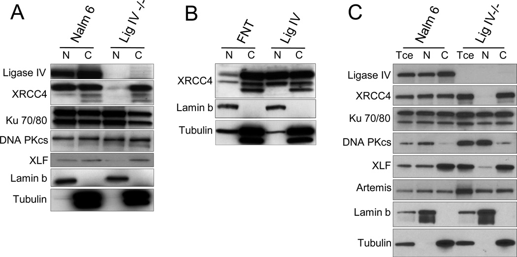
Figure 1. In the absence of Ligase IV, XRCC4 accumulates in the cytoplasm.
(A) Nuclear (N) and cytoplasmic (C) extracts from wild type human pre-B (Nalm 6) and N114P2 (Lig IV −/−) cells were prepared by differential fractionation using RSB buffer as described under Material and Methods. The subcellular localization of known factors involved in non-homologous end joining was analyzed by immunoblotting. Controls for the nuclear and cytoplasmic fractions are indicated, by lamin b and tubulin, respectively (in this and subsequent figures).
(B) Nuclear (N) and cytoplasmic (C) extracts from Lig IV −/− cells (N114P2) stably transfected with Flag tagged full length Ligase IV or empty vector FNT (Flag-NLS-thioredoxin) were prepared. Localization of XRCC4 was determined by immunoblotting.
(C) Nuclear (N) and cytoplasmic (C) extracts from wild type human pre-B (Nalm 6) and N114P2 (Lig IV −/−) cells were prepared by differential fractionation using the Dignam protocol as described under Material and Methods. The subcellular localization of known factors involved in non-homologous end joining was analyzed by immunoblotting. Total cell extract (Tce) was also analyzed in this experiment.
Flag Immunoprecipitation
Cell extracts were prepared in Buffer A (25 mM Tris-HCl pH 8.0, 150 mM KCl, 10% glycerol, 0.1% Triton X-100, 0.5 mM EDTA, 1 mM DTT) with protease inhibitors (HALT Protease Inhibitor Cocktail Mix (Fisher Scientific) and PMSF (Sigma) as described (28). Extracts were sonicated and incubated with 200 µg/ml Ethidium Bromide at 4°C for 30 minutes and centrifuged at 35,000 rpm for 45 minutes. The obtained supernatant was used for Flag IP with anti Flag M2-agarose beads (Sigma) and bound proteins were eluted with 0.2 mg/ml Flag peptide (Sigma) in Buffer C (25 mM Tris-HCl pH 8.0, 150 mM KCl, 20% glycerol). Elutions were used for western blott analysis.
Pulse chase
Was done as described (29). A brief description of the experimental approach is provided in the Supplementary information.
Immunofluorescence
Wild type human fibroblasts (NM-1 and HFF-1) and LIG4 Syndrome patient fibroblasts (GM16088 and GM17523B) were grown on coverslips. Cells were fixed and permeabilized for 10 minutes with cold 100% methanol at −20° C. Washes were carried out with 1X PBS + 0.1% Triton X-100 (twice, 10 minutes each) followed by 1X PBS wash (twice, 5 minutes each). Cells were blocked in 1X PBS + 10% goat serum (Sigma) overnight at 4° C and stained with an affinity purified anti XRCC4 (generated against full length XRCC4) diluted 1:500 in 1X PBS + 5% goat serum. Appropriate secondary antibodies were used. DNA was stained using Vectashield mounting media with DAPI.
Survival assay
Was done as described (30,31) with minor modifications detailed in the Supplementary information.
RESULTS
Absence of Ligase IV results in cytoplasmic accumulation of XRCC4, an effect that is not altered upon induction of DNA damage
Due to the reported defect in XRCC4 and XLF recruitment to chromatin in the absence of Ligase IV (14,15), we hypothesized that loss of Ligase IV would affect the nuclear localization of one or both of these core factors. To investigate our hypothesis, two human pre-B cell lines were utilized, Nalm 6 (WT) and the Ligase IV deficient line, N114P2 (Lig IV−/−) (22). Nuclear and cytoplasmic fractions were prepared from both WT and Lig IV−/− cells. These fractions were analyzed by immunoblot for nuclear and cytoplasmic levels of the known NHEJ factors including Ligase IV, XRCC4, XLF, DNA-PKcs, Artemis and the Ku70/80 heterodimer. In the absence of Ligase IV, XRCC4 was found only in the cytoplasm in contrast to WT cells, where it accumulates in both the nucleus and the cytoplasm (Fig. 1A). The nuclear localization of XRCC4 was recovered after expression of Ligase IV in Lig IV −/− cells (Fig. 1B). Additionally, in Lig IV−/− cells we observed a decrease in the nuclear accumulation of XLF (Fig. 1A and 1C). The hypotonic RSB buffer used to lysed the cells and prepare the nuclei in the experiments presented in Figures 1A and 1B contains 0.5% NP40. To rule out the possibility that this buffer might lead to the leakage of nuclear XRCC4 into the cytoplasm in the Ligase IV deficient cells, the Dignam method was utilized as an alternative approach for nuclei preparation. This protocol does not use detergent and was optimized for the isolation of transcription factors from the nuclear fraction (27). Importantly, using this alternative protocol, we continue to observe localization of XRCC4 in the cytoplasmic fraction as well as decreased recruitment of XLF to the nucleus in Ligase IV deficient cells (Fig. 1C). For subsequent experiments, the simpler approach involving the RSB buffer was utilized. In addition to changes in cellular distribution of XRCC4 and XLF in Ligase IV deficient cells and consistent with prior studies (15,32), we also observed a loss of interaction between XRCC4 and XLF in Lig IV −/− cells (Fig. S1A). Complementation with full length Ligase IV rescued the observed phenotypes (Fig. S1B).
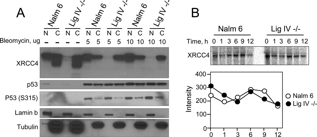
After DNA damage, NHEJ factors are known to rapidly localize to the nucleus and specifically to the site of the break (33–36). Since our initial studies on subcellular localization of NHEJ factors were performed in the absence of any overt DNA damage, we investigated whether inducing DNA damage, specifically DSBs, would alter the subcellular accumulation of XRCC4. To address this question, we treated WT and Lig IV −/− pre-B cells with the DNA damage inducing agent bleomycin. As a control, we included untreated cells (WT and Lig IV −/−). After 24 hours, the cells were fractionated into the nuclear and cytoplasmic fractions as done previously and the localization of XRCC4 was assessed using immunoblotting. We observed that XRCC4 in Lig IV −/− cells did not accumulate in the nucleus (Fig. 2A). However, in WT cells, there was a clear shift of XRCC4 from the cytoplasmic fraction to the nuclear fraction consistent with published data that have shown that these factors redistribute to the site of damage (14,15). To confirm induction of DNA damage in bleomycin treated cells, the levels of p53 and phosphorylated p53 were analyzed (37). Both p53 and phosphorylated p53 were induced by bleomycin treatment consistent with the induction of DNA damage and activation of the DNA damage response (Fig. 2A). To rule out the possibility that the absence of Ligase IV had an effect on the stability of XRCC4 that would account for the absence of its nuclear accumulation, we compared the stability of XRCC4 in WT and Lig IV −/− cells. As shown in Figure 2B, there was no change in the half-life of XRCC4 in Ligase IV −/− cells compared to WT when analyzed over a 12 hour time period.
Figure 2. DNA damage does not change the subcellular distribution of XRCC4 in Ligase IV deficient cells.
(A) Wild type Nalm 6 and Lig IV −/− cells were analyzed 24 hours after treatment with 0 µg, 5 µg or 10 µg/ml of Bleomycin. After treatment, cells were fractionated into nuclear (N) and cytoplasmic (C) extracts and localization of XRCC4 was determined by immunoblotting. Anti-p53 and anti-phospho-p53 (S315) were used as indicators of induction of DNA damage by Bleomycin treatment.
(B) XRCC4 showed comparable stability in wild type and Lig IV −/− cells. Nalm 6 and Lig IV −/− cells were incubated with 35S-methionine. Following labeling, cycloheximide at 10 ug/ml was added for the time periods indicated. Total cell extracts were immunoprecipitated with anti-XRCC4 antibody and labeled XRCC4 was detected by phosphorimaging. The lower graph represents the quantification of the bands shown in the immunoblot using ImageQuant software.
The C-terminal domain of Ligase IV is important for the subcellular localization of XRCC4
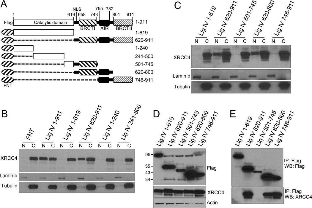
Ligase IV, at its N-terminus, contains the DNA binding domain (DBD), an adenylation domain that has the active site of the enzyme and an oligo-binding domain (OBD) (18). Interaction with the nuclease Artemis has been mapped to the Ligase IV DBD (20,38,39) and this interaction was shown to be important for V(D)J recombination (20). At its C-terminus, Ligase IV has two breast and ovarian cancer susceptibility protein C-terminus (BRCT) domains separated by a linker containing the XRCC4-interacting region (XIR) (40–42). Most recently, it was shown that at least one of the BRCT domains, BRCT II, is also required for efficient interaction of Ligase IV with XRCC4 (43). We hypothesized that these interactions not only stabilize the ligation complex, but are also involved in controlling the localization of these factors, in particular XRCC4, to the site of DNA damage by allowing localization and/or retention of XRCC4 into the nucleus. To determine whether the C-terminal region of Ligase IV played a role in nuclear accumulation of XRCC4, we tested a series of Ligase IV truncations, shown in Figure 3A, for their ability to support nuclear localization of XRCC4 when stably expressed in Lig IV −/− pre-B cells. Nuclear and cytoplasmic fractions were prepared from the stable cell lines and the localization of XRCC4 assessed by immunoblot. As previously demonstrated, expression of the full length Ligase IV resulted in the nuclear accumulation of XRCC4. In contrast, when we expressed the N-terminal domain of Ligase IV containing the DBD, AdD and OBD (amino acids (aa) 1–619), we found that the localization of XRCC4 was similar to that of Ligase IV deficient B cells, as it was found to accumulate primarily in the cytoplasm. This held true for the expression of constructs that contained aa 1–240 and 241–500. However, expression of the Ligase IV Cterminal, aa 620–911, was able to recover the accumulation of XRCC4 into the nucleus (Fig. 3B). This is consistent with the study that showed a role for the C terminal region of Ligase IV in directing the Ligase IV/XRCC4 complex to chromatin (16). Our finding and the recently published work suggest that both the BRCT domains, as well as the XIR of Ligase IV, are important in facilitating the localization of XRCC4 into the nucleus. To dissect this requirement, we generated and stably expressed shorter truncations of the Ligase IV C-terminus, which contained BRCT I (aa 501–745), BRCT I with the XIR (aa 620–800), and BRCT II with the XIR (aa 745–911) in the Lig IV −/− pre-B cells and determined whether any of these constructs would result in nuclear accumulation of XRCC4. We found that expression of a C-terminal fragment containing aa 620 to 800, which includes the BRCT I, the XIR, and a linker region between aa 620 and 659 containing the two in tandem NLS of Ligase IV (44), was sufficient to allow nuclear localization of XRCC4. We did not observe the same effect when we expressed the construct containing the XIR and BRCT II (aa 745–911). This result was unexpected, as it has been shown that the second BRCT domain of Ligase IV, in addition to the XIR, has an important contribution to maintaining the interaction between XRCC4 and Ligase IV. To further understand the relationship between the ability of the Ligase IV C-terminal fusion proteins to support the Ligase IV/XRCC4 interaction, as opposed to their ability to promote nuclear localization of XRCC4, all the fusion proteins presented in Figure 3C were immunoprecipitated from total cell extract and analyzed for their interaction with XRCC4 (Figs. 3D and E). Consistent with published data, we detected very efficient interaction of XRCC4 with the BRCT II and XIR fusion protein (746–911), as well as with the BRCTI and XIR fusion protein (620–800). However, from our localization studies, interaction of BRCTII-XIR fusion protein alone was not sufficient to enable accumulation of XRCC4 in the nucleus, even with an NLS, provided by the FNT (Fig. 3A). Our data suggest that a Ligase IV C terminal fragment, from aa 620 to 800, containing the BRCT I, XIR region and the Ligase IV NLS, has an important role in allowing nuclear localization of XRCC4. In addition, we also showed that interaction between XRCC4 and Ligase IV Cterminus is required but is not sufficient for nuclear localization of XRCC4. Interestingly, despite its ability to promote nuclear localization of XRCC4, expression of the Ligase IV C-terminal region (aa 620 to 911) had no effect on the survival defect of Ligase IV deficient cells (Supplementary Fig. 2). This result is consistent with published studies that demonstrate a requirement for Ligase IV core and C terminal region for in vivo plasmid repair (45).
Figure 3. The C-terminal domain of Ligase IV is critical for nuclear localization of XRCC4.
(A) Schematic representation of FNT (Flag-NLS (nuclear localization signal)-Thioredoxin) tagged Ligase IV deletions and Flag-tagged full length Ligase IV. The catalytic domain, NLS, BRCT I, XIR and BRCT II of Ligase IV are indicated.
(B) Lig IV −/− cells were transduced with a control (FNT) or the FNT tagged truncations of Ligase IV. After selection and expansion of the pools, nuclear (N) and cytoplasmic (C) extracts were prepared and localization of XRCC4 was determined by immunoblotting.
(C) Further mapping within the Ligase IV C-terminal domains indicates that the region of amino acids 620–800 is important for the nuclear localization of XRCC4.
(D) Expression of Ligase IV deletion-constructs were analyzed by immunoblotting of total cell extracts. Expression of XRCC4 and actin are also shown.
(E) Interaction of XRCC4 with Ligase IV C-terminal fragments was determined by Flag immunoprecipitation of Ligase IV truncations followed by western blot analysis for Flag-Ligase IV fusion proteins, using anti-Flag antibodies, and XRCC4 co-IP.
Decreased XRCC4 expression in human fibroblasts from LIG4 syndrome patients
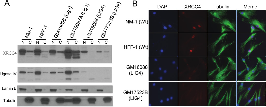
As we observed a change in the subcellular accumulation of XRCC4 in human pre-B cells, we wanted to determine whether this also applied to fibroblasts from LIG4 syndrome patients that carry hypomorphic mutations in Ligase IV. These mutations significantly reduced Ligase IV expression (12). To address this question, we obtained two untransformed patient fibroblast cell lines, GM16088, which has a R278H mutation and GM17523B, a C-terminal truncation mutant at R814X (24). While GM16088 has the mutation R278H lying within a highly conserved motif encompassing the active site of Ligase IV, the R814X mutation was of particular interest because of the loss of a portion of the Ligase IV C-terminal. This patient was considered Ligase IV deficient due to the significant impact of this truncation on protein expression (24). We fractionated these cells into nuclear and cytoplasmic fractions, and analyzed XRCC4 subcellular localization. The WT human fibroblast lines, NM-1 and HFF-1, as well as fibroblasts carrying mutations in the Ligase I gene, GM16096 and GM16097A (25), were included as controls. As depicted in Figure 4A, WT fibroblasts, as well as fibroblasts with mutations in Ligase I, showed that XRCC4 accumulated significantly in the nucleus. In contrast, primary human fibroblasts carrying hypomorphic Ligase IV mutations showed very low levels of XRCC4 (Figs. 4A and B). The large decreased in XRCC4 protein levels in Ligase IV mutant fibroblasts, as observed by western blot analysis and immunofluorescence (Fig. 4), suggests a strong regulatory role for Ligase IV on XRCC4 protein stability in this cellular system. It also highlights differences in the regulatory mechanism that controls NHEJ factors, which might be dependent on the cellular background. Early studies by Riballo et al., had shown a slight reduction in the level of XRCC4 in a Ligase IV mutant human fibroblasts (46), however, the decreased XRCC4 protein levels observed, by these investigators, was not nearly as pronounced as the decrease shown in Figure 4. An effect we observed even in the presence of protease inhibitors.
Figure 4. Hypomorphic LIG4 patient fibroblasts show decreased XRCC4 protein levels.
(A) Fibroblasts from LIG4 hypomorphs (GM16088 that carries an R278H mutation within the active site of Lig IV and GM17523B that carries a R814Ter mutation resulting in a C-terminal truncated protein); Ligase I hypomorphs (GM16096 and GM16097); wild type (WT) fibroblasts NM-1 and HFF-1 were fractionated into nuclear (N) and cytoplasmic (C) extracts. XRCC4 and Ligase IV localization was analyzed by immunoblotting.
(B) Immunofluorescence of XRCC4 in WT (NM-1 and HFF-1) and LIG4 hypomorphs (GM16088 and GM17523B) were analyzed. Nuclear staining with DAPI and cytoplasmic staining with tubulin are presented. The merged images are shown.
DISCUSSION
Ligase IV is essential for the final ligation of DSBs through the NHEJ pathway. Its co-factor, XRCC4, has been shown to be important for increasing its activity and stability. Interaction between these two factors have been mapped to the linker region between the two BRCT domains found in the Ligase IV C-terminal known as the XIR and a portion of the BRCT II, which is necessary to stabilize the interaction (41,43). In this work, we highlight a novel role for Ligase IV as an important regulator of the NHEJ reaction in controlling the nuclear localization of its co-factors XRCC4 and XLF. Ligase IV might facilitate efficient nuclear localization of XRCC4 by increasing its nuclear retention and/or by allowing its nuclear translocation. While future work will be required to establish the precise mechanism, our findings provide an explanation for the impaired recruitment of XRCC4 and XLF to chromatin that has been previously observed (14,15). We also highlight the role of Ligase IV BRCT I domain, along with the XIR, in the nuclear localization of XRCC4.
Our findings suggest that the Ligase IV/XRCC4 complex is formed in the cytoplasm and subsequently controls the organization of a repair complex within the nucleus. Structural and biochemical studies have revealed that together with XRCC4, XLF forms long filamentous structures that are important for the bridging of the two broken ends during ligation (47–49). Furthermore, DNA-PKcs serves to regulate this complex (50). Published work has shown that Ligase IV is essential for DNA-PKcs autophosphorylation and DNA end synapsis (17); these studies, together with our findings, suggest that Ligase IV has a critical, yet largely uncharacterized role in the assembly of the nuclear DNA repair complexes.
Two mechanisms have been suggested to control the nuclear localization of XRCC4, a nuclear localization signal present in XRCC4 (between aa 270 and 275) and the SUMO modification of the protein at lysine 210 (51,52). In this study, we show that Ligase IV allows nuclear localization of XRCC4 and in doing so, indirectly facilitates nuclear accumulation of XLF. Along with other NHEJ factors, such as DNA PKcs, this complex orchestrated by Ligase IV would promote repair via non-homologous end joining. Interestingly, in our cellular fractionation experiments, we observed a consistent difference in the migration pattern of the nuclear and cytoplasmic XRCC4 protein. Although earlier studies have demonstrated the phosphorylation and monoubiquitination of XRCC4, the role of these posttranslational modifications remains largely uncharacterized (21,53–56). We postulate that these modifications might also contribute to regulate the nuclear cytoplasmic distribution of XRCC4. How the various mechanisms that control nuclear localization and/or retention of XRCC4 in the nucleus work together to ensure efficient and regulated NHEJ will be the subject of future studies.
Human mutations in the Ligase IV gene have been identified in patients with LIG4 syndrome, as well as in patients with Dubowitz syndrome (12,13). These patients present a wide range of defects, including, high levels of chromosomal breaks, predisposition to tumor development and variable immune defects. The analysis of Ligase IV hypomorphs, presented in this manuscript, sheds light on the sensitivity to IR and defects in V(D)J recombination observed in patients with hypomorphic mutations within the Ligase IV gene. Our work shows that Ligase IV is not only critical for proper nuclear localization of XRCC4 but also appears to have a role in stabilizing XRCC4 in primary human fibroblasts. The difference in XRCC4 stability in human preB cells versus in human primary fibroblasts is still unclear but raises intriguing questions that deserve further investigation.
Our data directly implicate the C-terminal region of Ligase IV as important in facilitating the assembly of the Ligase IV/XRCC4/XLF complex and in allowing the accumulation of these factors in the nucleus, making them available to participate in DNA repair.
Supplementary Material
Highlights.
DNA Ligase IV controls the nuclear localization of XRCC4
XRCC4 localizes to the cytoplasm in DNA Ligase IV deficient lymphocytes
Decreased XRCC4 protein levels were observed in fibroblasts with Ligase IV mutations
ACKNOWLEDGEMENTS
We thank members of the Cortes laboratory for helpful discussions and reagents and Dr. Juan Carcamo for constructive suggestions and critical reading of the manuscript. We thank Dr. Michael R. Lieber for providing the Nalm 6 and N114P2 cells, Dr. Anna Villa for the control fibroblast NM-1. Dr. Tomas Lindahl and Dr. Michael Lieber provided the cDNA for human Ligase IV. The advice and equipment provided by the Mount Sinai Microscopy Shared Resource Facility were instrumental to perform the described studies. Work in the P.C. laboratory is supported by the National Institute of Health (R01 AI080755 and R01 AI070880 from NIH). D.B.F. was supported by pre-doctoral training grants given to the Immunology Institute by the National Institute of Health (T32-A1007605-09 and 5T32A1007605-10).
Footnotes
Publisher's Disclaimer: This is a PDF file of an unedited manuscript that has been accepted for publication. As a service to our customers we are providing this early version of the manuscript. The manuscript will undergo copyediting, typesetting, and review of the resulting proof before it is published in its final citable form. Please note that during the production process errors may be discovered which could affect the content, and all legal disclaimers that apply to the journal pertain.
Conflict of interest statement
The authors declare no conflict of interest.
References
- 1.Boboila C, Alt FW, Schwer B. Classical and alternative endjoining pathways for repair of lymphocyte-specific and general DNA double-strand breaks. Advances in immunology. 2012;116:1–49. doi: 10.1016/B978-0-12-394300-2.00001-6. [DOI] [PubMed] [Google Scholar]
- 2.Helmink BA, Sleckman BP. The response to and repair of RAG-mediated DNA double-strand breaks. Annual review of immunology. 2012;30:175–202. doi: 10.1146/annurev-immunol-030409-101320. [DOI] [PMC free article] [PubMed] [Google Scholar]
- 3.Lieber MR. The mechanism of double-strand DNA break repair by the nonhomologous DNA end-joining pathway. Annu Rev Biochem. 2010;79:181–211. doi: 10.1146/annurev.biochem.052308.093131. [DOI] [PMC free article] [PubMed] [Google Scholar]
- 4.Chapman JR, Taylor MR, Boulton SJ. Playing the end game: DNA double-strand break repair pathway choice. Mol Cell. 2012;47:497–510. doi: 10.1016/j.molcel.2012.07.029. [DOI] [PubMed] [Google Scholar]
- 5.Kass EM, Jasin M. Collaboration and competition between DNA double-strand break repair pathways. FEBS Lett. 2010;584:3703–3708. doi: 10.1016/j.febslet.2010.07.057. [DOI] [PMC free article] [PubMed] [Google Scholar]
- 6.Brandsma I, Gent DC. Pathway choice in DNA double strand break repair: observations of a balancing act. Genome Integr. 2012;3:9. doi: 10.1186/2041-9414-3-9. [DOI] [PMC free article] [PubMed] [Google Scholar]
- 7.Mahaney BL, Meek K, Lees-Miller SP. Repair of ionizing radiation-induced DNA double-strand breaks by non-homologous endjoining. Biochem J. 2009;417:639–650. doi: 10.1042/BJ20080413. [DOI] [PMC free article] [PubMed] [Google Scholar]
- 8.Malu S, Malshetty V, Francis D, Cortes P. Role of nonhomologous end joining in V(D)J recombination. Immunol Res. 2012;54:233–246. doi: 10.1007/s12026-012-8329-z. [DOI] [PubMed] [Google Scholar]
- 9.Frank KM, Sekiguchi JM, Seidl KJ, Swat W, Rathbun GA, Cheng HL, Davidson L, Kangaloo L, Alt FW. Late embryonic lethality and impaired V(D)J recombination in mice lacking DNA ligase IV. Nature. 1998;396:173–177. doi: 10.1038/24172. [DOI] [PubMed] [Google Scholar]
- 10.Revy P, Malivert L, de Villartay JP. Cernunnos-XLF, a recently identified non-homologous end-joining factor required for the development of the immune system. Current opinion in allergy and clinical immunology. 2006;6:416–420. doi: 10.1097/01.all.0000246623.72365.43. [DOI] [PubMed] [Google Scholar]
- 11.Gao Y, Sun Y, Frank KM, Dikkes P, Fujiwara Y, Seidl KJ, Sekiguchi JM, Rathbun GA, Swat W, Wang J, et al. A critical role for DNA end-joining proteins in both lymphogenesis and neurogenesis. Cell. 1998;95:891–902. doi: 10.1016/s0092-8674(00)81714-6. [DOI] [PubMed] [Google Scholar]
- 12.Chistiakov DA, Voronova NV, Chistiakov AP. Ligase IV syndrome. Eur J Med Genet. 2009;52:373–378. doi: 10.1016/j.ejmg.2009.05.009. [DOI] [PubMed] [Google Scholar]
- 13.Jayaram S, Ketner G, Adachi N, Hanakahi LA. Loss of DNA ligase IV prevents recognition of DNA by double-strand break repair proteins XRCC4 and XLF. Nucleic Acids Res. 2008;36:5773–5786. doi: 10.1093/nar/gkn552. [DOI] [PMC free article] [PubMed] [Google Scholar]
- 14.Drouet J, Delteil C, Lefrancois J, Concannon P, Salles B, Calsou P. DNA-dependent protein kinase and XRCC4-DNA ligase IV mobilization in the cell in response to DNA double strand breaks. J Biol Chem. 2005;280:7060–7069. doi: 10.1074/jbc.M410746200. [DOI] [PubMed] [Google Scholar]
- 15.Wu PY, Frit P, Malivert L, Revy P, Biard D, Salles B, Calsou P. Interplay between Cernunnos-XLF and nonhomologous endjoining proteins at DNA ends in the cell. Journal of Biological Chemistry. 2007;282:31937–31943. doi: 10.1074/jbc.M704554200. [DOI] [PubMed] [Google Scholar]
- 16.Liu S, Liu X, Kamdar RP, Wanotayan R, Sharma MK, Adachi N, Matsumoto Y. C-terminal region of DNA ligase IV drives XRCC4/DNA ligase IV complex to chromatin. Biochem Biophys Res Commun. 2013;439:173–178. doi: 10.1016/j.bbrc.2013.08.068. [DOI] [PubMed] [Google Scholar]
- 17.Cottarel J, Frit P, Bombarde O, Salles B, Negrel A, Bernard S, Jeggo PA, Lieber MR, Modesti M, Calsou P. A noncatalytic function of the ligation complex during nonhomologous end joining. The Journal of cell biology. 2013;200:173–186. doi: 10.1083/jcb.201203128. [DOI] [PMC free article] [PubMed] [Google Scholar]
- 18.Ellenberger T, Tomkinson AE. Eukaryotic DNA ligases: structural and functional insights. Annu Rev Biochem. 2008;77:313–338. doi: 10.1146/annurev.biochem.77.061306.123941. [DOI] [PMC free article] [PubMed] [Google Scholar]
- 19.Budman J, Kim SA, Chu G. Processing of DNA for nonhomologous end-joining is controlled by kinase activity and XRCC4/ligase IV. The Journal of biological chemistry. 2007;282:11950–11959. doi: 10.1074/jbc.M610058200. [DOI] [PubMed] [Google Scholar]
- 20.Malu S, De Ioannes P, Kozlov M, Greene M, Francis D, Hanna M, Pena J, Escalante CR, Kurosawa A, Erdjument-Bromage H, et al. Artemis C-terminal region facilitates V(D)J recombination through its interactions with DNA Ligase IV and DNA-PKcs. J Exp Med. 2012;209:955–963. doi: 10.1084/jem.20111437. [DOI] [PMC free article] [PubMed] [Google Scholar]
- 21.Koch CA, Agyei R, Galicia S, Metalnikov P, O'Donnell P, Starostine A, Weinfeld M, Durocher D. Xrcc4 physically links DNA end processing by polynucleotide kinase to DNA ligation by DNA ligase IV. EMBO J. 2004;23:3874–3885. doi: 10.1038/sj.emboj.7600375. [DOI] [PMC free article] [PubMed] [Google Scholar]
- 22.Grawunder U, Zimmer D, Fugmann S, Schwarz K, Lieber MR. DNA ligase IV is essential for V(D)J recombination and DNA double-strand break repair in human precursor lymphocytes. Mol Cell. 1998;2:477–484. doi: 10.1016/s1097-2765(00)80147-1. [DOI] [PubMed] [Google Scholar]
- 23.West KL, Singha NC, De Ioannes P, Lacomis L, Erdjument-Bromage H, Tempst P, Cortes P. A direct interaction between the RAG2 C terminus and the core histones is required for efficient V(D)J recombination. Immunity. 2005;23:203–212. doi: 10.1016/j.immuni.2005.07.004. [DOI] [PubMed] [Google Scholar]
- 24.O'Driscoll M, Cerosaletti KM, Girard PM, Dai Y, Stumm M, Kysela B, Hirsch B, Gennery A, Palmer SE, Seidel J, et al. DNA ligase IV mutations identified in patients exhibiting developmental delay and immunodeficiency. Molecular cell. 2001;8:1175–1185. doi: 10.1016/s1097-2765(01)00408-7. [DOI] [PubMed] [Google Scholar]
- 25.Webster AD, Barnes DE, Arlett CF, Lehmann AR, Lindahl T. Growth retardation and immunodeficiency in a patient with mutations in the DNA ligase I gene. Lancet. 1992;339:1508–1509. doi: 10.1016/0140-6736(92)91266-b. [DOI] [PubMed] [Google Scholar]
- 26.He KL, Ting AT. A20 inhibits tumor necrosis factor (TNF) alpha-induced apoptosis by disrupting recruitment of TRADD and RIP to the TNF receptor 1 complex in Jurkat T cells. Molecular and cellular biology. 2002;22:6034–6045. doi: 10.1128/MCB.22.17.6034-6045.2002. [DOI] [PMC free article] [PubMed] [Google Scholar]
- 27.Dignam JD, Lebovitz RM, Roeder RG. Accurate transcription initiation by RNA polymerase II in a soluble extract from isolated mammalian nuclei. Nucleic Acids Res. 1983;11:1475–1489. doi: 10.1093/nar/11.5.1475. [DOI] [PMC free article] [PubMed] [Google Scholar]
- 28.Ma Y, Pannicke U, Schwarz K, Lieber MR. Hairpin opening and overhang processing by an Artemis/DNA-dependent protein kinase complex in nonhomologous end joining and V(D)J recombination. Cell. 2002;108:781–794. doi: 10.1016/s0092-8674(02)00671-2. [DOI] [PubMed] [Google Scholar]
- 29.Couedel C, Roman C, Jones A, Vezzoni P, Villa A, Cortes P. Analysis of mutations from SCID and Omenn syndrome patients reveals the central role of the Rag2 PHD domain in regulating V(D)J recombination. J Clin Invest. 2010;120:1337–1344. doi: 10.1172/JCI41305. [DOI] [PMC free article] [PubMed] [Google Scholar]
- 30.Uckun FM, Mitchell JB, Obuz V, Park CH, Waddick K, Friedman N, Oubaha L, Min WS, Song CW. Radiation sensitivity of human B-lineage lymphoid precursor cells. Int J Radiat Oncol Biol Phys. 1991;21:1553–1560. doi: 10.1016/0360-3016(91)90332-x. [DOI] [PubMed] [Google Scholar]
- 31.Holt SM, Scemama JL, Panayiotidis MI, Georgakilas AG. Compromised repair of clustered DNA damage in the human acute lymphoblastic leukemia MSH2-deficient NALM-6 cells. Mutat Res. 2009;674:123–130. doi: 10.1016/j.mrgentox.2008.09.014. [DOI] [PubMed] [Google Scholar]
- 32.Malivert L, Callebaut I, Rivera-Munoz P, Fischer A, Mornon JP, Revy P, de Villartay JP. The C-terminal domain of Cernunnos/XLF is dispensable for DNA repair in vivo. Molecular and cellular biology. 2009;29:1116–1122. doi: 10.1128/MCB.01521-08. [DOI] [PMC free article] [PubMed] [Google Scholar]
- 33.Koike M, Yutoku Y, Koike A. Accumulation of Ku70 at DNA double-strand breaks in living epithelial cells. Experimental cell research. 2011;317:2429–2437. doi: 10.1016/j.yexcr.2011.07.018. [DOI] [PubMed] [Google Scholar]
- 34.Reynolds P, Anderson JA, Harper JV, Hill MA, Botchway SW, Parker AW, O'Neill P. The dynamics of Ku70/80 and DNAPKcs at DSBs induced by ionizing radiation is dependent on the complexity of damage. Nucleic acids research. 2012;40:10821–10831. doi: 10.1093/nar/gks879. [DOI] [PMC free article] [PubMed] [Google Scholar]
- 35.Kim JS, Krasieva TB, Kurumizaka H, Chen DJ, Taylor AM, Yokomori K. Independent and sequential recruitment of NHEJ and HR factors to DNA damage sites in mammalian cells. The Journal of cell biology. 2005;170:341–347. doi: 10.1083/jcb.200411083. [DOI] [PMC free article] [PubMed] [Google Scholar]
- 36.Mari PO, Florea BI, Persengiev SP, Verkaik NS, Bruggenwirth HT, Modesti M, Giglia-Mari G, Bezstarosti K, Demmers JA, Luider TM, et al. Dynamic assembly of end-joining complexes requires interaction between Ku70/80 and XRCC4. Proceedings of the National Academy of Sciences of the United States of America. 2006;103:18597–18602. doi: 10.1073/pnas.0609061103. [DOI] [PMC free article] [PubMed] [Google Scholar]
- 37.Blaydes JP, Luciani MG, Pospisilova S, Ball HM, Vojtesek B, Hupp TR. Stoichiometric phosphorylation of human p53 at Ser315 stimulates p53-dependent transcription. The Journal of biological chemistry. 2001;276:4699–4708. doi: 10.1074/jbc.M003485200. [DOI] [PubMed] [Google Scholar]
- 38.De Ioannes P, Malu S, Cortes P, Aggarwal AK. Structural basis of DNA ligase IV-Artemis interaction in nonhomologous end-joining. Cell reports. 2012;2:1505–1512. doi: 10.1016/j.celrep.2012.11.004. [DOI] [PMC free article] [PubMed] [Google Scholar]
- 39.Ochi T, Gu X, Blundell TL. Structure of the Catalytic Region of DNA Ligase IV in Complex with an Artemis Fragment Sheds Light on Double-Strand Break Repair. Structure. 2013;21:672–679. doi: 10.1016/j.str.2013.02.014. [DOI] [PMC free article] [PubMed] [Google Scholar]
- 40.Grawunder U, Zimmer D, Kulesza P, Lieber MR. Requirement for an interaction of XRCC4 with DNA ligase IV for wild-type V(D)J recombination and DNA double-strand break repair in vivo. J Biol Chem. 1998;273:24708–24714. doi: 10.1074/jbc.273.38.24708. [DOI] [PubMed] [Google Scholar]
- 41.Grawunder U, Zimmer D, Leiber MR. DNA ligase IV binds to XRCC4 via a motif located between rather than within its BRCT domains. Curr Biol. 1998;8:873–876. doi: 10.1016/s0960-9822(07)00349-1. [DOI] [PubMed] [Google Scholar]
- 42.Critchlow SE, Bowater RP, Jackson SP. Mammalian DNA double-strand break repair protein XRCC4 interacts with DNA ligase IV. Curr Biol. 1997;7:588–598. doi: 10.1016/s0960-9822(06)00258-2. [DOI] [PubMed] [Google Scholar]
- 43.Wu PY, Frit P, Meesala S, Dauvillier S, Modesti M, Andres SN, Huang Y, Sekiguchi J, Calsou P, Salles B, et al. Structural and functional interaction between the human DNA repair proteins DNA ligase IV and XRCC4. Mol Cell Biol. 2009;29:3163–3172. doi: 10.1128/MCB.01895-08. [DOI] [PMC free article] [PubMed] [Google Scholar]
- 44.Girard PM, Kysela B, Harer CJ, Doherty AJ, Jeggo PA. Analysis of DNA ligase IV mutations found in LIG4 syndrome patients: the impact of two linked polymorphisms. Human molecular genetics. 2004;13:2369–2376. doi: 10.1093/hmg/ddh274. [DOI] [PubMed] [Google Scholar]
- 45.Teo SH, Jackson SP. Lif1p targets the DNA ligase Lig4p to sites of DNA double-strand breaks. Curr Biol. 2000;10:165–168. doi: 10.1016/s0960-9822(00)00317-1. [DOI] [PubMed] [Google Scholar]
- 46.Riballo E, Critchlow SE, Teo SH, Doherty AJ, Priestley A, Broughton B, Kysela B, Beamish H, Plowman N, Arlett CF, et al. Identification of a defect in DNA ligase IV in a radiosensitive leukaemia patient. Curr Biol. 1999;9:699–702. doi: 10.1016/s0960-9822(99)80311-x. [DOI] [PubMed] [Google Scholar]
- 47.Hammel M, Rey M, Yu Y, Mani RS, Classen S, Liu M, Pique ME, Fang S, Mahaney BL, Weinfeld M, et al. XRCC4 protein interactions with XRCC4-like factor (XLF) create an extended grooved scaffold for DNA ligation and double strand break repair. The Journal of biological chemistry. 2011;286:32638–32650. doi: 10.1074/jbc.M111.272641. [DOI] [PMC free article] [PubMed] [Google Scholar]
- 48.Hammel M, Yu Y, Fang S, Lees-Miller SP, Tainer JA. XLF regulates filament architecture of the XRCC4.ligase IV complex. Structure. 2010;18:1431–1442. doi: 10.1016/j.str.2010.09.009. [DOI] [PMC free article] [PubMed] [Google Scholar]
- 49.Ropars V, Drevet P, Legrand P, Baconnais S, Amram J, Faure G, Marquez JA, Pietrement O, Guerois R, Callebaut I, et al. Structural characterization of filaments formed by human Xrcc4-Cernunnos/XLF complex involved in nonhomologous DNA end-joining. Proceedings of the National Academy of Sciences of the United States of America. 2011;108:12663–12668. doi: 10.1073/pnas.1100758108. [DOI] [PMC free article] [PubMed] [Google Scholar]
- 50.Roy S, Andres SN, Vergnes A, Neal JA, Xu Y, Yu Y, Lees-Miller SP, Junop M, Modesti M, Meek K. XRCC4's interaction with XLF is required for coding (but not signal) end joining. Nucleic Acids Res. 2012;40:1684–1694. doi: 10.1093/nar/gkr1315. [DOI] [PMC free article] [PubMed] [Google Scholar]
- 51.Yurchenko V, Xue Z, Sadofsky MJ. SUMO modification of human XRCC4 regulates its localization and function in DNA doublestrand break repair. Mol Cell Biol. 2006;26:1786–1794. doi: 10.1128/MCB.26.5.1786-1794.2006. [DOI] [PMC free article] [PubMed] [Google Scholar]
- 52.Li Z, Otevrel T, Gao Y, Cheng HL, Seed B, Stamato TD, Taccioli GE, Alt FW. The XRCC4 gene encodes a novel protein involved in DNA double-strand break repair and V(D)J recombination. Cell. 1995;83:1079–1089. doi: 10.1016/0092-8674(95)90135-3. [DOI] [PubMed] [Google Scholar]
- 53.Yu Y, Wang W, Ding Q, Ye R, Chen D, Merkle D, Schriemer D, Meek K, Lees-Miller SP. DNA-PK phosphorylation sites in XRCC4 are not required for survival after radiation or for V(D)J recombination. DNA Repair (Amst) 2003;2:1239–1252. doi: 10.1016/s1568-7864(03)00143-5. [DOI] [PubMed] [Google Scholar]
- 54.Lee KJ, Jovanovic M, Udayakumar D, Bladen CL, Dynan WS. Identification of DNA-PKcs phosphorylation sites in XRCC4 and effects of mutations at these sites on DNA end joining in a cell-free system. DNA Repair (Amst) 2004;3:267–276. doi: 10.1016/j.dnarep.2003.11.005. [DOI] [PubMed] [Google Scholar]
- 55.Mahaney BL, Hammel M, Meek K, Tainer JA, Lees-Miller SP. XRCC4 and XLF form long helical protein filaments suitable for DNA end protection and alignment to facilitate DNA double strand break repair. Biochem Cell Biol. 2013;91:31–41. doi: 10.1139/bcb-2012-0058. [DOI] [PMC free article] [PubMed] [Google Scholar]
- 56.Foster RE, Nnakwe C, Woo L, Frank KM. Monoubiquitination of the nonhomologous end joining protein XRCC4. Biochem Biophys Res Commun. 2006;341:175–183. doi: 10.1016/j.bbrc.2005.12.166. [DOI] [PubMed] [Google Scholar]
Associated Data
This section collects any data citations, data availability statements, or supplementary materials included in this article.


