Abstract
Background:
The prevalence of hip dysplasia is 1 in 1000. Several pelvic osteotomy methods have been developed to prevent early osteoarthritis, such as triple osteotomy. In this study we are going to introduce our new technique that was done on 4 patients with favorable short-term results.
Methods:
Four patients underwent triple osteotomy and fixation using a reconstruction plate and early weight bearing was started.
Results:
The Harris Hip Score, limb length, center-edge angle, and acetabular inclination showed improvement.
Conclusion:
This modified technique is suggested for corrective surgery on adult dysplastic hips.
Keywords: Triple pelvic, Osteotomy, Hip hypoplasia.
Introduction
Hip dysplasia, with a prevalence of 1:1000 (1), is a debilitating disorder characterized by poor acetabular development and shallow acetabulum with an abnormal slop (2-4). Moreover, there is a positive correlation between severity of dysplasia and osteoarthritis development (5). If it is treated as soon as it is diagnosed, especially in the first 6 weeks after birth, the affected hip can be remodeled into a normal one (6). Otherwise, it will result in an abnormal weight bearing surface of acetabulum, leading to early cartilage degeneration and secondary osteoarthritis (3, 4).
Hip dysplasia in young adults is described by the malpositioning of the proximal femur due to deficient acetabular coverage (7). Several pelvic osteotomy methods have been developed to prevent early osteoarthritis, such as triple osteotomy, periacetabular osteotomy and spherical osteotomy (8-11). More simple modalities are suitable for patients with open triradiate cartilage and more complicated techniques such as steel triple osteotomy are suitable for more severe dysplasia in adults (12).
In this study, we are going to introduce our modified technique on 4 patients with favorable short-term results.
Materials and Methods
Between 2009 and 2012 ,4 female patients were admitted to our hospital. Their chief complaint was pain and claudication. Standard pelvic radiography showed hip dysplasia in all 4 patients. With consideration to their age and range of motion, we planned a triple osteotomy plus internal fixation and early motion.
One of the patients had a bilateral hypoplasia;on the left side we used the classic method of triple osteotomy and spica cast and on the right hip we used the modified technique of osteotomy plus internal fixation and early motion.
Initially, musculoskeletal and teratological disorders were ruled out at first. All of the patients were suffering from pain for more than 6 months. Two patients received an osteotomy on their right side and the other two received an osteotomy on their left side.
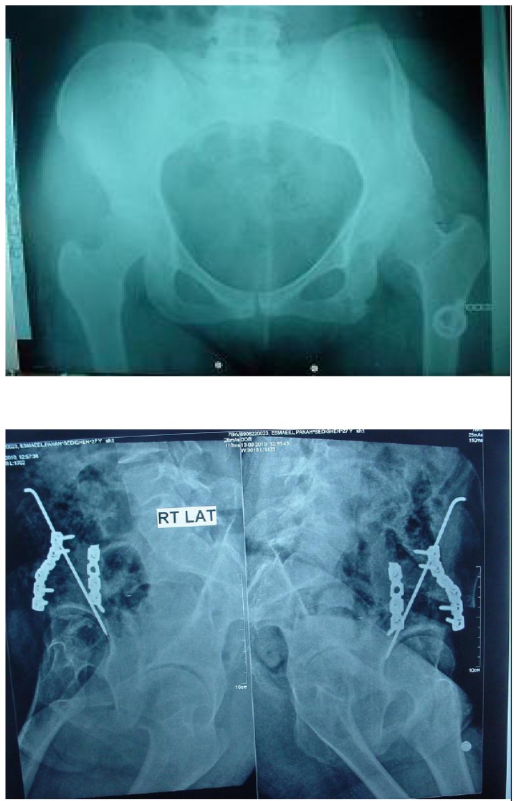
Our indications for surgery were the center edge angle of less than 20 degrees, acetabular inclination (Tonnis angle) of more than 30 degrees, coverage of less than 70% of the head, and painful hip. Harris Hip score was filled out to evaluate their pre-operative and 6 months post-operative status as well as pre- and post operative limb length measuring. (Table 1; Figure 1a, b)
Table 1.
Operated side and chief complaint of the patients
| Age | Operated side | Chief complaint | |
|---|---|---|---|
| Patient 1 | 23 | Right | Pain |
| Patient 2 | 29 | Right | Pain |
| Patient 3 | 27 | Left | Pain+ LLD |
| Patient 4 | 26 | Left | Pain+ LLD |
| Limb length discrepancy | |||
Figure 1a, b.
a pre- and b post operative radiography of the modified triple osteotomy.
Operation technique
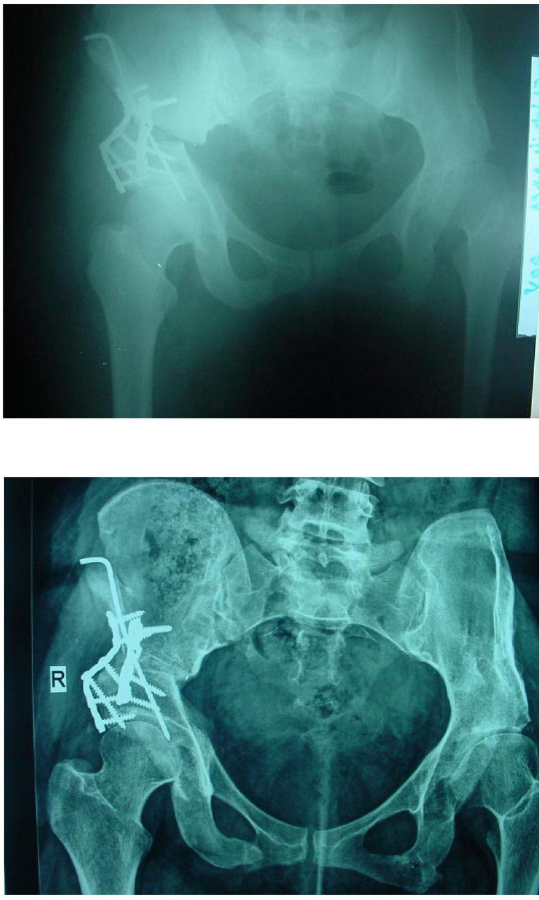
This operation was performed in the supine position with the iliofemoral (Smith-Peterson) approach. After performing osteotomies of the superior pubic ramus, ischium, supra-acetabular iliac and posterior column, acetabulum was rotated by a Schanz pin as a handle to achieve proper coverage for the head in both the anteroposterior and false-profile views. The coverage was desirable if the sourcil was roughly horizontal, the femoral head was covered properly and the posterior acetabular wall was on the center of the femoral head. Moreover, we checked the range of motion. Finally, the osteotomy sites were fixed by two reconstruction plates and screws on the anterior and posterior columns by inserting a three cortical and three angular bone graft removed from the iliac bone, between the osteotomized supra acetabular bone and the surgical plans were repaired as anatomical (Figure 2a, b).
Figure 2a, b.
a A 26-year-old female after triple pelvic osteotomy. b. union acheived and acetabular index improved after operation.
After the operation no immobilization was needed and the patients were able to start walking with crutches and bear weight on the operated limb by toe touching after 48 h postoperatively. In all of the patients we administered cephalexin for infection prophylaxis 24 h and enoxaparin for DVT prophylaxis for 2 weeks postoperatively. All of the patients were discharged 3 days postoperatively with no complications. The patients were allowed to bear full weight 10-12 weeks postoperatively with the union of their osteotomies. Additionally, the Harris Hip Score was assessed preoperatively and 6 months postoperatively.
Results
Demographic data of the 4 patients is shown in Table 1. Comparison of pre- and postoperative measurements are shown in Table 2.
Table 2.
pre- and post-operative measurements
| Patient 1 | Patient 2 | Patient 3 | Patient4 | ||
|---|---|---|---|---|---|
| Limb length increase | 10 mm | 10 mm | 7 mm | 12 mm | |
| Center edge | pre-operative | 0 | 12 | 11 | 50 |
| post-operative | 20 | 45 | 45 | 40 | |
| Acetabular index | pre-operative | 35 | 30 | 45 | 40 |
| post-operative | 20 | 5 | 10 | 10 | |
| Harris Hip Score | pre-operative | 74 | 68 | 81 | 74 |
| post-operative | 89 | 83 | 93 | 90 | |
Discussion
Since this technique does not need any postoperative cast, early weight bearing is possible; thus we are preventing muscular atrophy and facilitating patient rehabilitation.. Also, the increase in limb length will be assumed favorable considering the preoperative shortening of the involved limbs.
Hip dysplasia involves a wide age spectrum and clinical presentation varies from complete dislocation or subluxation in a new born to just an acetabular hypoplasia in older patients (13). Usually, the femoral head is covered insufficiently on the anterior and superior-lateral side, thus any excessive pressure burdened on the femoral head concentrates on a small contact surface. The uncovered portion increases the contact stress and leads to cartilage damage and osteoarthritis (14). The prevalence of secondary osteoarthritis due to hip dysplasia has been reported to be 25 % to 58 % (3, 4). Before the presentation of other osteoarthritis symptoms, the patients might suffer from mild to moderate pain related to fatigue or acetabular labrum damage (15). In order to prevent the degenerative process, different surgical techniques were introduced to keep the acetabular cavity near to normal depending on the patients’ age and stage of osteoarthritis. Surgical intervention for hip hypoplasia in young patients with no signs of cartilage degeneration includes different types of osteotomies to enlarge the weight bearing surface of the acetabulum and afford better femoral head coverage (7).
The best zone for correction of the deformity is the periacetabular region (15). Triple osteotomy was introduced in 1973 for the first time and several studies showed that it is a successful technique for adult hip dysplasia (6-8). Kleuver reported improvement in the general condition of patients after triple osteotomy in 39 out of 48 (81%) patients in a 5-year follow-up study (7). In a 7-year followup study, Faciszewski reported favorable outcomes after triple osteotomy in 53 out of 56 (94%) patients (16). This rate was reported in 10 out of 11 in Guille`s study after a 12-year follow up (17). Temporary complications consists of transient sciatic nerve palsy, paresthesia in the lateral femoral cutaneous nerve region, deep vein thrombosis, infection, and nonunion (7). In the original technique, pin fixation was proposed to be done at the sites of the osteotomy and the spica cast was applied at the end of surgery (8). In our modified technique, we did not apply the spica cast for any of the 4 patients since the osteotomy sites were fixed rigidly by a plate.
Therefore, in our patients no complications were reported related to spica casting, such as perineal hygiene problems, ileus due to loss of motility , and skin necrosis. Besides, there was no need to administer anticoagulant agents for a long period. The patients did not report temporary complications either. Rigid fixation at the osteotomy sites can lead to earlier weight bearing and thus early union can occur.
According to pre- and postoperative radiography, the possibility of early weight-bearing and higher patient satisfaction is obvious. We suggest this modified technique, which uses reconstruction plates as an alternative method, for the management of adult patients with hip dysplasia
References
- 1.Sankar WN, Weiss J, Skaggs DL. Orthopaedic conditions in the newborn. J Am Acad Orthop Surg. 2009; 17(2 ):112–22. doi: 10.5435/00124635-200902000-00007. [DOI] [PubMed] [Google Scholar]
- 2.Cooperman DR, Wallensten R, Stulberg SD. Acetabular dysplasia in the adult. Clin Orthop Relat Res. 1983. pp. 79–85. [PubMed]
- 3.Wedge JH, Wasylenko MJ. The natural history of congenital disease of the hip. J Bone Joint Surg Br. 1979; 61-B(3 ):334–8. doi: 10.1302/0301-620X.61B3.158025. [DOI] [PubMed] [Google Scholar]
- 4.Wiberg G. Studies on dysplastic acetabulum and congenital subluxation of the hip joint with special reference to the complications of osteoarthritis. Acta Chir Scand Suppl. 1939;83:1–135. [Google Scholar]
- 5.Harris NH. Acetabular growth potential in congenital dislocation of the hip and some factors upon which it may depend. Clin Orthop Relat Res. 1976. pp. 99–106. [PubMed]
- 6.Hailer NP, Soykaner L, Ackermann H, Rittmeister M. Triple osteotomy of the Pelvis for acetabular dysplasia. J Bone Joint Surg Am. 2005;87(12):1622–26. doi: 10.1302/0301-620X.87B12.15482. [DOI] [PubMed] [Google Scholar]
- 7.Kleuver M, Kooijman MAP, Pavlov PW, Veth RPH. Triple osteotomy of the pelvis for acetabular dysplasia. J Bone Joint Surg. 1997 Mar;79(2 ):225–9. doi: 10.1302/0301-620x.79b2.7167. [DOI] [PubMed] [Google Scholar]
- 8.Steel HH. Triple osteotomy of the innominate bone. J Bone Joint Surg Am. 1973;55(2 ):343–50. [PubMed] [Google Scholar]
- 9.Ganz R, Klaue K, Vinh TS, Mast JW. A new periacetabular osteotomy for the treatment of hip dysplasias: technique and preliminary results. Clin Orthop. 2004;418:3–8. [PubMed] [Google Scholar]
- 10.Schramm M, Hohmann D, Radespiel-Troger M, Pitto RP. The wagner spherical osteotomy of the acetabulum.sergical technique. J Bone Joint Surg Am. 2004 Mar;86-A (Suppl 1 ):73–80. doi: 10.2106/00004623-200403001-00010. [DOI] [PubMed] [Google Scholar]
- 11.Ninomiya S, Tagawa H. Rotational acetabular osteotomy for the dysplastic hip. J Bone Joint Surg Am. 1984;66:430–436. [PubMed] [Google Scholar]
- 12.Steel HH. Triple osteotomy of the innominate bone. A procedure to accomplish coverage of the dislocated or subluxated femoral head in the older patient. Clin Orthop Relat Res. 1977. pp. 116–27. [PubMed]
- 13.Shlens M. congenital hip dysplasia. Calif Med. 1973 Mar;118(3 ):33. [PMC free article] [PubMed] [Google Scholar]
- 14.Hadley NA, Brown TD, Weinstein SL. The effect of contact pressure elevation and aseptic necrosis on the long-term outcome of congenital hip dislocation. J Orthop Res. 1990;8:504–13. doi: 10.1002/jor.1100080406. [DOI] [PubMed] [Google Scholar]
- 15.Millis MB, Kim YJ. Rationale of osteotomy and related procedures for hip preservation: a review. Clin Orthop Relat Res. 2002;405:108–21. doi: 10.1097/00003086-200212000-00013. [DOI] [PubMed] [Google Scholar]
- 16.Faciszewski T, Coleman SS, Biddulph G. Triple innominate osteotomy for acetabular dysplasia. J Pediatric Orthop. 1993;13(4 ):426–30. doi: 10.1097/01241398-199307000-00002. [DOI] [PubMed] [Google Scholar]
- 17.Guille J T, Forlin E, Kumar SJ, Mac-Even GD. Triple osteotomy of the innominate bone in treatment of developemental dysplasia of the hip. J Pediatric Orthop. 1992;12 (6 ):718–21. doi: 10.1097/01241398-199211000-00003. [DOI] [PubMed] [Google Scholar]


