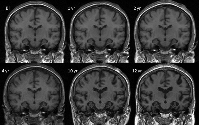
Figure 1.

Mid-temporal axial volumetric T1-weighted MR images acquired at initial presentation and subsequent repeat visits (1-, 2-, 4-, 10-, and 12-year follow-up). All repeat images have undergone 12 degrees of freedom registration to spatially align them to the baseline (Bl).
