Abstract
DIVA (differentiating infected from vaccinated animals) vaccines have proven extremely useful for control and eradication of infectious diseases in livestock. We describe here the characterization of a serologic marker epitope, so-called epitope-M201, which can be a potential target for development of a live-attenuated DIVA vaccine against porcine reproductive and respiratory syndrome virus (PRRSV). Epitope-M201 is located at the carboxyl terminus (residues 161-174) of the viral M protein. The epitope is highly immunodominant and well-conserved among type-II PRRSV isolates. Rabbit polyclonal antibodies prepared against this epitope are non-neutralizing; thus, the epitope does not seem to contribute to the protective immunity against PRRSV infection. Importantly, the immunogenicity of epitope-M201 can be disrupted through the introduction of a single amino acid mutation which does not adversely affect the viral replication. All together, our results provide an important starting point for the development of a live-attenuated DIVA vaccine against type-II PRRSV.
Keywords: PRRSV, M protein, Serological marker epitope, DIVA vaccines
1. Introduction
Porcine reproductive and respiratory syndrome (PRRS) was first reported in the U.S. in late 1980s [1]. Since then, the disease has been widespread in most swine-producing countries worldwide, causing substantial economic losses to the swine producers. Typical clinical signs associated with the disease include late-term reproductive failure in pregnant sows and respiratory distress in young pigs [2]. Recently, a highly pathogenic form of PRRS (HP-PRRS) has emerged in Asia, causing death in pigs of all ages with the mortality up to 100% [3]. The etiologic agent of PRRS is an RNA virus that belongs to the family Arteriviridae, the order Nidovirales, and is referred to as PRRS virus (PRRSV) [4–6]. The PRRSV genome is a linear, positive sense and single stranded RNA molecule of about 15 kb in length, which is flanked by a methyl-cap and a poly (A) tail at its 5′ and 3′ end, respectively [7–9]. It encodes a total of 14 non-structural and 8 structural proteins [7–9], none of which are known to be dispensable for the viral replication cycle.
Vaccines for protection against PRRSV infection have been commercially available since 1994. Two types of PRRSV vaccines are licensed for clinical applications including inactivated and live-attenuated vaccines. No subunit PRRSV vaccines are available at the present. It has been demonstrated that live-attenuated vaccines are much more effective than inactivated vaccines [10,11]. Under field conditions, the use of live-attenuated PRRSV vaccines improves pigs′ performance but co-circulation of field virus and vaccine virus has been reported to occur in the vaccinated herds [12,13]. Under experimental conditions, current live-attenuated vaccines protect the vaccinated pigs from development of clinical disease but they often fail to prevent infection and shedding of the challenge viruses [11,14–16]. One major limitation of the current live-attenuated PRRSV vaccines is that they do not allow serological discrimination between naturally infected and vaccinated animals, a property designated as DIVA (differentiating infected from vaccinated animals).
DIVA vaccines are vaccines that lack at least one antigenic component (so-called serologic marker antigen) when compared to the corresponding wild-type viruses [17]. Therefore, only wild-type virus infected, but not vaccinated, animals will develop antibodies to the marker antigen. Consequently, serological assays that detect antibodies specific to the marker antigen can be used to identify wild-type virus infected animals in the vaccinated population. The use of DIVA vaccines is preferred or even mandatory in animal health campaigns aiming toward control and eradication of important animal diseases [17–19]. Classically, live-attenuated DIVA vaccines are generated through deletion of an entire gene encoding an immunogenic, non-essential protein [17]. While technically straightforward in the case of some double-stranded DNA viruses like Pseudorabies Virus (Suid Herpesvirus 1), it is very difficult to delete an entire gene of a smaller RNA genome virus like PRRSV, whose genes are all essential for the productive viral infection [20–23]. An alternative approach to develop live-attenuated DIVA vaccines for RNA viruses is to selectively eliminate a small protein fragment or an epitope, instead of deleting the whole protein [24–26]. Previously, we identified several immunodominant B-cell epitopes located in non-structural protein 2 (nsp2) and in different structural proteins of a type-II PRRSV strain FL12 [27]. In the present study, we provide detailed characterization of a conserved and immunodominant epitope located at the carboxyl-terminus of PRRSV M protein and propose to use this epitope as a serologic marker for development of live-attenuated DIVA vaccines against type-II PRRSV.
2. Materials and methods
2.1. Antibodies and viruses
A hybridoma cell line expressing monoclonal antibody (MAb) specific to the peptide-M201 (161AVKQGVVRLVKYAK174) [27] was produced through a contract with the Immunological Resource Center (Urbana, IL). The peptide was conjugated with Keyhole Lipet Hemocyanin (KLH) and used to immunized mice. After the hybridoma cell line was achieved, ascites fluid production and antibody purification and HRP-conjugation were done through a contract with GenScript USA Inc. (Piscataway, NJ). MAb SDOW-17 specific for PRRSV nucleocapsid protein was purchased from National Veterinary Services Laboratories (Ames, IA). PRRSV negative and positive control serum samples were collected from different experiments previously conducted in our laboratory [28,29]. These included sera from 36 pigs that had been infected with PRRSV strain FL12 or its derivative mutants and 28 pigs that had been infected with PRRSV strain PRRSV-01 or its derivative mutants. These mutant viruses carry different N-linked glycosylation sites in their GP3 and GP5 but they all have intact M protein sequence [28,29]. The PRRSV vaccine strain Prime Pac has been obtained from a commercial batch of that vaccine received in our laboratory in 1997 and maintained through several passages in MARC-145 cells [30]. The collection of 82 type-II PRRSV field-isolates used in this study were collected from Midwestern states (NE, IA and IL) of the U.S.
2.2. Plasmid construction
For alanine-scanning mutagenesis, PRRSV ORF6 carrying alanine substitutions within epitope-M201 region was prepared by site-directed mutagenesis using synthesized mutagenic primers (Table 1). The resulting PCR products were cloned into the pIHA plasmid [31] at the XhoI and NotI restriction enzyme sites. The megaprimer PCR method [32] was used to prepare mutant pFL12 plasmids that contain mutations in the epitope-M201 region of the PRRSV strain FL12 [33].
Table 1. Primers used in this study.
| Primers | Nucleotide sequence (5′ → 3′) |
|---|---|
| For construction of pIHA plasmids encoding wild-type or mutant ORF6 | |
| ORF6For | ATATATCTCGAGGGGTCGTCTTTAGACGACTTTTG |
| ORF6Rev | ATATATGCGGCCGCTTATTTGGCATATTTGACAAG |
| Mut-1Rev | ATATATGCGGCCGCTTATTTGGCATATTTGACAAGGTTTACCACTCCTGCTGCTGCAGCTTTTCTGCCA |
| Mut-2Rev | ATATATGCGGCCGCTTATTTGGCATATTTGACAAGGTTTGCTGCTGCCTGTTTAACAGCTTTTC |
| Mut-3Rev | ATATATGCGGCCGCTTATTTGGCATATTTGGCAGCGGCTACCACTCCCTGTTTAAC |
| Mut-4Rev | ATATATGCGGCCGCTTAAGCGGCAGCAGCGACAAGGTTTACCACTCC |
| Mut-5Rev | ATATATGCGGCCGCTTATTTGGCATATTTGACAAGGTTTACCACTCCCGCAGCAACAGCTTTTCTGCC |
| Mut-6Rev | ATATATGCGGCCGCTTAGGCGGCATAGGCGACAAGGGCTACCACTCCCTGTTTAAC |
| For construction of full-length pFL12 plasmids containing different mutations in epitope-M201 | |
| Mut-1For | TGGCAGAAAAGCTGCAGCAGCAGGAGTGGTAAACCTTG |
| Mut-2For | GAAAAGCTGTTAAACAGGCAGCAGCAAACCTTGTCAAATATG |
| Mut-3For | AAAGCTGTTAAACAGGGAGTGGTAGCCGCTGCCAAATATGCCAAATAACAAC |
| Mut-5For | GTTGGGTGGCAGAAAAGCTGTTGCTGCTGGAGTGGTAAACCTTG |
| Mut-6For | CAGGGAGTGGTAGCCCTTGTCGCCTATGCCGCCTAACAACGGCAAGCAGCA |
| N168RFor | CAGGGAGTGGTAAGGCTTGTCAAATATGCC |
| Q164RFor | GTGTTGGGTGGCAAGCGCGCTGTTAAAAGAGGAGTGGTAAAC |
| 11691For | GCAACAGAAGAGTTGTCGGGTCC |
| A70Rev | CACACTTAATTAACGT(70)AATTTCGGCCGCATG |
Mutated nucleotides are underlined. Restriction enzyme sites incorporated into the primers for cloning purposes are shown in bold.
2.3. Recovery and in vitro characterization of mutant viruses
Recovery of mutant viruses from the full-genome cDNA clones was done as previously described [33,34]. Multiple step growth kinetics were performed in MARC-145 [35]. Reactivity of the mutant viruses with different MAbs was analyzed by indirect immunofluorescent assay [30].
2.4. Blocking ELISA
Details of the bELISA protocol are described in the supplemental section.
2.5. Animal experiments
All pigs used in this research were housed and handled following the protocols approved by the Institutional Animal Care Committee at University of Nebraska-Lincoln. The pigs were purchased from a swine herd with certified records of absence of PRRSV infection and were accommodated in an isolated bio-safety level-2 facility. The pigs were about 3–4 weeks of age at the beginning of the experiment. The pigs were separately infected with one of the following PRRSV strains: PP-virus, Q164R and N168R. In all cases, the pigs were inoculated intramuscularly with 105.0 TCID50 of virus diluted in 2 ml of cell culture medium. Serum samples were collected from all animals before infection and periodically after infection and stored in small aliquots at −80 °C for serological and virological analysis.
3. Results
3.1. Epitope-M201 is conserved and immunodominant
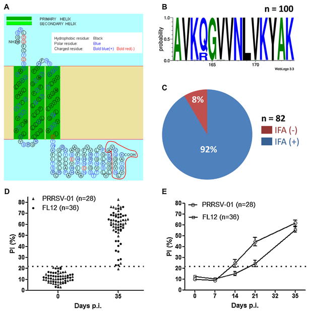
Epitope-M201, spanning the residues 161–174 of M protein (Fig. 1A) is well conserved among type-II PRRSV isolates. Analysis of 100 type-II PRRSV M protein sequences collected from GenBank revealed that all of the amino acid residues of epitope-M201 were identical except the residue at position 164, where 16% of the analyzed sequences had a substitution from glutamine to arginine (Fig. 1B). The monoclonal antibody specific to epitope-M201 (MAb-201) recognized 92% (n = 82) PRRSV field-isolates originating from the Midwestern states of the U.S. (Fig. 1C), confirming the conservation of this epitope. Through pepscan analysis, we reported previously that 100% (n = 15) of pigs infected with the highly virulent PRRSV strain FL12 developed antibodies against epitope-M201 [27]. To further confirm the immunogenicity of this epitope, we developed a bELISA that allowed us to measure antibodies to epitope-M201 more specifically. Using this bELISA we showed that 63 out of 64 pigs (98.4%) infected pigs had antibodies specific to epitope-M201 at 35 days p.i. (Fig. 1D). Detailed analysis revealed that antibodies to epitope-M201 appeared at about 14 days p.i. (Fig. 1E). Together, the results presented here confirm that epitope-M201 is highly immunodominant as previously anticipated [27].
Fig. 1.

Epitope-M201 is highly conserved and immunodominant. (A) Prediction of transmembrane helixes and topology of the PRRSV strain FL12 M protein. The figure was generated through the use of the web based application SOSUI engine ver. 1.11. Epitope-M201 is outlined in red at the carboxyl-terminus of the protein. (B) Sequence logo of epitope-M201 generated from 100 type-II PRRSV M protein sequences collected from GenBank. The relative positions of the amino acids are indicated in the X-axis. The height of symbols within the stack indicates the relative frequency of each amino at that position. The figure was generated through the use of the web based application WebLogo 3.3. (C) Reactivity of MAb-201 with 82 type-II PRRSV strains/isolates as determined by indirect immunofluorescence assay (IFA). (D) Validation of the bELISA. The positive and negative control sera were collected from 64 pigs before infection and at 35 days p.i. (details of the serum samples are described in Section 2). (E) Kinetics of seroconversion to epitope-M201 as determined by bELISA. The sera were collected from the same pigs mentioned in panel (D). The horizontal dotted line represents the cut-off of the assay. (For interpretation of the references to color in this figure legend, the reader is referred to the web version of the article.)
3.2. Core amino acid sequence of epitope-M201
To determine the core amino acid residues of the epitope-M201, we constructed an expression vector encoding the M protein of PRRSV strain FL12. Using this vector as a backbone, we performed alanine-scanning mutagenesis where amino acids of the epitope-M201 were replaced by alanine (Table 2). Cells expressing the Mut-4 construct carrying alanine substitutions at the last 4 amino acids of epitope-M201 were still recognized by MAb-201. By contrast, cells transfected with mutant plasmids carrying substitutions within the first 10 amino acids were not recognized by MAb-201. The results indicated that the core amino acid sequence of epitope-M201 resided within its first 10 amino acids (residue 161–170).
Table 2. Mutagenesis to disrupt the antigenicity of epitope-M201.
| Constructs | Sequence | IFAa | Virus recovery |
|---|---|---|---|
| Wil d type | 161AVKQGVVNLVKYAK174 | +++ | +++ |
| Mut-1 | .AAA.......... | - | - |
| Mut-2 | ....AAA....... | - | - |
| Mut-3 | .......AAA.... | - | - |
| Mut-4 | ..........AAAA | +++ | NDb |
| Mut-5 | ..AA.......... | - | - |
| Mut-6 | .......A..A..A | - | - |
IFA: indirect immunofluorescence assay with the MAb-201.
ND, not done.
3.3. Generation and characterization of N168R mutant virus
Mutations that disrupt the antigenicity of epitope-M201, as determined by transient expression, were incorporated into the pFL12 plasmid [33]. At 48 h post-electroporation with the mutant RNA transcripts, cells were immunostained with MAb SDOW-17 specific to viral N protein. Specific signal was detected from cells electroporated with all mutant RNA transcripts (data not shown), suggesting that mutations within epitope-M201 did not affect viral sub-genomic mRNA transcription. However, we could not recover viable mutant viruses from any of those constructs (Table 2), indicating that amino acids within the epitope-M201 region were essential for productive infection.
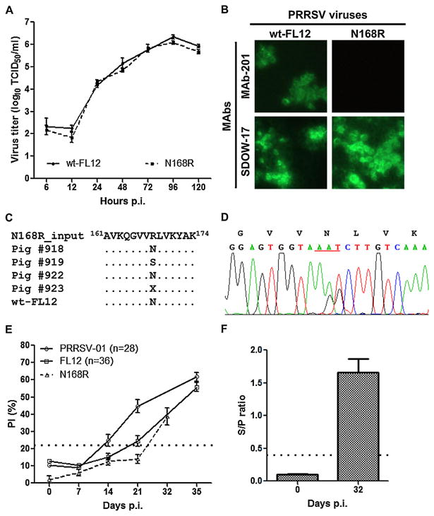
By comparison between Mut-4 and Mut-6 constructs (Table 2), it appeared that a single amino acid substitution from asparagine to alanine at position 168 was sufficient to abolish the antigenicity of epitope-M201. Further analysis revealed that 5 out of 16 amino acids at the carboxyl-terminus of the FL12 M protein, including 3 residues inside epitope-M201 and 2 residues immediately upstream of epitope-M201, were positively charged. Consequently, a mutant virus N168R was generated by mutating asparagine 168 to arginine, a positive charge amino acid, instead of alanine. The N168R mutant virus replicated as efficiently as the parental wild-type FL12 in cell culture (Fig. 2A). More importantly, the N168R mutant did not react with MAb-201 (Fig. 2B), indicating that antigenicity of epitope-M201 was successfully disrupted.
Fig. 2.

Characterization of the N168R mutant virus. (A) Multiple step growth curves of the indicated viruses upon infection of MARC-145 cells. Viral titers are expressed as mean and standard error of mean (SEM) of data obtained from three independent experiments. (B) Reactivity of the indicated virus with different MAbs. (C) ORF6 region of viruses in the serum samples collected at 7 days p.i. was amplified by RT-PCR and subjected to DNA sequencing. The consensus sequences of the serum-derived viruses were aligned with that of input virus (before inoculation). Only the epitope-M201 region is shown. The conserved residues in the aligned sequences are indicated by (.). The X represents the ambiguous sequence. (D) Sequencing chromatograms of one representative serum-derived virus (from pig # 918) at 7 days p.i. Only region covering the mutation sites is shown. The triplet codon 168 is underlined. (E) Seroconversion to epitope-M201 of pigs infected with N168R mutant virus as measured by bELISA. Data are expressed as mean and SEM of 4 infected pigs. For the comparison purposes, kinetics of seroconversion to epitope-M201 of pigs infected with FL12 and PRRSV-01 (Fig. 1E) are shown. (F) Antibodies to N protein of pigs infected with N168R mutant virus as determined by the IDEXX ELISA. Data are expressed as mean and SEM of 4 infected pigs. The horizontal dotted lines in panels (E) and (F) indicate the cut-off of the assays.
To study the stability and immunogenicity of the N168R mutation 4 recently weaned pigs were infected with N168R mutant virus. Analysis of the consensus sequences of the viruses from serum samples collected at 7 days p.i. revealed that two viruses exhibited arginine to asparagine mutation; one virus displayed arginine to serine mutation and one had an ambiguous sequence (Fig. 2C). Detailed analysis of the sequencing chromatograms indicated that all the serum-derived viruses carried a mix of nucleotide sequences at codon 168 (Fig. 2D). All pigs infected with N168R mutant were tested positive by the bELISA at 32 days p.i. (Fig. 2E). The results further confirmed that reversion mutation had occurred in all 4 infected pigs. As expected, all infected pigs were tested positive by the IDEXX ELISA (Fig. 2F).
3.4. Generation and characterization of Q164R mutant virus
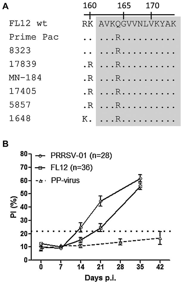
To seek a more stable mutant deprived of epitope-M201 immunogenicity, we investigated the M protein sequences of PRRSV strains/isolates that were naturally not recognized by MAb-201 (Fig. 1C) and found that they all carried a single amino acid substitution within the epitope-M201 region, from glutamine to arginine at position 164 (Q164R) (Fig. 3A). To test whether the Q164R substitution was sufficient to abolish the antibody response to epitope-M201, a group of 10 pigs were inoculated with the attenuated PRRSV vaccine strain Prime Pac (herein designated as PP-virus) [36], one of the PRRSV strains that were not recognized by MAb-201 (Fig. 3A). Serum samples collected from pigs infected with PP-virus were tested negative by the bELISA (Fig. 3B), indicating that the Q164R substitution eliminates the immunogenicity of epitope-M201.
Fig. 3.

A single amino acid substitution Q164R is sufficient to disrupt epitope-M201. (A) Multiple sequence alignment of the epitope-M201 region of the PRRSV isolates that were naturally not recognized by MAb-201. Conserved residues in the aligned sequences are indicated by (.). The gray box depicts epitope-M201 region. (B) Seroconversion to epitope-M201 of pigs infected with the attenuated PRRSV vaccine strain Prime Pac (PP-virus) as measured by bELISA. Data are expressed as mean and SEM of 10 infected pigs. Kinetics of seroconversion to epitope-M201 of pigs infected with FL12 and PRRSV-01 (Fig. 1E) are shown for the comparison purposes. The horizontal dotted line indicates the cut-off of the assay.
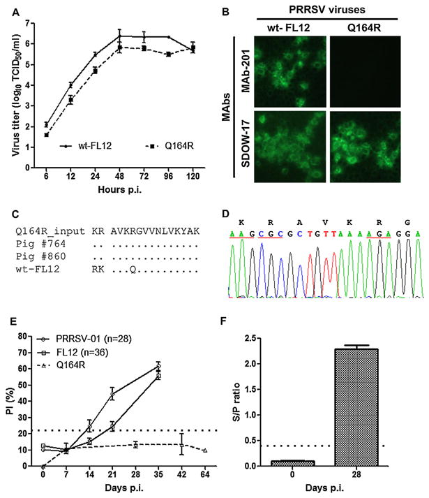
Based on this finding, we constructed the Q164R mutant virus by mutating glutamine 164 to arginine. In addition, we also incorporated into the Q164R mutant virus two additional mutations (arginine 159 to lysine and lysine 160 to arginine) that are located immediately upstream of epitope-M201. We reasoned that the incorporation of these two additional mutations may help stabilize the Q164R mutation because they were commonly found in the epitope-M201 negative PRRSV isolates (Fig. 3A). The growth kinetics of Q164R mutant virus was slightly reduced as compared to the wild-type FL12 strain (Fig. 4A). Importantly, the Q164R mutant virus was no longer recognized by MAb-201 (Fig. 4B), indicating that antigenicity of epitope-M201 was successfully disrupted.
Fig. 4.

Characterization of the Q164R mutant virus. The experiments in this figure were done similar to those described in Fig. 3. (A) Multiple step growth curves. (B) Reactivity with different MAbs. (C) Multiple sequence alignment of the epitope-M201 regions of the viruses from serum samples collected at 14 days p.i. (D) Sequencing chromatograms of one representative serum-derived virus (from pig #860) at 14 days p.i. The triplet codons where amino acid mutations were introduced are underlined. (E) Seroconversion to epitope-M201 of pigs infected with Q164R mutant virus measured by bELISA. (F) Antibodies to N protein of pigs infected with Q164R mutant virus as determined by the commercial IDEXX ELISA.
Two pigs were inoculated with Q164R mutant virus to study the stability and immunogenicity of the virus. ORF6 region of the virus in serum samples collected at 14 days p.i. was sequenced, which confirmed that all 3 mutations of the Q164R mutant virus were stably maintained at this time point (Fig. 4C and D). As expected, antibodies to epitope-M201 were absent in pigs infected with the Q164R mutant virus at all time-points analyzed (Fig. 4E). Conversely, all infected pigs were tested positive by the IDEXX ELISA (Fig. 4F). Together, the results confirm that that the Q164R mutation eliminates the immunogenicity of epitope-M201.
4. Discussion
Since the initial application of DIVA vaccines to eradicate Pseudorabies Virus in pigs, epidemiological and regulatory considerations dictate that a DIVA vaccine should be designed based on a “negative” serologic marker antigen [17,19]. A serologic marker antigen is a viral protein or an epitope that is absent (thus “negative”) from the vaccine strains but consistently present in the corresponding wild-type viruses. Therefore, only animals that have been infected with wild-type virus will develop antibodies against the serologic marker antigen while the vaccinated animals will not. The most critical step in the development of a live-attenuated DIVA vaccine is the identification of a potential serologic marker antigen. Since a DIVA vaccine is only useful if it is used in conjunction with a companion diagnostic test, the marker antigen should be conserved and immunodominant to ensure that the companion diagnostic test based on this marker antigen will produce reliable diagnostic results. In addition, the marker antigen should be dispensable for the viral life cycle but should not contribute significantly to the overall immunogenicity of the vaccine. These two criteria are to ensure that elimination of the marker antigen from the viral genome will not adversely affect the viral replication and its ability to elicit protective immunity. The PRRSV genome encodes 14 non-structural and 8 structural proteins, several of which are well conserved and/or highly immunogenic [37–40]. Up to now, efforts to delete an entire gene of PRRSV have not been successful [20–23]. Therefore, the most feasible approach to develop a live-attenuated DIVA vaccine against PRRSV would be selectively eliminating a small protein fragment or an epitope, instead of deleting an entire protein. Thus far, efforts to develop a DIVA vaccine for PRRSV have been focused on deleting different immunogenic fragments of the viral nsp2 because this protein can tolerate large deletions [41–43]. However, the biggest shortcoming of the nsp2-mutant viruses, in the context of a DIVA vaccine, is that the differential peptide-based ELISA used in conjunction with the DIVA vaccine has very limited diagnostic sensitivity [42], mainly due to the substantial genetic variation of the nsp2 [44,45].
Epitope-M201 can be a potential serologic marker candidate for development of a live-attenuated DIVA vaccine against PRRSV. The epitope is highly immunodominant and well-conserved among PRRSV isolates. In addition, the epitope does not seem to contribute significantly to the protective immunity against PRRSV infection because a polyclonal antibody specific to this epitope, as well as MAb-201, lacks PRRSV-neutralizing ability (unpublished results). Initially, we attempted to delete the whole or portions of epitope-M201 because this would minimize the chance of reversion to wild-type sequence. Unfortunately, we could not recover any mutant viruses carrying deletions in the epitope-M201 region (unpublished results). Similarly, we could not recover any viruses that carried multiple amino acid substitutions within epitope-M201 (Table 2). The results indicate the important functions of this protein segment in the viral replication cycle. Epitope-M201 is located at the cytoplasmic tail of M protein, which has been suggested to interact with N protein [46]. It is possible that deletions or substitutions of multiple amino acids within epitope-M201 may interfere with the interaction between M and N proteins, thus, being detrimental to the virus. Through studying PRRSV isolates that were naturally not recognized by MAb-201, we identified that a single amino acid substitution Q164R is sufficient to abolish the immunogenicity of epitope-M201. We demonstrated that the Q164R mutation can be introduced into the genome of the PRRSV strain FL12 without severely impairing the viral replication. Importantly, the Q164R mutant virus stably maintained its mutations and lost its capacity to elicit antibodies to epitope-M210 when it was inoculated into pigs. It has been previously reported that reversion to wild-type sequence occurred after the PRRSV mutant viruses were inoculated into pigs [47]. In the present study, we observed that the N168R mutant virus, but not the Q164R mutant virus, quickly reverted to wild-type sequence after the first week of inoculation into the pigs. We believe that the Q164R mutant virus was stable in vivo because it carries mutations mimicking the natural PRRSV sequences that have already been selected by nature.
Validation data based on a set of control serum samples collected from pigs experimentally infected with different PRRSV strains indicated that our bELISA could correctly detect 98.4% (n = 64) infected animals (Fig. 1D). However, before the definitive adoption of this bELISA as a companion serological test to be used with the epitope-M201-negative DIVA vaccine, it would be essential to validate the performance of this test with a large collection of clinical serum samples. Our results indicate that eitope-M201 is absent in approximately 10–15% of PRRSV field isolates (Fig. 1B and C). This will potentially affect the diagnostic sensitivity of our bELISA. In the scenario that our bELISA has significantly low diagnostic sensitivity when it is used with clinical serum samples, the test may need to be used for herd diagnosis instead of individual animal diagnosis. This way, the low diagnostic sensitivity of the test may be overcome by increasing the test samples collected from each herds. Alternatively, we could consider the possibility of eliminating one additional marker epitope from the genome of the Q164R mutant virus to produce a double negative marker PRRSV strain that simultaneously lacks 2 immunodominant epitopes in its genome. This is based on the contention that the occurrence of PRRSV field strains that simultaneously lack two immunodominant marker epitopes would be negligible.
In summary, we report here the characterization of a potential serological marker epitope located in the M protein of PRRSV. We demonstrate that the immunogenicity of this epitope can be eliminated through the introduction of a single amino acid mutation which does not severely affect the viral replication. We believe that this mutation can be easily introduced into the genome of conventional live-attenuated type-II PRRSV vaccine strains in order to convert them into vaccine with DIVA capability.
Supplementary Material
Acknowledgments
This research has been supported by grants from the University of Nebraska Life Sciences Competitive Grants Program (Facilitating UNL and Industry Partnerships 2011–2013) and from the National Pork Board of the U.S. (NPB 08-248). We thank Dr. Kyoung-Jin Yoon at Iowa State University – Veterinary Diagnostic Laboratory and by Dr. Tony Goldberg at School of Veterinary Medicine, University of Wisconsin-Madison for providing us the PRRSV field isolates.
Footnotes
Appendix A. Supplementary data: Supplementary data associated with this article can be found, in the online version, at doi:10.1016/j.vaccine.2013.07.020.
References
- 1.Hill H. Overview and history of mystery swine disease (swine infertility and respiratory syndrome) 1990:29–30. [Google Scholar]
- 2.Rossow KD. Porcine reproductive and respiratory syndrome. Veterinary Pathology. 1998 Jan;35(1):1–20. doi: 10.1177/030098589803500101. [DOI] [PubMed] [Google Scholar]
- 3.Tian K, Yu X, Zhao T, Feng Y, Cao Z, Wang C, et al. Emergence of fatal PRRSV variants: unparalleled outbreaks of atypical PRRS in China and molecular dissection of the unique hallmark. PLoS One. 2007;2(6):e526. doi: 10.1371/journal.pone.0000526. [DOI] [PMC free article] [PubMed] [Google Scholar]
- 4.Wensvoort G, Terpstra C, Pol JM, ter Laak EA, Bloemraad M, de Kluyver EP, et al. Mystery swine disease in The Netherlands: the isolation of Lelystad virus. Veterinary Quarterly. 1991 Jul;13(3):121–30. doi: 10.1080/01652176.1991.9694296. [DOI] [PubMed] [Google Scholar]
- 5.Cavanagh D. Nidovirales: a new order comprising Coronaviridae and Arteriviridae. Archives of Virology. 1997;142(3):629–33. [PubMed] [Google Scholar]
- 6.Collins JE, Benfield DA, Christianson WT, Harris L, Hennings JC, Shaw DP, et al. Isolation of swine infertility and respiratory syndrome virus (isolate ATCC VR-2332) in North America and experimental reproduction of the disease in gnotobiotic pigs. Journal of Veterinary Diagnostic Investigation. 1992 Apr;4(2):117–26. doi: 10.1177/104063879200400201. [DOI] [PubMed] [Google Scholar]
- 7.Meulenberg JJ, Petersen den Besten A, de Kluyver E, van Nieuwstadt A, Wensvoort G, Moormann RJ. Molecular characterization of Lelystad virus. Veterinary Microbiology. 1997 Apr;55(1/4):197–202. doi: 10.1016/S0378-1135(96)01335-1. [DOI] [PMC free article] [PubMed] [Google Scholar]
- 8.Conzelmann KK, Visser N, Van Woensel P, Thiel HJ. Molecular characterization of porcine reproductive and respiratory syndrome virus, a member of the arterivirus group. Virology. 1993 Mar;193(1):329–39. doi: 10.1006/viro.1993.1129. [DOI] [PMC free article] [PubMed] [Google Scholar]
- 9.Snijder E, Spaan WJ. Ateriviruses. In: Knipe DM, Howley PM, editors. Fields virology. 5th. Philadelphia: Lippincott Williams & Wilkins; 2007. pp. 1337–55. [Google Scholar]
- 10.Osorio FA, Zuckermann F, Wills R, Meier W, Christian S, Galeota J, et al. PRRSV: comparison of commercial vaccines in their ability to induce protection against current PRRSV strains of high virulence. Allen D Leman Swine Conference. 1998;25:176–82. [Google Scholar]
- 11.Zuckermann FA, Garcia EA, Luque ID, Christopher-Hennings J, Doster A, Brito M, et al. Assessment of the efficacy of commercial porcine reproductive and respiratory syndrome virus (PRRSV) vaccines based on measurement of serologic response, frequency of gamma-IFN-producing cells and virological parameters of protection upon challenge. Veterinary Microbiology. 2007 Jul 20;123(1/3):69–85. doi: 10.1016/j.vetmic.2007.02.009. [DOI] [PubMed] [Google Scholar]
- 12.Alexopoulos C, Kritas SK, Kyriakis CS, Tzika E, Kyriakis SC. Sow performance in an endemically porcine reproductive and respiratory syndrome (PRRS)-infected farm after sow vaccination with an attenuated PRRS vaccine. Veterinary Microbiology. 2005 Dec 20;111(3/4):151–7. doi: 10.1016/j.vetmic.2005.10.007. [DOI] [PubMed] [Google Scholar]
- 13.Pejsak Z, Markowska-Daniel I. Randomised, placebo-controlled trial of a live vaccine against porcine reproductive and respiratory syndrome virus in sows on infected farms. Veterinary Record. 2006 Apr 8;158(14):475–8. doi: 10.1136/vr.158.14.475. [DOI] [PubMed] [Google Scholar]
- 14.Prieto C, Alvarez E, Martinez-Lobo FJ, Simarro I, Castro JM. Similarity of European porcine reproductive and respiratory syndrome virus strains to vaccine strain is not necessarily predictive of the degree of protective immunity conferred. Veterinary Journal. 2008 Mar;175(3):356–63. doi: 10.1016/j.tvjl.2007.01.021. [DOI] [PubMed] [Google Scholar]
- 15.Diaz I, Darwich L, Pappaterra G, Pujols J, Mateu E. Different European-type vaccines against porcine reproductive and respiratory syndrome virus have different immunological properties and confer different protection to pigs. Virology. 2006 Aug 1;351(2):249–59. doi: 10.1016/j.virol.2006.03.046. [DOI] [PubMed] [Google Scholar]
- 16.Christopher-Hennings J, Nelson EA, Nelson JK, Benfield DA. Effects of a modified-live virus vaccine against porcine reproductive and respiratory syndrome in boars. American Journal of Veterinary Research. 1997 Jan;58(1):40–5. [PubMed] [Google Scholar]
- 17.van Oirschot JT. Diva vaccines that reduce virus transmission. Journal of Biotechnology. 1999 Aug 20;73(2/3):195–205. doi: 10.1016/s0168-1656(99)00121-2. [DOI] [PubMed] [Google Scholar]
- 18.Capua I, Schmitz A, Jestin V, Koch G, Marangon S. Vaccination as a tool to combat introductions of notifiable avian influenza viruses in Europe 2000 to 2006. Revue Scientifique et Technique. 2009 Apr;28(1):245–59. doi: 10.20506/rst.28.1.1861. [DOI] [PubMed] [Google Scholar]
- 19.van Oirschot JT, Kaashoek MJ, Rijsewijk FA, Stegeman JA. The use of marker vaccines in eradication of herpesviruses. Journal of Biotechnology. 1996 Aug 20;44(2/3):75–81. doi: 10.1016/0168-1656(95)00129-8. [DOI] [PubMed] [Google Scholar]
- 20.Welch SK, Jolie R, Pearce DS, Koertje WD, Fuog E, Shields SL, et al. Construction and evaluation of genetically engineered replication-defective porcine reproductive and respiratory syndrome virus vaccine candidates. Veterinary Immunology and Immunopathology. 2004 Dec 8;102(3):277–90. doi: 10.1016/j.vetimm.2004.09.022. [DOI] [PubMed] [Google Scholar]
- 21.Lee C, Yoo D. The small envelope protein of porcine reproductive and respiratory syndrome virus possesses ion channel protein-like properties. Virology. 2006 Nov 10;355(1):30–43. doi: 10.1016/j.virol.2006.07.013. [DOI] [PMC free article] [PubMed] [Google Scholar]
- 22.Sun L, Li Y, Liu R, Wang X, Gao F, Lin T, et al. Porcine reproductive and respiratory syndrome virus ORF5a protein is essential for virus viability. Virus Research. 2013 Jan;171(1):178–85. doi: 10.1016/j.virusres.2012.11.005. [DOI] [PubMed] [Google Scholar]
- 23.Wissink EH, Kroese MV, van Wijk HA, Rijsewijk FA, Meulenberg JJ, Rottier PJ. Envelope protein requirements for the assembly of infectious virions of porcine reproductive and respiratory syndrome virus. Journal of Virology. 2005 Oct;79(19):12495–506. doi: 10.1128/JVI.79.19.12495-12506.2005. [DOI] [PMC free article] [PubMed] [Google Scholar]
- 24.Uddowla S, Hollister J, Pacheco JM, Rodriguez LL, Rieder E. A safe foot-and-mouth disease vaccine platform with two negative markers for differentiating infected from vaccinated animals. Journal of Virology. 2012 Nov;86(21):11675–85. doi: 10.1128/JVI.01254-12. [DOI] [PMC free article] [PubMed] [Google Scholar]
- 25.Castillo-Olivares J, Wieringa R, Bakonyi T, de Vries AA, Davis-Poynter NJ, Rottier PJ. Generation of a candidate live marker vaccine for equine arteritis virus by deletion of the major virus neutralization domain. Journal of Virology. 2003 Aug 15;77:8470–80. doi: 10.1128/JVI.77.15.8470-8480.2003. [DOI] [PMC free article] [PubMed] [Google Scholar]
- 26.Holinka LG, Fernandez-Sainz I, O'Donnell V, Prarat MV, Gladue DP, Lu Z, et al. Development of a live attenuated antigenic marker classical swine fever vaccine. Virology. 2009 Feb 5;384(1):106–13. doi: 10.1016/j.virol.2008.10.039. [DOI] [PubMed] [Google Scholar]
- 27.de Lima M, Pattnaik AK, Flores EF, Osorio FA. Serologic marker candidates identified among B-cell linear epitopes of Nsp2 and structural proteins of a North American strain of porcine reproductive and respiratory syndrome virus. Virology. 2006;353:410–21. doi: 10.1016/j.virol.2006.05.036. [DOI] [PubMed] [Google Scholar]
- 28.Das PB, Vu HL, Dinh PX, Cooney JL, Kwon B, Osorio FA, et al. Glycosylation of minor envelope glycoproteins of porcine reproductive and respiratory syndrome virus in infectious virus recovery, receptor interaction, and immune response. Virology. 2011 Feb 20;410(2):385–94. doi: 10.1016/j.virol.2010.12.002. [DOI] [PubMed] [Google Scholar]
- 29.Vu HL, Kwon B, Yoon KJ, Laegreid WW, Pattnaik AK, Osorio FA. Immune evasion of porcine reproductive and respiratory syndrome virus through glycan shielding involves both glycoprotein 5 as well as glycoprotein 3. Journal of Virology. 2012 Jun;85(11):5555–64. doi: 10.1128/JVI.00189-11. [DOI] [PMC free article] [PubMed] [Google Scholar]
- 30.Kwon B, Ansari IH, Osorio FA, Pattnaik AK. Infectious clone-derived viruses from virulent and vaccine strains of porcine reproductive and respiratory syndrome virus mimic biological properties of their parental viruses in a pregnant sow model. Vaccine. 2006 Nov 30;24(49/50):7071–80. doi: 10.1016/j.vaccine.2006.07.010. [DOI] [PubMed] [Google Scholar]
- 31.Beura LK, Sarkar SN, Kwon B, Subramaniam S, Jones C, Pattnaik AK, et al. Porcine reproductive and respiratory syndrome virus nonstructural protein 1beta modulates host innate immune response by antagonizing IRF3 activation. Journal of Virology. 2010 Feb;84(3):1574–84. doi: 10.1128/JVI.01326-09. [DOI] [PMC free article] [PubMed] [Google Scholar]
- 32.Sarkar G, Sommer SS. The “megaprimer” method of site-directed mutagenesis. Biotechniques. 1990 Apr;8(4):404–7. [PubMed] [Google Scholar]
- 33.Truong HM, Lu Z, Kutish GF, Galeota J, Osorio FA, Pattnaik AK. A highly pathogenic porcine reproductive and respiratory syndrome virus generated from an infectious cDNA clone retains the in vivo virulence and transmissibility properties of the parental virus. Virology. 2004 Aug 1;325(2):308–19. doi: 10.1016/j.virol.2004.04.046. [DOI] [PMC free article] [PubMed] [Google Scholar]
- 34.Ansari IH, Kwon B, Osorio FA, Pattnaik AK. Influence of N-linked glycosylation of porcine reproductive and respiratory syndrome virus GP5 on virus infectivity, antigenicity, and ability to induce neutralizing antibodies. Journal of Virology. 2006 Apr;80(8):3994–4004. doi: 10.1128/JVI.80.8.3994-4004.2006. [DOI] [PMC free article] [PubMed] [Google Scholar]
- 35.Subramaniam S, Beura LK, Kwon B, Pattnaik AK, Osorio FA. Amino acid residues in the non-structural protein 1 of porcine reproductive and respiratory syndrome virus involved in down-regulation of TNF-alpha expression in vitro and attenuation in vivo. Virology. 2012 Oct 25;432(2):241–9. doi: 10.1016/j.virol.2012.05.014. [DOI] [PubMed] [Google Scholar]
- 36.Hesse R, Couture L, Lau M, Wunder K, Wasmoen T. Efficacy of Prime Pac PRRS in controlling PRRS reproductive disease: heterologous challenge. 1996:107–10. [Google Scholar]
- 37.Brown E, Lawson S, Welbon C, Gnanandarajah J, Li J, Murtaugh MP, et al. Antibody response of nonstructural proteins: implications for diagnostic detection and differentiation of Type I and Type II porcine reproductive and respiratory syndrome viruses. Clinical and Vaccine Immunology. 2009 Mar;4(16):628–35. doi: 10.1128/CVI.00483-08. [DOI] [PMC free article] [PubMed] [Google Scholar]
- 38.Yoon KJ, Zimmerman JJ, Swenson SL, McGinley MJ, Eernisse KA, Brevik A, et al. Characterization of the humoral immune response to porcine reproductive and respiratory syndrome (PRRS) virus infection. Journal of Veterinary Diagnostic Investigation. 1995 Jul;7(3):305–12. doi: 10.1177/104063879500700302. [DOI] [PubMed] [Google Scholar]
- 39.Kapur V, Elam MR, Pawlovich TM, Murtaugh MP. Genetic variation in porcine reproductive and respiratory syndrome virus isolates in the Midwestern United States. Journal of General Virology. 1996 Jun;77(Pt 6):1271–6. doi: 10.1099/0022-1317-77-6-1271. [DOI] [PubMed] [Google Scholar]
- 40.Johnson CR, Yu W, Murtaugh MP. Cross-reactive antibody responses to nsp1 and nsp2 of Porcine reproductive and respiratory syndrome virus. Journal of General Virology. 2007 Apr;88(Pt 4):1184–95. doi: 10.1099/vir.0.82587-0. [DOI] [PubMed] [Google Scholar]
- 41.Kim DY, Kaiser TJ, Horlen K, Keith ML, Taylor LP, Jolie R, et al. Insertion and deletion in a non-essential region of the nonstructural protein 2 (nsp2) of porcine reproductive and respiratory syndrome (PRRS) virus: effects on virulence and immunogenicity. Virus Genes. 2009 Feb;38(1):118–28. doi: 10.1007/s11262-008-0303-4. [DOI] [PubMed] [Google Scholar]
- 42.de Lima M, Kwon B, Ansari IH, Pattnaik AK, Flores EF, Osorio FA. Development of a porcine reproductive and respiratory syndrome virus differentiable (DIVA) strain through deletion of specific immunodominant epitopes. Vaccine. 2008 Jul 4;26(29/30):3594–600. doi: 10.1016/j.vaccine.2008.04.078. [DOI] [PubMed] [Google Scholar]
- 43.Fang Y, Christopher-Hennings J, Brown E, Liu H, Chen Z, Lawson SR, et al. Development of genetic markers in the non-structural protein 2 region of a US type 1 porcine reproductive and respiratory syndrome virus: implications for future recombinant marker vaccine development. Journal of General Virology. 2008 Dec;89(Pt 12):3086–96. doi: 10.1099/vir.0.2008/003426-0. [DOI] [PubMed] [Google Scholar]
- 44.Han J, Wang Y, Faaberg KS. Complete genome analysis of RFLP 184 isolates of porcine reproductive and respiratory syndrome virus. Virus Research. 2006 Dec;122(1/2):175–82. doi: 10.1016/j.virusres.2006.06.003. [DOI] [PubMed] [Google Scholar]
- 45.Yoshii M, Okinaga T, Miyazaki A, Kato K, Ikeda H, Tsunemitsu H. Genetic polymorphism of the nsp2 gene in North American type – porcine reproductive and respiratory syndrome virus. Archives of Virology. 2008;153(7):1323–34. doi: 10.1007/s00705-008-0098-6. [DOI] [PubMed] [Google Scholar]
- 46.Spilman MS, Welbon C, Nelson E, Dokland T. Cryo-electron tomography of porcine reproductive and respiratory syndrome virus: organization of the nucleocapsid. Journal of General Virology. 2009 Mar;90(Pt 3):527–35. doi: 10.1099/vir.0.007674-0. [DOI] [PubMed] [Google Scholar]
- 47.Beura LK, Subramaniam S, Vu HL, Kwon B, Pattnaik AK, Osorio FA. Identification of amino acid residues important for anti-IFN activity of porcine reproductive and respiratory syndrome virus non-structural protein 1. Virology. 2012 Nov 25;433(2):431–9. doi: 10.1016/j.virol.2012.08.034. [DOI] [PMC free article] [PubMed] [Google Scholar]
Associated Data
This section collects any data citations, data availability statements, or supplementary materials included in this article.


