Abstract
Optic nerve head drusen (ONHD) are incidental ophthalmologic finding in the optic nerve. Patients with ONHD are often asymptomatic, but sometimes present with transient visual obscuration's (TVO), the reported incidence of which is 8.6%. Optic nerve head drusen are of two types: Superficial; visible and deep. The deep-buried drusen mimic papilledema. Because of the varied presentation deep-buried drusen pose a diagnostic challenge to the ophthalmologists. In young patients, they are mistaken for papilledema as it is clinically difficult to detect a buried drusen in the optic nerve head, but are seen on the surface with aging as the retinal nerve fiber layer thins out. They are observed as pale yellow lesions more often located towards the poles. Clinical examination aided with diagnostic tests like computed tomography (CT) orbits and ultrasound B scan can help establish the diagnosis. Herein, we report a rare case of optic nerve head drusen in a young lady, who presented with loss of vision and clinical evaluation and investigations suggested ONHD with anterior ischemic optic neuropathy.
Keywords: Optic nerve head drusen, optic neuropathy, pseudopapilledema
ONHD can mimic papilledema and present very rarely as anterior ischemic optic neuropathy. As clinicians we should be aware of this rare presentation to prevent unnecessary neurologic workup. Optic nerve head drusen (ONHD) are acellular deposits of laminated hyaline bodies, which progressively calcify in the optic nerve head.[1] Sometimes, ONHD show an inheritance pattern, which is usually autosomal dominant.[2] They are 75 to 80% of times bilateral in occurrence. Reported incidence is 3.4 to 4.9 per 1000 individuals. Higher incidence of 20.4 per 1000 is reported in autopsy studies.[3] ONHD can sometimes be mistaken for papilledema. Patients’ with ONHD can present with visual acuity loss and field loss. The incidence of field defects in ONHD is to the extent of 71% in patients with visible drusen.[4,5] ONHD can also cause anterior ischemic optic neuropathy due to crowding and compression on the axons.[6] The largest reported series which established the association of ONHD and anterior ischemic optic neuropathy is by Purvin V et al., which involved 24 eyes. Here we report one such rare occurrence of anterior ischemic optic neuropathy with ONHD. Certain conditions like retinitis pigmentosa, angioid streaks, usher syndrome show an increased incidence of optic nerve head drusen. The primary pathology of optic disc drusen is most likely an inherited dysplasia of the disc and its blood supply with smaller disc size being an additional risk factor.
Case Report
A 28-year-old lady presented to us with history of decreased vision in her right eye of one month duration. Patient had episodes of headache associated with her visual complaints. Rest of the patient's complaint history was not contributory. One month prior to her presentation to our clinic, the patient had visited two different ophthalmologists, who diagnosed her as having papilledema. Her vision in the right eye was 20/200 (refractive error of − 1.00 spherical − 1.50 cylinder axis 180) and the left eye vision was 20/40 (−1.25 spherical − 2.00 cylinder axis 165) at the time of presentation. Slit lamp biomicroscopy of the anterior segment was unremarkable in both eyes. She had a relative afferent pupillary defect in her right eye. Dilated pupillary examination in the right eye showed optic disc edema, more obvious in the inferior half of the disc with faint splinter hemorrhage in the parapapilary area. The arterioles over the disc showed attenuation in their caliber. Further refined examination of the right optic disc same disc showed a circumscribed pale yellow lesion buried in the disc suggestive of disc drusen [Fig. 1a]. The optic disc in the left eye showed an irregular disc margin with minimal elevation and disc vessels showed anomalous branching pattern [Fig. 1b]. Visual fields in the right eye showed superior altitudinal defect [Fig. 2] and left eye was normal. Ultrasound B scan confirmed the presence of ONHD in both eyes [Fig. 3]. A diagnosis of ONHD in both eyes, with anterior ischemic optic neuropathy in the right eye was made. Patient was treated for her headache (tension type) symptomatically. Our patient was seen again after 6 weeks and right eye disc showed pallor suggestive of optic atrophy [Fig. 4] and spectral domain optical coherence tomography also confirmed the presence of ONHD with retinal nerve fiber layer (RNFL) loss [Fig. 5]. Visual acuity remained at 20/200 in the right eye.
Figure 1.
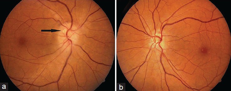
At presentation (a) Right eye disc showing edema and blurring of disc margins, attenuation of peripapillary arterioles and evidence of drusen (black arrow) in the superior pole of the disc margins (b) Left eye disc showing anomalous branching pattern of the vessels and faintly visible drusen at the superior pole
Figure 2.
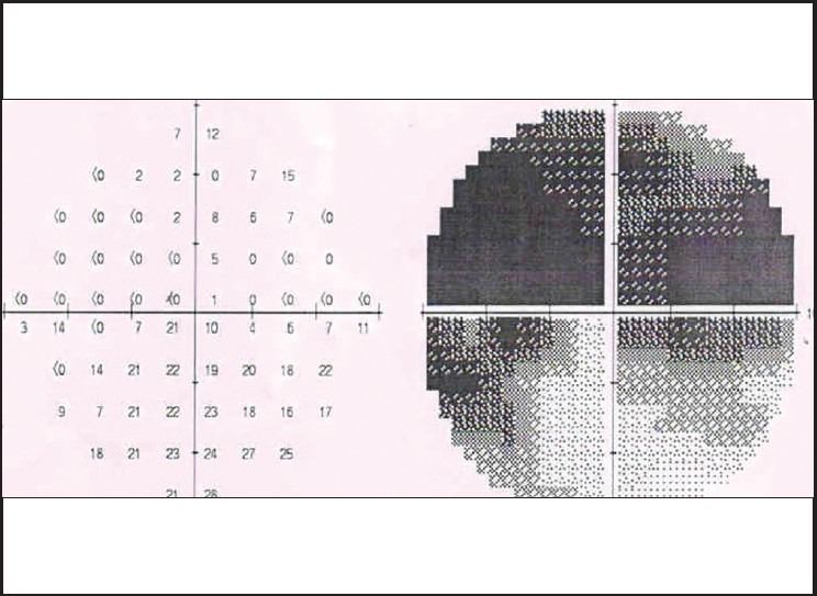
Superior altitudinal defect corresponding to the severe disc edema in the inferior pole of the right disc
Figure 3.
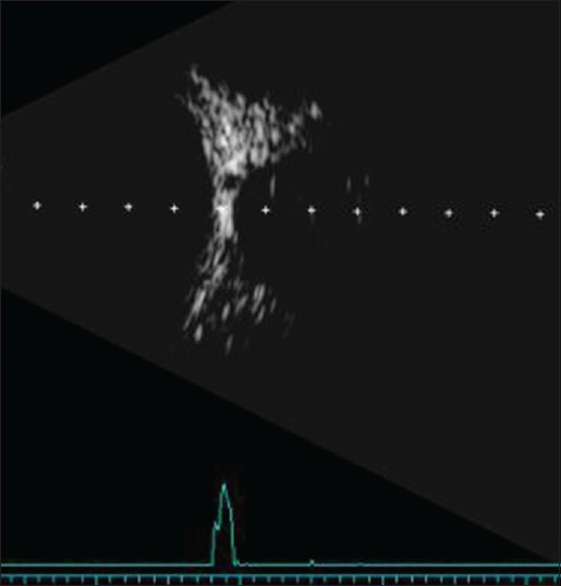
Ultrasound B scan showing drusen
Figure 4.
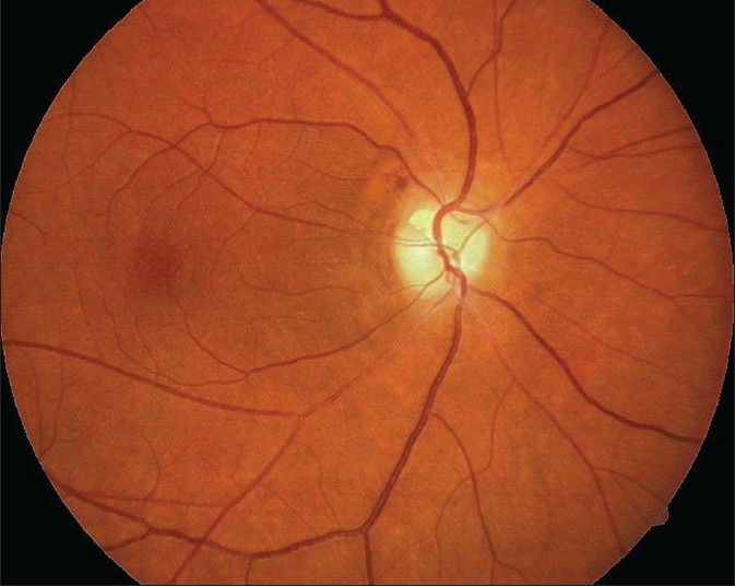
After 6 weeks, right eye disc showing optic atrophy and attenuation of the arterioles
Figure 5.
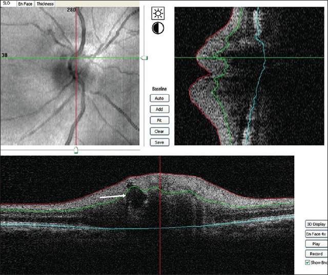
Spectral domain OCT showing the internal dome shaped contour of the drusen (white arrow) and abrupt end of the pigment epithelium and photoreceptor layer
Discussion
ONHD pose a diagnostic dilemma especially in young patients who concomitantly have headache as a presenting complaint. Our patient had earlier consulted two different ophthalmologists who had made diagnosis of papilledema in both eyes, with resulting optic atrophy in the right eye. The patient was further referred to a neurologist where both magnetic resonance imaging (MRI) and MRI venogram were normal. Cerebrospinal fluid manometry was also normal.
Optic nerve head drusen mimic papilledema especially in young patients. Clinical symptoms and disc examination offer clues that can help differentiate the two entities. Chronic severe headache associated with vomiting may often be the presenting symptoms in papilledema. Tortuosity of optic disc venules, fiery looking disc vessels associated with congestion and hemorrhagic capillaries will favor a diagnosis of papilledema. In ONHD, the disc vessels are usually normal in caliber and disc elevation is not associated with congestion and hyperemia of the disc. The presence of disc edema in ONHD suggests the presence of ischemia. Long-standing papilledema can also lead to optic atrophy, which can be confusing at times. Our patient had visual loss only in her right eye; the optic disc showed features suggestive of anterior ischemic optic neuropathy. Visual field testing showed altitudinal defect in the right eye and was normal in the left eye. Young patients with ONHD may develop ischemic optic neuropathy in a way similar to the elderly age group patients because of the crowding and compressive effect on the axons by the drusen.[6,7,8] Disc swelling in our patient's right eye resolved without any treatment after 3 months, but the visual field defect persisted with supporting RNFL thinning on optical coherence tomography (OCT).
A high index of suspicion is needed when young patients present with disc edema and loss of vision. Ultrasound B scan is a sensitive tool to detect ONHD.[9] Spectral domain optical coherence tomography (SDOCT) is helpful in imaging ONHD because of its better axial resolution and it quantifies the RNFL loss with greater reproducibility.[7,10] OCT features observed in ONHD are subretinal hyporeflective space within 0.7 mm radius of the disc and smooth dome shaped lesions in the disc with a lumpy internal contour and a hyporeflective space between the retinal pigmented epithelium and photoreceptor layers that has an abrupt end. Computed tomography (CT) of the orbits can also help in establishing the diagnosis [Fig. 6]. ONHD can be a rare cause of ischemic optic neuropathy in young patients.[6,8] This case highlights the fact that establishing the diagnosis of disc drusen sooner in this patient could have avoided the unnecessary neurological workup.
Figure 6.
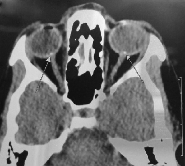
Computed tomographic scans of orbits: Bilateral drusen (indicated by the arrows)
Footnotes
Source of Support: Nil
Conflict of Interest: None declared
References
- 1.Tso MO. Pathology and pathogenesis of drusen of the optic nerve head. Ophthalmology. 1981;88:1066–80. doi: 10.1016/s0161-6420(81)80038-3. [DOI] [PubMed] [Google Scholar]
- 2.Antcliff RJ, Spalton DJ. Are the optic disc drusen inherited? Ophthalmology. 1999;106:1278–81. doi: 10.1016/S0161-6420(99)00708-3. [DOI] [PubMed] [Google Scholar]
- 3.Friedman AH, Gartner S, Modi SS. Drusen of the optic disc. A retrospective study in cadaver eyes. Br J Ophthalmol. 1975;59:413–21. doi: 10.1136/bjo.59.8.413. [DOI] [PMC free article] [PubMed] [Google Scholar]
- 4.Lee AG, Zimmerman MB. The rate of visual field loss in optic nerve head drusen. Am J Ophthalomol. 2005;139:1062–6. doi: 10.1016/j.ajo.2005.01.020. [DOI] [PubMed] [Google Scholar]
- 5.Wilkins JM, Pomeranz HD. Visual manifestations of visible and buried optic disc drusen. J Neuroophthalmol. 2004;24:125–9. doi: 10.1097/00041327-200406000-00006. [DOI] [PubMed] [Google Scholar]
- 6.Purvin V, King R, Kawasaki A, Yee R. Anterior ischemic optic neuropathy in eyes with optic disc drusen. Arch Ophthalmol. 2004;122:48–53. doi: 10.1001/archopht.122.1.48. [DOI] [PubMed] [Google Scholar]
- 7.Auw-Haedrich C, Staubach F, Witschel H. Optic disc drusen. Surv Ophthalmol. 2002;47:515–32. doi: 10.1016/s0039-6257(02)00357-0. [DOI] [PubMed] [Google Scholar]
- 8.Jonas JB, Gusek GC, Naumann GO. Anterior ischemic optic neuropathy: Nonarteritic form in small normal sized optic disc. Int Ophthalmol. 1988;12:119–25. doi: 10.1007/BF00137137. [DOI] [PubMed] [Google Scholar]
- 9.Boldt HC, Byrne SF, DiBernardo C. Echographic evaluation of the optic disc drusen. J Clin Neuroophthalmol. 1991;11:85–91. [PubMed] [Google Scholar]
- 10.Davis PL, Jay WM. Optic nerve head drusen. Semin Ophthalmol. 2003;18:222–42. doi: 10.1080/08820530390895244. [DOI] [PubMed] [Google Scholar]


