

Abstract
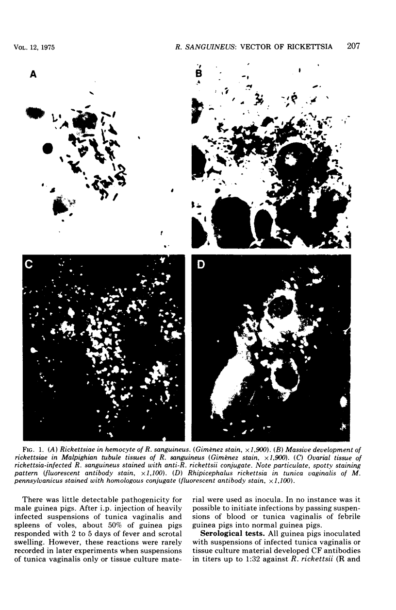
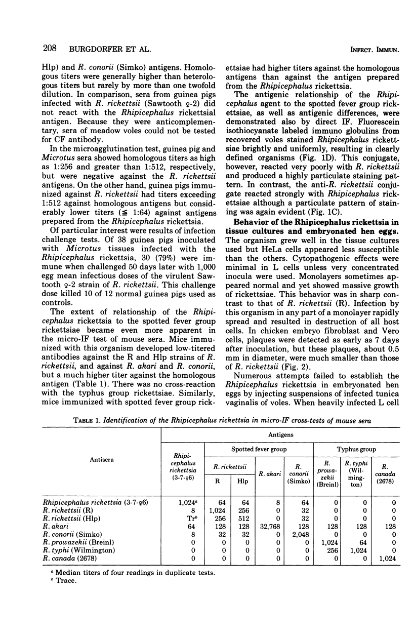
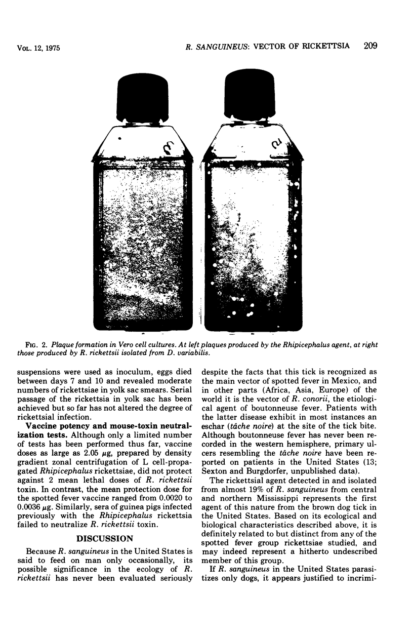
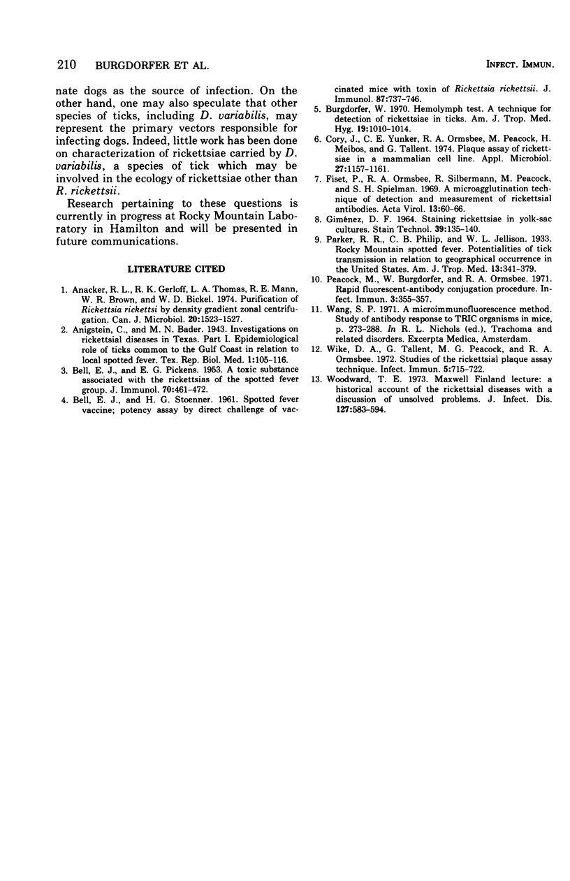
A rickettsia related to but distinct from the spotted fever agent, Rickettsia rickettsii, has been detected in 167 (18.9%) of 884 Rhipicephalus sanguineus taken off dogs in central and northern Mississippi. The organisms could readily be isolated in male meadow voles (Microtus pennsylvanicus), where it produced massive infections in the tissues of tunica vaginalis. It was practically nonpathogenic for male guinea pigs, although inoculation of these animals with infected tunica vaginalis of voles afforded in 30 of 38 instances solid immunity to challenge with virulent R. rickettsii. The Rhipicephalus rickettsia grew well in monolayers of chicken embryo fibroblast, Vero, mouse L, and HeLa cells. Cytopathogenic effects were minimal unless large concentrations of rickettsiae were used as inocula. It also could be established in embryonated hen eggs but only after injection of massive doses of L cell-propagated organisms. Serological tests (complement fixation, microagglutination and/or micro immunofluorescence) indicated that the newly described Rickettsia belongs to the spotted fever group but differs from R. rickettsii, R. akari, and R. conorii. Antigenic differences were also demonstrated by direct fluorescence microscopy as well as by vaccine potency and mouse-toxin neutralization tests.
Full text
PDF
Images in this article
Selected References
These references are in PubMed. This may not be the complete list of references from this article.
- Anacker R. L., Gerloff R. K., Thomas L. A., Mann R. E., Brown W. R., Bickel W. D. Purification of Rickettsia rickettsi by density-gradient zonal centrifugation. Can J Microbiol. 1974 Nov;20(11):1523–1527. doi: 10.1139/m74-238. [DOI] [PubMed] [Google Scholar]
- BELL E. J., PICKENS E. G. A toxic substance associated with the rickettsias of the spotted fever group. J Immunol. 1953 May;70(5):461–472. [PubMed] [Google Scholar]
- BELL E. J., STOENNER H. G. Spotted fever vaccine; potency assay by direct challenge of vaccinated mice with toxin of Rickettsia rickettsii. J Immunol. 1961 Dec;87:737–746. [PubMed] [Google Scholar]
- Burgdorfer W. Hemolymph test. A technique for detection of rickettsiae in ticks. Am J Trop Med Hyg. 1970 Nov;19(6):1010–1014. [PubMed] [Google Scholar]
- Cory J., Yunker C. E., Ormsbee R. A., Peacock M., Meibos H., Tallent G. Plaque assay of rickettsiae in a mammalian cell line. Appl Microbiol. 1974 Jun;27(6):1157–1161. doi: 10.1128/am.27.6.1157-1161.1974. [DOI] [PMC free article] [PubMed] [Google Scholar]
- Fiset P., Ormsbee R. A., Silberman R., Peacock M., Spielman S. H. A microagglutination technique for detection and measurement of rickettsial antibodies. Acta Virol. 1969 Jan;13(1):60–66. [PubMed] [Google Scholar]
- GIMENEZ D. F. STAINING RICKETTSIAE IN YOLK-SAC CULTURES. Stain Technol. 1964 May;39:135–140. doi: 10.3109/10520296409061219. [DOI] [PubMed] [Google Scholar]
- Peacock M., Burgdorfer W., Ormsbee R. A. Rapid fluorescent-antibody conjugation procedure. Infect Immun. 1971 Feb;3(2):355–357. doi: 10.1128/iai.3.2.355-357.1971. [DOI] [PMC free article] [PubMed] [Google Scholar]
- Wike D. A., Tallent G., Peacock M. G., Ormsbee R. A. Studies of the rickettsial plaque assay technique. Infect Immun. 1972 May;5(5):715–722. doi: 10.1128/iai.5.5.715-722.1972. [DOI] [PMC free article] [PubMed] [Google Scholar]
- Woodward T. E. A historical account of the rickettsial diseases with a discussion of unsolved problems. J Infect Dis. 1973 May;127(5):583–594. doi: 10.1093/infdis/127.5.583. [DOI] [PubMed] [Google Scholar]