

Abstract
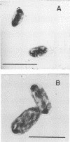
Coxiella burneti phase I, purified from a formalin-inactivated yolk-sac vaccine, was separated into two bands of morphologically distinct cell types when subjected to sucrose gradient centrifugation. Recycling of the less dense, rod-shaped cells in unbuffered sucrose gradients (pH 5.5 to 6.0) resulted in the formation of bands having the location and appearance of the original two bands. Recycling of the denser band of larger ovoid-shaped cells yielded a single band, suggesting that the larger cell type arose from the smaller cell. In contrast to vaccine-derived rickettsiae, live, cell culture-propagated phase I organisms formed a single band in unbuffered sucrose gradients, at the same density as the upper band of the vaccine preparation. Centrifugation of cell culture-derived rickettsiae for 26 to 48 h in sucrose gradients of pH 5.5 resulted in the formation of a second band, at the same density as the lower band of the vaccine preparation. This did not occur in gradients of pH 7.0. Treatment of cell culture-propagated rickettsiae with formalin or germicidal ultraviolet radiation induced a total shift of the less dense cell population to a zone of higher density when centrifuged isopycnically in CsC1 gradients. This density change did not occur in sucrose gradients, suggesting a difference in the effect of these treatments on the permeability of the cell membrane to sucrose and CsC1.
Full text
PDF


Images in this article
Selected References
These references are in PubMed. This may not be the complete list of references from this article.
- ANACKER R. L., FUKUSHI K., PICKENS E. G., LACKMAN D. B. ELECTRON MICROSCOPIC OBSERVATIONS OF THE DEVELOPMENT OF COXIELLA BURNETII IN THE CHICK YOLK SAC. J Bacteriol. 1964 Oct;88:1130–1138. doi: 10.1128/jb.88.4.1130-1138.1964. [DOI] [PMC free article] [PubMed] [Google Scholar]
- Berman S., Gochenour R. B., Cole G., Lowenthal J. P., Benenson A. S. METHOD FOR THE PRODUCTION OF A PURIFIED DRY Q FEVER VACCINE. J Bacteriol. 1961 May;81(5):794–799. doi: 10.1128/jb.81.5.794-799.1961. [DOI] [PMC free article] [PubMed] [Google Scholar]
- Brezina R. Advances in rickettsial research. Curr Top Microbiol Immunol. 1969;47:20–39. doi: 10.1007/978-3-642-46160-6_2. [DOI] [PubMed] [Google Scholar]
- Burton P. R., Kordová N., Paretsky D. Electron microscopic studies of the rickettsia Coxiella burneti: entry, lysosomal response, and fate of rickettsial DNA in L-cells. Can J Microbiol. 1971 Feb;17(2):143–150. doi: 10.1139/m71-025. [DOI] [PubMed] [Google Scholar]
- CASEY H. L. STANDARDIZED DIAGNOSTIC COMPLEMENT FIXATION METHOD AND ADAPTATION TO MICRO TEST. I. LABORATORY BRANCH COMPLEMENT FIXATION METHOD BY LABORATORY BRANCH TASK FORCE. II. ADAPTATION OF LBCF METHOD TO MICRO TECHNIQUE. Public Health Monogr. 1965;74:1–34. [PubMed] [Google Scholar]
- Canonico P. G., Van Zwieten M. J., Christmas W. A. Purification of large quantities of coxiella burnetii rickettsia by density gradient zonal centrifugation. Appl Microbiol. 1972 May;23(5):1015–1022. doi: 10.1128/am.23.5.1015-1022.1972. [DOI] [PMC free article] [PubMed] [Google Scholar]
- Fiset P., Ormsbee R. A., Silberman R., Peacock M., Spielman S. H. A microagglutination technique for detection and measurement of rickettsial antibodies. Acta Virol. 1969 Jan;13(1):60–66. [PubMed] [Google Scholar]
- GIMENEZ D. F. STAINING RICKETTSIAE IN YOLK-SAC CULTURES. Stain Technol. 1964 May;39:135–140. doi: 10.3109/10520296409061219. [DOI] [PubMed] [Google Scholar]
- LOWRY O. H., ROSEBROUGH N. J., FARR A. L., RANDALL R. J. Protein measurement with the Folin phenol reagent. J Biol Chem. 1951 Nov;193(1):265–275. [PubMed] [Google Scholar]
- Moulder J. W. The contribution of model systems to the understanding of infectious diseases. Perspect Biol Med. 1971 Spring;14(3):486–502. doi: 10.1353/pbm.1971.0024. [DOI] [PubMed] [Google Scholar]
- Myers W. F., Provost P. J., Wisseman C. L., Jr Permeability properties of Rickettsia mooseri. J Bacteriol. 1967 Mar;93(3):950–960. doi: 10.1128/jb.93.3.950-960.1967. [DOI] [PMC free article] [PubMed] [Google Scholar]
- Nermut M. V., Schramek S., Brezina R. Electron microscopy of Coxiella burneti phase I and II. Acta Virol. 1968 Sep;12(5):446–452. [PubMed] [Google Scholar]
- Ormsbee R. A. Rickettsiae (as organisms). Annu Rev Microbiol. 1969;23:275–292. doi: 10.1146/annurev.mi.23.100169.001423. [DOI] [PubMed] [Google Scholar]
- ROSENBERG M., KORDOVA N. Study of intracellular forms of Coxiella burneti in the electron microscope. Acta Virol. 1960 Jan;4:52–55. [PubMed] [Google Scholar]
- SHARP D. G., OVERMAN J. R. Enumeration of vaccinia virus particles in crude extracts of infected tissues by electron microscopy. Proc Soc Exp Biol Med. 1958 Nov;99(2):409–413. doi: 10.3181/00379727-99-24366. [DOI] [PubMed] [Google Scholar]
- Wiebe M. E., Burton P. R., Shankel D. M. Isolation and characterization of two cell types of Coxiella burneti phase I. J Bacteriol. 1972 Apr;110(1):368–377. doi: 10.1128/jb.110.1.368-377.1972. [DOI] [PMC free article] [PubMed] [Google Scholar]