Abstract
Study Objectives:
Mandibular advancement splints (MAS) are often preferred to CPAP treatment for OSA but are not always equally efficacious. High therapeutic CPAP pressure has been associated with MAS treatment failure in a Japanese population. We sought to assess the relationship between CPAP pressure and MAS treatment response in an Australian population.
Methods:
Therapeutic CPAP pressure and MAS treatment response were obtained from a one-month crossover trial of both treatments. Predictive utility of CPAP pressure to identify MAS treatment response was assessed.
Results:
Seventy-eight OSA patients were included (age 49.3 ± 11.1 years, BMI 29.1 ± 5.8 kg/m2) with predominantly moderate-severe OSA (AHI 30.0 ± 12.7/h). CPAP pressure was lower in MAS responders (MAS AHI < 10/h) 9.7 ± 1.6 vs. 11.7 ± 2.4 cm H O, p < 0.01, with area under ROC curve of 0.74 (95% CI 0.63-0.86), p < 0.01. The best cutoff value of 10.5 cm H O useful for discriminating MAS responders and non-responders in the previous Japanese population, was inadequate for prediction in the current population (0.47 negative predictive value [NPV]). However a cutoff of 13 cm H O identified MAS non-responders (1.0 NPV). Multivariate regression identified CPAP pressure (odds ratio [95% confidence interval] 0.53 [0.33-0.87], age (0.93 [0.87-0.99]) and AHI (0.92 [0.86-0.97]) as predictors of MAS treatment response (model r2 = 0.54, p < 0.001).
Conclusions:
In Australian patients, the majority of whom are Caucasian, a higher therapeutic CPAP pressure requirement in conjunction with age and OSA severity characteristics may be useful to indicate likelihood of success with MAS as an alternative therapy.
Citation:
Sutherland K, Phillips CL, Davies A, Srinivasan VK, Dalci O, Yee BJ, Darendeliler MA, Grunstein RR, Cistulli PA. CPAP pressure for prediction of oral appliance treatment response in obstructive sleep apnea. J Clin Sleep Med 2014;10(9):943-949.
Keywords: obstructive sleep apnea, oral appliance, continuous positive airway pressure, treatment response
Continuous positive airway pressure (CPAP) is the standard treatment for obstructive sleep apnea (OSA). Although highly efficacious, CPAP is often hindered by poor tolerance and suboptimal adherence,1 limiting its effectiveness in the real world. Mandibular advancement splints (MAS) are an alternative option recommended as a first-line treatment for mild-moderate OSA.2 We have recently found that health outcome improvements, including sleepiness, are similar with MAS and CPAP treatments in patients with moderate-severe OSA.3 Superior patient adherence appears to offset any inferiority of MAS efficacy,3 and MAS may be considered a viable alternative for many patients.
However despite similar health benefits between treatments, approximately one-third of OSA patients will not respond to MAS.4–7 This is of significant concern in terms of resource wastage and treatment delays. Much attention has been given to understanding patient phenotypes which relate to MAS response such as gender, obesity, craniofacial structure, and type and severity of OSA.8 However, none of these factors are universal, and hence there is an unresolved need for reliable indicators of MAS treatment response.
BRIEF SUMMARY
Current Knowledge/Study Rationale: CPAP pressure has been shown to predict oral appliance treatment response in Japanese male OSA patients and could be a simple and useful clinical predictor for some OSA patients. We sought to assess the relationship between CPAP pressure and oral appliance treatment response in a predominantly Caucasian population.
Study Impact: We confirm a relationship between lower CPAP pressure and oral appliance treatment response, although not as strong as in the Japanese population and requiring a higher CPAP pressure cutoff value for best predictive utility. CPAP pressure requirement, in conjunction with patient characteristics of age and OSA severity, may be useful in indicating oral appliance treatment response in Caucasian OSA populations.
A recent Japanese study has identified pressure requirement in CPAP users as a predictor of MAS response.9 In established CPAP users, a prescribed pressure of above 10.5 cm H2O indicated poor response to subsequent MAS therapy. This prediction method is, of course, restricted to patients who have used CPAP and wish to try MAS. However CPAP pressure would represent a simple predictor, either alone or possibly in combination with other patient characteristics to further improve prediction. This would be clinically useful in patients who have failed or are non-adherent to CPAP and would support implementation of MAS therapy as an alternative in such patients.
In this study, we aimed to firstly confirm a relationship between therapeutic CPAP pressure and MAS treatment response in treatment-naive OSA patients, and secondly to investigate the utility of therapeutic CPAP pressure as an indicator of MAS treatment outcome in an Australian population, predominantly comprised of Caucasians.
METHODS
Patients
OSA patients were participants in a previously published randomized crossover trial of one month of CPAP versus MAS to compare health effects.3 Inclusion criteria for this trial were a new diagnosis of OSA, aged ≥ 20 years, apnea-hypopnea index (AHI) > 10 events/h, ≥ 2 symptoms of OSA, and willingness to use both CPAP and MAS. No limits for body mass index (BMI) were set for inclusion. Exclusion criteria were central sleep apnea, previous OSA treatment, requiring immediate treatment, contraindications to MAS therapy, regular sedative or narcotic use, preexisting lung disease, or psychiatric disease. Self-reported ethnicity data was not collected in this study; however, the majority of patients likely have Caucasian ancestry.
Study Protocol
Patients underwent an acclimatization phase to both CPAP and MAS in a randomized order to optimize both treatments before the study. Subsequently patients were randomized to one month each of CPAP and MAS, with treatment response determined by polysomnography at end of each treatment. The study protocol in Figure 1 illustrates the acquisition of data utilized in this analysis.
Figure 1. Flowchart for CPAP and MAS data acquisition.
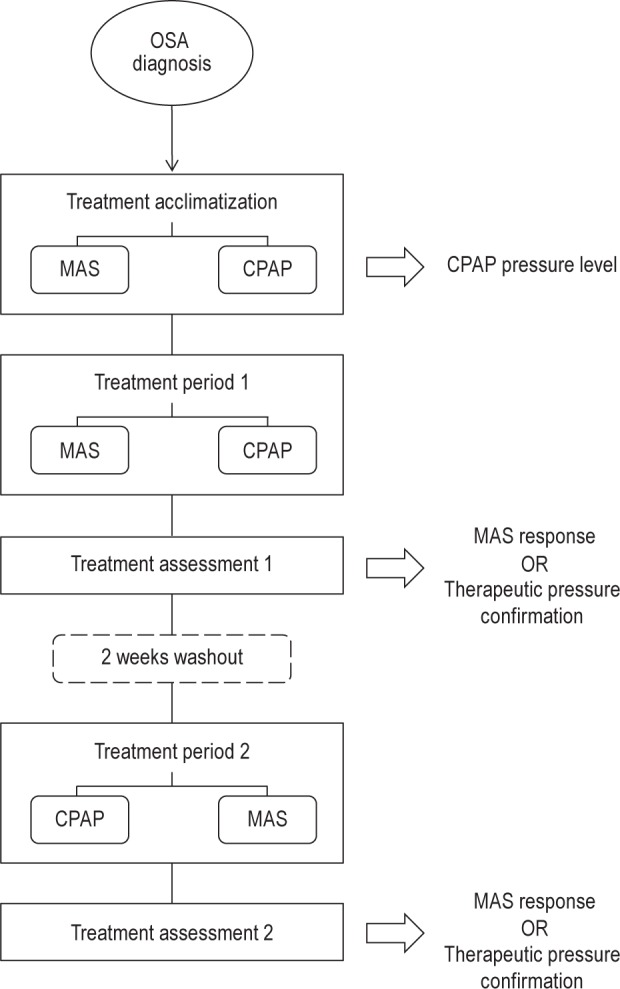
Patients were participants in a randomized crossover trial to compare health outcomes of one-month optimal treatment of CPAP and MAS. Patients underwent an acclimatization period to both MAS and CPAP (in a randomized order) before being randomized to one month of full treatment with either device. Therapeutic CPAP pressure was determined during the CPAP acclimatization period. Treatment efficacy was assessed by overnight polysomnography at the end of each treatment period. Patients underwent a 2-week washout period of no treatment before commencing one month of the alternate treatment. Arrows indicated the points at which data in this analysis was acquired.
CPAP
All patients used the same CPAP device (ResMed Autoset S8, ResMed, Bella Vista, Australia). Patients were given the device to use in Autoset mode at home. Therapeutic pressure was determined by the 95th percentile pressure from usage exceeding 4 hours. Therapeutic CPAP pressure was confirmed by overnight polysomnography on CPAP treatment. Only participants who achieved AHI < 5 events/h on this night were included in the analysis as a stringent definition of therapeutic CPAP pressure.
MAS
The MAS used was a titratable two-piece customized device (SomnoDent, SomnoMed Ltd, Australia) with previously established clinical efficacy.4,10 Patients underwent a 6-week acclimatization period to incrementally advance the device until maximal comfortable jaw protrusion was reached and confirmed by the treating orthodontist.
Treatment Response Definitions
In order to assess generalizability, we used 3 definitions of treatment outcome that are used variably in clinical practice and to allow comparison with previous findings.9 MAS treatment response was most stringently defined as complete resolution of OSA defined by a treatment AHI < 5 events/h (definition 1). As baseline AHI was > 10/h in this sample, this also reflects a > 50% decrease in all patients. Secondly, treatment response was defined as a MAS treatment AHI < 10 events/h and > 50% reduction in AHI from baseline (definition 2). Thirdly, a more liberal definition of response was defined as ≥ 50% reduction in AHI from baseline regardless of the final AHI achieved (definition 3). These 3 alternate definitions of MAS treatment response are summarized in Table 1.
Table 1.
Mandibular advancement splint (MAS) treatment response definitions.

Statistical Analysis
Statistical analyses were performed using statistical software (SPSS version 21.0 for Windows; SPSS, Inc., IL, USA). Continuous variables (optimal CPAP pressure and other baseline characteristics) were compared between MAS treatment response groups using independent t-test and categorical variables with χ2 test. Univariate logistic regression was used to assess the predictive value of CPAP pressure for MAS response. Multivariate logistic regression analysis was used to identify the best prediction model from patient variables and therapeutic CPAP pressure. Predicted values of the models were assessed by receiver operating characteristic (ROC) curve analysis using the area under curve (AUC).
RESULTS
Patient Characteristics
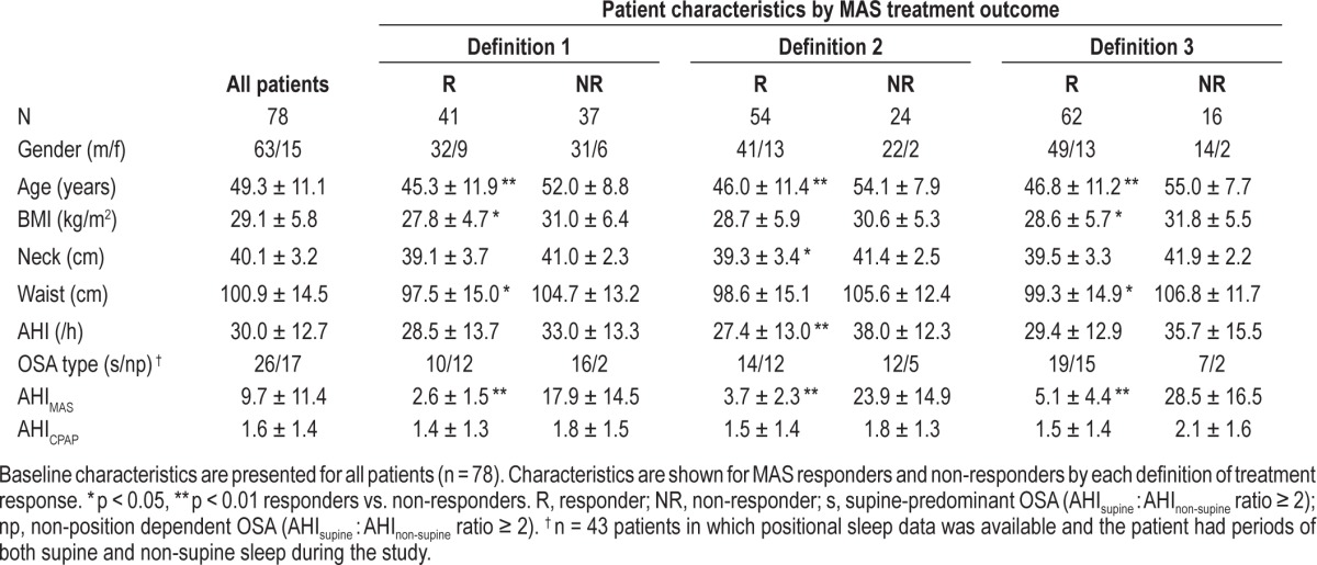
Seventy-eight patients who completed both MAS and CPAP treatment arms were confirmed to have AHI < 5/h on CPAP and were therefore included in the analysis. Excluded patients (CPAP AHI > 5/h) did not differ in age, BMI, neck or waist circumference, AHI, MAS response, or CPAP pressure requirement compared to those included in the analysis. Eight patients had mild OSA (AHI 10-14.9/h), 33 moderate (AHI 15-29.9/h), and 37 had severe OSA (AHI > 30/h). Patients were predominantly Caucasian, mostly male (81%), middle-aged, and over-weight; 52.6 percent of patients had a complete response to MAS treatment (AHI < 5/h, definition 1). Baseline characteristics are shown in Table 2. MAS treatment responders were significantly younger and less obese with a tendency towards a lower baseline AHI than non-responders. However gender proportions and supine-predominant OSA frequency did not differ between responders and non-responders in this sample.
Table 2.
Patient characteristics.
Therapeutic CPAP Pressure and MAS Treatment Response
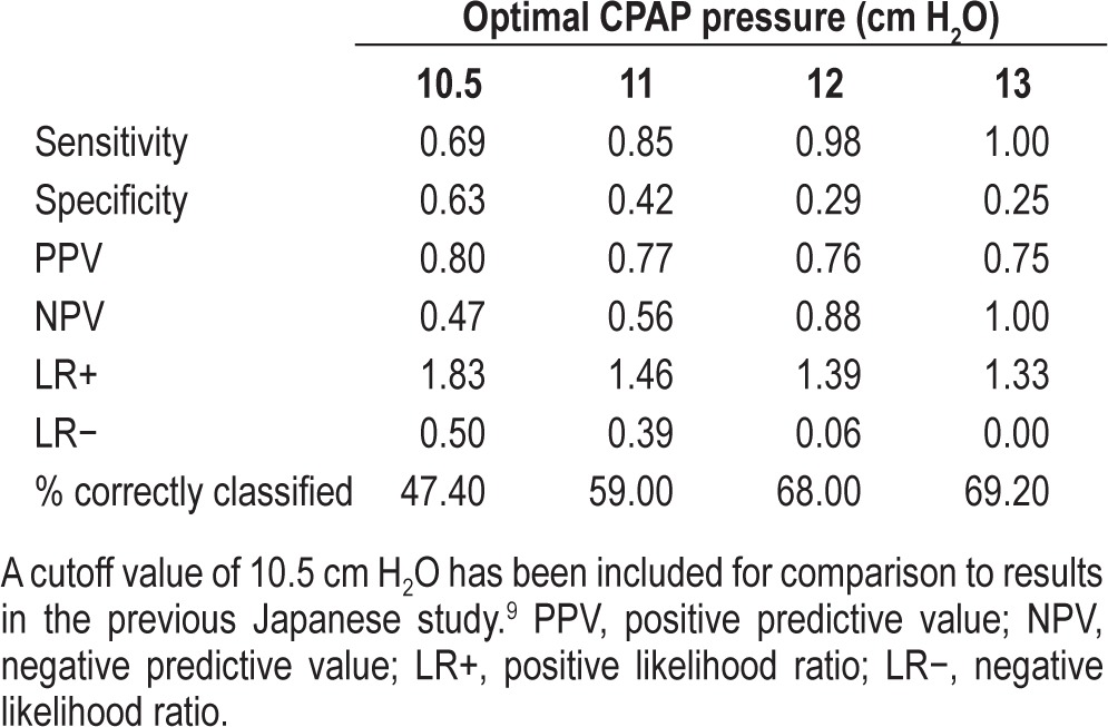
Mean CPAP pressure was 10.4 ± 2.1 (± SD), with a range of 4-18 cm H2O. CPAP pressure did not significantly differ between responders and non-responders by definition 1 (10.0 ± 1.4 vs. 10.8 ± 2.6 cm H2O, p = 0.09). By definition 2, responders (AHI < 10/h on MAS) had a lower CPAP pressure requirement than non-responders (9.7 ± 1.6 vs. 11.7 ± 2.4 cm H2O, p < 0.01). Responders defined by ≥ 50% reduction in AHI (definition 3) also had a lower CPAP pressure (10.0 ± 2.0 vs. 11.6 ± 2.3 cm H2O, p < 0.05). CPAP pressures for responders and non-responders, by all 3 definitions, are shown in Figure 2. In univariate analysis CPAP pressure had predictive value in discriminating MAS treatment responders and non-responders by definitions 2 (AUC [95% CI] 0.74 [0.63-0.86], p < 0.01) and 3 (0.70 [0.55-0.84], p < 0.05) (Table 3). As post-treatment AHI < 10/h (definition 2) is probably the most clinically useful, we explored CPAP pressure cutoff values to correctly classify patients using this model (Table 4). A pressure cutoff value of 13 cm H2O most accurately identified non-responders to MAS therapy (negative predictive value = 1). However, patients requiring pressures ≥ 13 cm H2O represented < 10% of this patient sample. This cutoff value correctly classified 69.2% of patients as MAS responders or non-responders. Below 13 cm H2O there was much overlap in pressures between responders and non-responders making CPAP pressure alone inadequate to discriminate between these patients.
Figure 2. Therapeutic CPAP pressure in responders and non-responders.
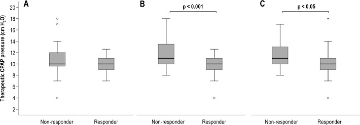
CPAP pressures are shown in box plots for MAS responders and non-responders by the 3 treatment response definitions. (A) Responder = AHI < 5/h with MAS (definition 1), (B) Responder = AHI < 10/h with MAS (definition 2), (C) Responder = ≥ 50% reduction in AHI from baseline with MAS (definition 3). Therapeutic CPAP pressure requirement was lower in MAS treatment responders by definitions 2 and 3. Box plot: Median values represented by horizontal line within the box, interquartile range by box edges. T-bars represent minimum and maximum values within range. Open circles represent outlier cases (values lying between one and a half to three box lengths above or below box edges) and the asterisk represents an extreme case (value more than 3 box lengths from either end of the box).
Table 3.
Univariate logistic regression analyses for prediction of MAS treatment response with optimal CPAP pressure (cm H2O) as the predictor variable.

Table 4.
Performance of various CPAP pressure values as cutoff values for prediction of MAS treatment response (defined as treatment AHI < 10/h, definition 2).
Prediction of MAS Treatment Response
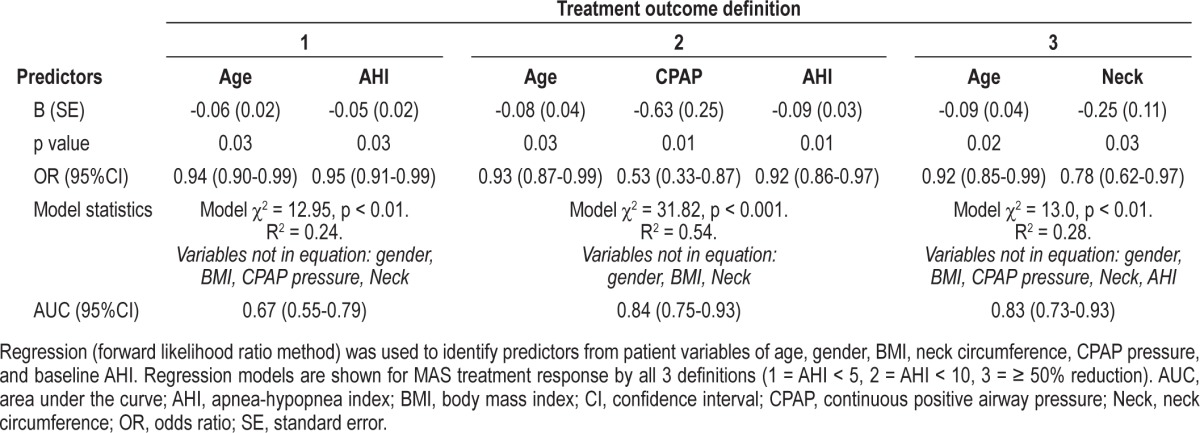
Multivariate logistic regression was performed to assess the utility of baseline characteristics (age, gender, BMI, neck circumference, baseline AHI, in combination with CPAP pressure) in the prediction of MAS treatment response (Table 5). In the model for MAS response by definition 1 (MAS AHI < 5/h) only baseline AHI and age were significant predictors. In predicting MAS response by definition 2 (MAS AHI < 10/h), the combination of baseline AHI, age, and CPAP pressure were significant, with 54% of the variance in MAS response explained by the model. This multivariate model correctly classified more patients than the prediction model based on CPAP pressure alone (AUC [95%CI] 0.84[0.75-0.93], p < 0.001). By definition 3 of MAS response (≥ 50% AHI reduction), only age and neck circumference, but not CPAP pressure, had predictive value.
Table 5.
Logistic regression analyses for prediction of MAS treatment response using baseline variables and therapeutic CPAP
DISCUSSION
This is the largest study to assess the relationship between therapeutic CPAP pressure and MAS treatment response and the first study in a treatment-naive and a non-Japanese population. Our findings lend support to the previously reported relationship between CPAP pressure and MAS treatment response,9 and extend these findings by identifying a much higher CPAP pressure cutoff for negative prediction of MAS response in this population. The implication is that there may be population-specific characteristics that influence the cutoff pressure values for which CPAP is best predictive of MAS response. This could be attributed to differences in obesity and craniofacial phenotypes between Japanese and Australian populations.
Our data support the notion that there is some relationship between MAS treatment response and CPAP pressure requirement with MAS non-responders requiring higher pressures. However this relationship seems not to be as pronounced as in the previous Japanese study, with higher pressures only observed in nonresponders by definitions 2 and 3 (MAS AHI < 10/h and > 50% AHI reduction). The median difference in pressures between responders and non-responders was also much narrower in the current study at 1 cm H2O, compared to ≥ 4 cm H2O in the previous study. In the Japanese study, a CPAP pressure cutoff value of 10.5 cm H2O most reliably classified patients in terms of MAS response, with pressures higher than this generally indicating a negative response to MAS.9 This value was inadequate for use in our patient sample and correctly classified only 47% of patients as non-responders, due to a large overlap of MAS treatment responders and non-responders with therapeutic CPAP pressures in the < 12 cm H2O range. Our results indicate that application of this method of prediction to an Australian population requires a higher cutoff value of 13 cm H2O for best discrimination, with 100% of patients above this level correctly classified as non-responders and 75% of the patients below this level as responders. This substantial difference in CPAP cutoff values to best classify MAS responders and nonresponders between these two populations suggests that there may be an influence of ethnicity factors on the relationship between CPAP pressure and MAS treatment response.
There are recognized differences between ethnicities in craniofacial and obesity risk factors associated with OSA. For the same level of OSA severity, Caucasians have been shown to have more obesity compared to Asians with OSA, whereas Asians show a greater restriction in craniofacial skeletal measurements associated with OSA, such as restricted maxillary and dimensions and retro-positioning, compared to Caucasians.11–14 Therefore both populations appear to have an anatomical imbalance contributing to upper airway collapsibility,15,16 but this is primarily driven by excess soft tissues in Caucasians and bony restriction in Asians. Differences in the relationship between CPAP pressure and MAS response may relate to these different hard and soft tissue proportions. BMI was lower in MAS responders in our study and was a predictor of response in univariate analyses (data not shown); however, no such relationship was evident in the Japanese study,9 suggesting obesity was less of a factor in MAS treatment response. BMI and neck circumference also relate to CPAP pressure in Caucasian populations.17,18 Craniofacial measurements have additionally contributed to CPAP pressure determination in a Japanese study, whereas only soft palate length had any association with CPAP pressure in a French study.19,20 Therefore craniofacial/obesity factors may have also differentially contributed to MAS response and/or CPAP pressure requirements between the two populations, although craniofacial factors were not assessed in either study.
Differences in the relationship between CPAP pressure and MAS treatment response between these two studies may additionally relate to other factors. There was also a difference in gender between the two studies, with the study of Tsuiki and colleagues including only males. Nineteen percent of subjects in the current study population were female. However there was still not adequate numbers to determine if gender has an influence on the MAS response/CPAP pressure relationship, although there was no difference in pressure requirement between genders (data not shown). Treatment success rates were much higher in the current study, with a greater proportion of patients achieving AHI < 5/h with MAS (47.6% vs. 29%), which may relate to differences in MAS devices. In the current study a titratable, two-piece appliance was used which allows the jaw to be advanced incrementally over time to maximize efficacy.21,22 Our treatments were implemented as part of a one-month crossover trial of optimal forms of both MAS and CPAP treatment with a 2-week treatment washout period in between. This differs to the previous Japanese study in which long-term compliant CPAP users were invited to participate and try MAS therapy.9 Previous and consistent use of CPAP may have some effect on the subsequent relationship with MAS treatment outcome, as it is possible that long-term CPAP use may influence the efficacy of MAS therapy through changes in upper airway and soft tissues and craniofacial skeletal structure.23,24
Our study found CPAP pressure combined with patient age and OSA severity (AHI) in a multivariate model provided the best discrimination of MAS treatment responders and nonresponders in this OSA population. Patient factors such as younger age, less obesity, female gender, and supine-dependent OSA have variously been associated with MAS treatment success.6,25–28 A significant limitation of MAS therapy is the inability to pre-identify patients with a good treatment response. Overall it seems unlikely that MAS response can be determined by single patient characteristics alone. MAS response is influenced by multiple factors relating to both structural and functional aspects of the upper airway.29 Objectively validated tests of MAS treatment function may ultimately be required to accurately predict treatment response.10,30–33 For example a single-night titration study of mandibular advancement using an available commercial remotely controlled titration device or assessment of upper airway response to mandibular advancement via nasendoscopy to observe the airway response during drug-induced sleep or even wakefulness.34–36 However in CPAP failure patients with known therapeutic pressure, this information in conjunction with age and OSA severity characteristics, may be useful to give an indication of the likelihood of success with MAS as an alternative therapy.
This study has extended investigation of a relationship between therapeutic CPAP pressure and MAS treatment response in a large sample of Australian OSA patients. However, potential study limitations include that although there was a range of pressures in the sample (4-18 cm H2O), only a minority of the sample (10%) required pressures higher than 13 cm H2O. Therefore we cannot confirm whether our negative predictive value would remain as high with the inclusion of more patients in the higher range. However, these pressures were confirmed to be therapeutic by polysomnography and they are within the range of commonly prescribed pressures. Furthermore, we were able to adequately demonstrate that the lower pressure cutoff value of 10.5 cm H2O is unsuitable for the studied population. Craniofacial factors are also implicated in MAS treatment response, but craniofacial assessment was not included in this analysis; however, a comprehensive cephalometric study in a similar OSA population suggests that craniofacial factors alone are not highly predictive of MAS response,37 and these can be difficult to assess in routine clinical practice. Finally, although our sample population likely included mostly patients with Caucasian ancestry, no ethnicity data was collected in this study.
In conclusion, therapeutic CPAP pressure was higher in MAS treatment non-responders compared to responders (depending on the definition of response used). CPAP pressure did have predictive utility in discriminating MAS treatment responders and non-responders in this sample of Australian OSA patients. However, the previously determined CPAP pressure threshold to identify MAS non-responders in a Japanese population was found to be inadequate for reliable prediction. Our results suggest CPAP pressures above 13 cm H2O are likely to indicate non-responsiveness to MAS treatment in the studied population. However prospective validation of CPAP pressure as a predictor of MAS response is still required. A combination of age, OSA severity, and CPAP pressure provided the best estimation of MAS treatment response, illustrating that one single patient variable is unlikely to provide a definitive indication in all patients. This study highlights the need to test reported prediction methods in different OSA populations in which relevant factors such as obesity and craniofacial phenotypes are likely to differ.
DISCLOSURE STATEMENT
ResMed Inc donated all continuous positive airway pressure equipment for the trial. SomnoMed Ltd. donated all oral appliances for the trial. Dr. Cistulli is a chief investigator on sponsored clinical trials in obstructive sleep apnea for ResMed Inc and Exploramed Inc. His department receives equipment support for oral appliance research from SomnoMed Ltd, and he has a pecuniary interest in the company from previous involvement in product development. He is a medical advisor to Exploramed Inc (a US medical device incubator) and Zephyr Sleep Technologies. He has received speaker fees/travel support from ResMed Inc Fisher & Paykel Healthcare. The other authors have indicated no financial conflicts of interest.
REFERENCES
- 1.Kribbs NB, Pack AI, Kline LR, et al. Objective measurement of patterns of nasal CPAP use by patients with obstructive sleep apnea. Am Rev Respir Dis. 1993;147:887–95. doi: 10.1164/ajrccm/147.4.887. [DOI] [PubMed] [Google Scholar]
- 2.Kushida CA, Morgenthaler TI, Littner MR, et al. Practice parameters for the treatment of snoring and obstructive sleep apnea with oral appliances: an update for 2005. Sleep. 2006;29:240–3. doi: 10.1093/sleep/29.2.240. [DOI] [PubMed] [Google Scholar]
- 3.Phillips CL, Grunstein RR, Darendeliler MA, et al. Health outcomes of continuous positive airway pressure versus oral appliance treatment for obstructive sleep apnea: a randomized controlled trial. Am J Respir Crit Care Med. 2013;187:879–87. doi: 10.1164/rccm.201212-2223OC. [DOI] [PubMed] [Google Scholar]
- 4.Chan AS, Sutherland K, Schwab RJ, et al. The effect of mandibular advancement on upper airway structure in obstructive sleep apnoea. Thorax. 2010;65:726–32. doi: 10.1136/thx.2009.131094. [DOI] [PubMed] [Google Scholar]
- 5.Gotsopoulos H, Kelly JJ, Cistulli PA. Oral appliance therapy reduces blood pressure in obstructive sleep apnea: a randomized, controlled trial. Sleep. 2004;27:934–41. doi: 10.1093/sleep/27.5.934. [DOI] [PubMed] [Google Scholar]
- 6.Mehta A, Qian J, Petocz P, Darendeliler MA, Cistulli PA. A randomized, controlled study of a mandibular advancement splint for obstructive sleep apnea. Am J Respir Crit Care Med. 2001;163:1457–61. doi: 10.1164/ajrccm.163.6.2004213. [DOI] [PubMed] [Google Scholar]
- 7.Pitsis AJ, Darendeliler MA, Gotsopoulos H, Petocz P, Cistulli PA. Effect of vertical dimension on efficacy of oral appliance therapy in obstructive sleep apnea. Am J Respir Crit Care Med. 2002;166:860–4. doi: 10.1164/rccm.200204-342OC. [DOI] [PubMed] [Google Scholar]
- 8.Sutherland K, Cistulli P. Mandibular advancement splints for the treatment of sleep apnea syndrome. Swiss Med Wkly. 2011;141:w13276. doi: 10.4414/smw.2011.13276. [DOI] [PubMed] [Google Scholar]
- 9.Tsuiki S, Kobayashi M, Namba K, et al. Optimal positive airway pressure predicts oral appliance response to sleep apnoea. Eur Respir J. 2010;35:1098–105. doi: 10.1183/09031936.00121608. [DOI] [PubMed] [Google Scholar]
- 10.Zeng B, Ng AT, Qian J, Petocz P, Darendeliler MA, Cistulli PA. Influence of nasal resistance on oral appliance treatment outcome in obstructive sleep apnea. Sleep. 2008;31:543–7. doi: 10.1093/sleep/31.4.543. [DOI] [PMC free article] [PubMed] [Google Scholar]
- 11.Lee RW, Vasudavan S, Hui DS, et al. Differences in craniofacial structures and obesity in Caucasian and Chinese patients with obstructive sleep apnea. Sleep. 2010;33:1075–80. doi: 10.1093/sleep/33.8.1075. [DOI] [PMC free article] [PubMed] [Google Scholar]
- 12.Li KK, Kushida C, Powell NB, Riley RW, Guilleminault C. Obstructive sleep apnea syndrome: a comparison between Far-East Asian and white men. Laryngoscope. 2000;110:1689–93. doi: 10.1097/00005537-200010000-00022. [DOI] [PubMed] [Google Scholar]
- 13.Liu Y, Lowe AA, Zeng X, Fu M, Fleetham JA. Cephalometric comparisons between Chinese and Caucasian patients with obstructive sleep apnea. Am J Orthod Dentofacial Orthop. 2000;117:479–85. doi: 10.1016/s0889-5406(00)70169-7. [DOI] [PubMed] [Google Scholar]
- 14.Sutherland K, Lee RW, Cistulli PA. Obesity and craniofacial structure as risk factors for obstructive sleep apnoea: impact of ethnicity. Respirology. 2012;17:213–22. doi: 10.1111/j.1440-1843.2011.02082.x. [DOI] [PubMed] [Google Scholar]
- 15.Watanabe T, Isono S, Tanaka A, Tanzawa H, Nishino T. Contribution of body habitus and craniofacial characteristics to segmental closing pressures of the passive pharynx in patients with sleep-disordered breathing. Am J Respir Crit Care Med. 2002;165:260–5. doi: 10.1164/ajrccm.165.2.2009032. [DOI] [PubMed] [Google Scholar]
- 16.Tsuiki S, Isono S, Ishikawa T, Yamashiro Y, Tatsumi K, Nishino T. Anatomical balance of the upper airway and obstructive sleep apnea. Anesthesiology. 2008;108:1009–15. doi: 10.1097/ALN.0b013e318173f103. [DOI] [PubMed] [Google Scholar]
- 17.Hoffstein V, Mateika S. Predicting nasal continuous positive airway pressure. Am J Respir Crit Care Med. 1994;150:486–8. doi: 10.1164/ajrccm.150.2.8049834. [DOI] [PubMed] [Google Scholar]
- 18.Loredo JS, Berry C, Nelesen RA, Dimsdale JE. Prediction of continuous positive airway pressure in obstructive sleep apnea. Sleep Breath. 2007;11:45–51. doi: 10.1007/s11325-006-0082-x. [DOI] [PubMed] [Google Scholar]
- 19.Akashiba T, Kosaka N, Yamamoto H, Ito D, Saito O, Horie T. Optimal continuous positive airway pressure in patients with obstructive sleep apnoea: role of craniofacial structure. Respir Med. 2001;95:393–7. doi: 10.1053/rmed.2001.1058. [DOI] [PubMed] [Google Scholar]
- 20.Sforza E, Krieger J, Bacon W, Petiau C, Zamagni M, Boudewijns A. Determinants of effective continuous positive airway pressure in obstructive sleep apnea. Role of respiratory effort. Am J Respir Crit Care Med. 1995;151:1852–6. doi: 10.1164/ajrccm.151.6.7767530. [DOI] [PubMed] [Google Scholar]
- 21.Aarab G, Lobbezoo F, Hamburger HL, Naeije M. Effects of an oral appliance with different mandibular protrusion positions at a constant vertical dimension on obstructive sleep apnea. Clin Oral Investig. 2010;14:339–45. doi: 10.1007/s00784-009-0298-9. [DOI] [PubMed] [Google Scholar]
- 22.Walker-Engstrom ML, Ringqvist I, Vestling O, Wilhelmsson B, Tegelberg A. A prospective randomized study comparing two different degrees of mandibular advancement with a dental appliance in treatment of severe obstructive sleep apnea. Sleep Breath. 2003;7:119–30. doi: 10.1007/s11325-003-0119-3. [DOI] [PubMed] [Google Scholar]
- 23.Ryan CF, Lowe AA, Li D, Fleetham JA. Magnetic resonance imaging of the upper airway in obstructive sleep apnea before and after chronic nasal continuous positive airway pressure therapy. Am Rev Respir Dis. 1991;144:939–44. doi: 10.1164/ajrccm/144.4.939. [DOI] [PubMed] [Google Scholar]
- 24.Tsuda H, Almeida FR, Tsuda T, Moritsuchi Y, Lowe AA. Craniofacial changes after 2 years of nasal continuous positive airway pressure use in patients with obstructive sleep apnea. Chest. 2010;138:870–4. doi: 10.1378/chest.10-0678. [DOI] [PubMed] [Google Scholar]
- 25.Chung JW, Enciso R, Levendowski DJ, Morgan TD, Westbrook PR, Clark GT. Treatment outcomes of mandibular advancement devices in positional and nonpositional OSA patients. Oral Surg Oral Med Oral Pathol Oral Radiol Endod. 2010;109:724–31. doi: 10.1016/j.tripleo.2009.11.031. [DOI] [PMC free article] [PubMed] [Google Scholar]
- 26.Hoekema A, Doff MH, de Bont LG, et al. Predictors of obstructive sleep apneahypopnea treatment outcome. J Dent Res. 2007;86:1181–6. doi: 10.1177/154405910708601208. [DOI] [PubMed] [Google Scholar]
- 27.Liu Y, Lowe AA, Fleetham JA, Park YC. Cephalometric and physiologic predictors of the efficacy of an adjustable oral appliance for treating obstructive sleep apnea. Am J Orthod Dentofacial Orthop. 2001;120:639–47. doi: 10.1067/mod.2001.118782. [DOI] [PubMed] [Google Scholar]
- 28.Marklund M, Stenlund H, Franklin KA. Mandibular advancement devices in 630 men and women with obstructive sleep apnea and snoring: tolerability and predictors of treatment success. Chest. 2004;125:1270–8. doi: 10.1378/chest.125.4.1270. [DOI] [PubMed] [Google Scholar]
- 29.Chan AS, Lee RW, Srinivasan VK, Darendeliler MA, Cistulli PA. Use of flow-volume curves to predict oral appliance treatment outcome in obstructive sleep apnea: a prospective validation study. Sleep Breath. 2011;15:157–62. doi: 10.1007/s11325-010-0395-7. [DOI] [PubMed] [Google Scholar]
- 30.Bosshard V, Masse JF, Series F. Prediction of oral appliance efficiency in patients with apnoea using phrenic nerve stimulation while awake. Thorax. 2011;66:220–5. doi: 10.1136/thx.2010.150334. [DOI] [PubMed] [Google Scholar]
- 31.De Backer JW, Vanderveken OM, Vos WG, et al. Functional imaging using computational fluid dynamics to predict treatment success of mandibular advancement devices in sleep-disordered breathing. J Biomech. 2007;40:3708–14. doi: 10.1016/j.jbiomech.2007.06.022. [DOI] [PubMed] [Google Scholar]
- 32.Zeng B, Ng AT, Darendeliler MA, Petocz P, Cistulli PA. Use of flow-volume curves to predict oral appliance treatment outcome in obstructive sleep apnea. Am J Respir Crit Care Med. 2007;175:726–30. doi: 10.1164/rccm.200608-1205OC. [DOI] [PubMed] [Google Scholar]
- 33.Zhao M, Barber T, Cistulli P, Sutherland K, Rosengarten G. Computational fluid dynamics for the assessment of upper airway response to oral appliance treatment in obstructive sleep apnea. J Biomech. 2013;46:142–50. doi: 10.1016/j.jbiomech.2012.10.033. [DOI] [PubMed] [Google Scholar]
- 34.Remmers JE, Charkhandeh S, Grosse J, et al. Remotely controlled mandibular protrusion during sleep predicts therapeutic success with oral appliances in patients with obstructive sleep apnea. Sleep. 2013;36:1517–25. doi: 10.5665/sleep.3048. [DOI] [PMC free article] [PubMed] [Google Scholar]
- 35.Vroegop AV, Vanderveken OM, Dieltjens M, et al. Sleep endoscopy with simulation bite for prediction of oral appliance treatment outcome. J Sleep Res. 2013;22:348–55. doi: 10.1111/jsr.12008. [DOI] [PubMed] [Google Scholar]
- 36.Chan AS, Lee RW, Srinivasan VK, Darendeliler MA, Grunstein RR, Cistulli PA. Nasopharyngoscopic evaluation of oral appliance therapy for obstructive sleep apnoea. Eur Respir J. 2010;35:836–42. doi: 10.1183/09031936.00077409. [DOI] [PubMed] [Google Scholar]
- 37.Ng AT, Darendeliler MA, Petocz P, Cistulli PA. Cephalometry and prediction of oral appliance treatment outcome. Sleep Breath. 2012;16:47–58. doi: 10.1007/s11325-011-0484-2. [DOI] [PubMed] [Google Scholar]


