Abstract
Pericardial cysts are rare congenital abnormalities with a reported incidence rate of 1/100,000 males. The most common locations of these cysts are middle mediastinum and right cardiophrenic angle. Pericardial cysts are usually asymptomatic unless a complication or rapid growth occurs. Diagnosis is generally made incidentally or using combined radiological X-ray, contrast Computed Tomography (CT), and echocardiography imaging. Various forms of treatments include observation, needle aspiration, and surgical excision of the cysts. Recurrence of the disease is common when aspiration approach is selected. Herein, we present a case of pericardial cyst affecting right cardiophrenic angle in a 36 year-old man with an atypical right chest pain and chronic cough. Finally, the patient was treated successfully using surgical approach.
Keywords: Pericardium, Cyst, Cough, Surgery
INTRODUCTION
Pericardial cysts are considered as a congenital abnormality. They are usually located in middle mediastinum and are mostly found incidentally. Pericardial cysts rarely cause symptoms (1). Occasionally, they are accompanied by complications including infection, tamponade, hemorrhage and respiratory symptoms which necessitate treatment (2–4). Herein, we report a patient complaining of non-productive chronic cough who was diagnosed with pericardial cyst and consequently treated successfully using surgical approach.
CASE SUMMARIES
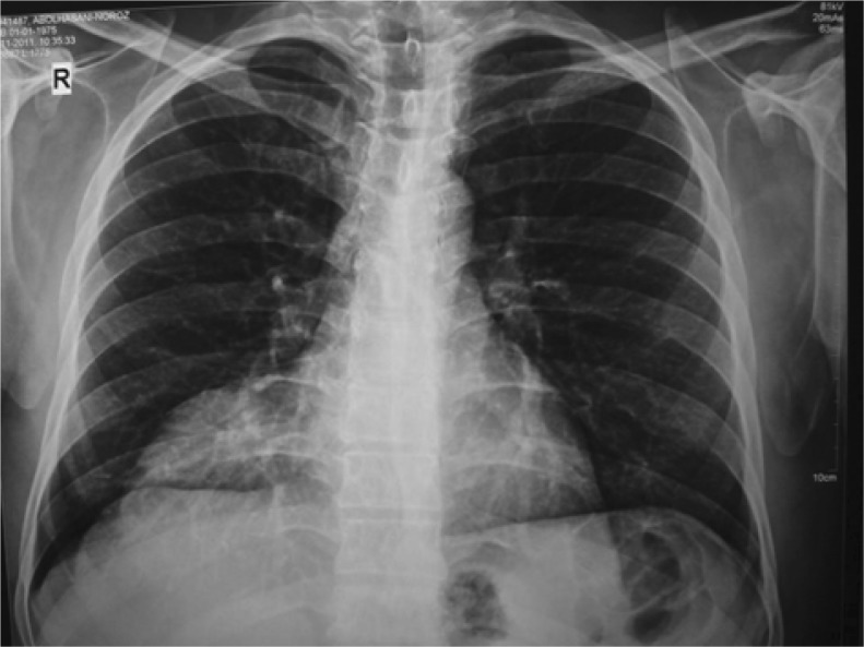
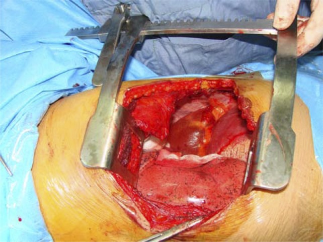
In March 2010, a 36 year-old man presented to our referral hospital with chronic non-productive cough and right chest pain which did not alter by respiration but deteriorated by positioning to either side. His chronic cough started three years ago without sputum. Cardiopulmonary examination and vital signs were normal. Chest X-ray examination revealed a homogenous density in the right cardiophrenic angle (Figure 1). Abdominal Ultrasound revealed loculated fluid in the medial base of right pulmonary area and a grade II fatty liver. On Computed Tomography (CT) a sharply-marginated cystic mass with dimensions of 14×10×7 cm and attenuation value of 40 HU was observed. With probable diagnoses of pericardial or thymus cyst and Morgagni's hernia, he was prepared for surgery. Through right standard posterolateral thoracotomy and 6th intercostal space, the pleura lying on the cystic mass in the cardiophrenic triangle was dissected from the pericardium in the anterior, posterior and inferior aspects of the cyst. The vascular connections of the cyst with pericardium were easily ligated and the cyst was resected completely from the pericardium while no connection to the pericardial cavity was observed (Figure 2).
Figure 1.
Chest X-ray showing a large mass in the right cardiophrenic triangle
Figure 2.
A large pericardial cyst seen in the right cardiophrenic triangle
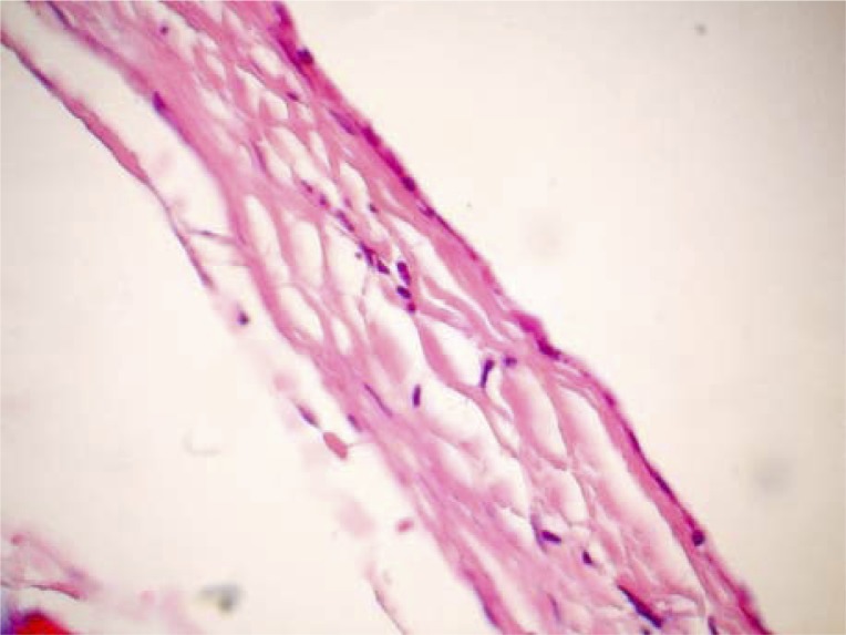
After insertion of a chest tube into the pleural cavity the chest was closed. Pathologic examinations revealed a single layer of mesothelial cells and fluid-filled cyst compatible with pericardial cyst (Figure 3). No complications observed during or after surgery and in the one-year follow up period of the patient.
Figure 3.
Pathological view of a sample from the cyst
DISCUSSION
Pericardial cysts are rare benign congenital abnormalities (5). They may; however, be acquired after cardiothoracic surgeries iatrogenically (1, 6). Their sizes are usually reported to be 2 to 28 cm and giant cysts have been reported sporadically as well (6, 7). Our patient had a 14 cm slow-growing congenital cyst. Their estimated prevalence is 1/100,000 males. Pericardial cysts are usually located in the right cardiophrenic triangle (75%), and less frequently in the left cardiophrenic triangle (22%) or other parts of the mediastinum (8). The cyst in the present case was located in the right cardiophrenic triangle which is the most commonly involved area. Although pericardial cysts are asymptomatic, some patients may experience chest pain, dyspnea, pericardial tamponade (2), hemorrhage, infection, atrial fibrillation and heart failure due to hemorrhage (3, 4, 8). Our patient presented with chronic non-productive cough and atypical chest pain, a case similar to a patient reported by Patel et al (6). Some pericardial cysts resolve spontaneously and can rupture into pleural or pericardial spaces (4).
CT and Echocardiography are the choice diagnostic procedures (5, 6). Ultrasound and CT scan can often distinguish between mediastinal cysts and masses as observed in our patient (1).
Observation is one of the management approaches toward pericardial cysts especially in high risk patients or when the cysts are not enlarged. However, there is no safety guarantee. Percutaneous drainage and aspiration of the cyst are another approach of treatment associated with a 30% recurrence. Resection is indicated for symptomatic enlarged pericardial cysts, those with malignancy potential and for prevention of complications (1, 4, 9). Today, VATS (video-assisted thoracoscopic surgery) and robotic resection are well-accepted procedures in most centers (9); however, we did not select this procedure due to the probability of other differential diagnoses like Morgagni's hernia. VATS procedure is associated with low operative risks and therefore is recommended for all healthy patients with pericardial cyst. It has numerous advantages including less pain, shorter post-operative recovery and better cosmetic results. However, in complicated pericardial cysts or cases with doubtful diagnosis, VATS procedure is not recommended as in our case.
CONCLUSION
Pericardial cysts are usually asymptomatic. However, resection is indicated when they are enlarged or accompanied by complications and symptoms.
REFERENCES
- 1.Stoller JK, Shaw C, Matthay RA. Enlarging, atypically located pericardial cyst. Recent experience and literature review. Chest. 1986;89(3):402–6. doi: 10.1378/chest.89.3.402. [DOI] [PubMed] [Google Scholar]
- 2.Komodromos T, Lieb D, Baraboutis J. Unusual presentation of a pericardial cyst. Heart Vessels. 2004;19(1):49–51. doi: 10.1007/s00380-003-0716-x. [DOI] [PubMed] [Google Scholar]
- 3.Hoque M, Siripurapu S. Methicillin-resistant Staphylococcus aureus-infected pericardial cyst. Mayo Clin Proc. 2005;80(9):1116. doi: 10.4065/80.9.1116. [DOI] [PubMed] [Google Scholar]
- 4.Shiraishi I, Yamagishi M, Kawakita A, Yamamoto Y, Hamaoka K. Acute cardiac tamponade caused by massive hemorrhage from pericardial cyst. Circulation. 2000;101(19):E196–7. doi: 10.1161/01.cir.101.19.e196. [DOI] [PubMed] [Google Scholar]
- 5.Demos TC, Budorick NE, Posniak HV. Benign mediastinal cysts: pointed appearance on CT. J Comput Assist Tomogr. 1989;13(1):132–3. doi: 10.1097/00004728-198901000-00030. [DOI] [PubMed] [Google Scholar]
- 6.Patel J, Park C, Michaels J, Rosen S, Kort S. Pericardial cyst: case reports and a literature review. Echocardiography. 2004;21(3):269–72. doi: 10.1111/j.0742-2822.2004.03097.x. [DOI] [PubMed] [Google Scholar]
- 7.Satur CM, Hsin MK, Dussek JE. Giant pericardial cysts. Ann Thorac Surg. 1996;61(1):208–10. doi: 10.1016/0003-4975(95)00720-2. [DOI] [PubMed] [Google Scholar]
- 8.Borges AC, Gellert K, Dietel M, Baumann G, Witt C. Acute right-sided heart failure due to hemorrhage into a pericardial cyst. Ann Thorac Surg. 1997;63(3):845–7. doi: 10.1016/s0003-4975(96)01373-2. [DOI] [PubMed] [Google Scholar]
- 9.Bacchetta MD, Korst RJ, Altorki NK, Port JL, Isom OW, Mack CA. Resection of a symptomatic pericardial cyst using the computer-enhanced da Vinci Surgical System. Ann Thorac Surg. 2003;75(6):1953–5. doi: 10.1016/s0003-4975(02)05008-7. [DOI] [PubMed] [Google Scholar]


