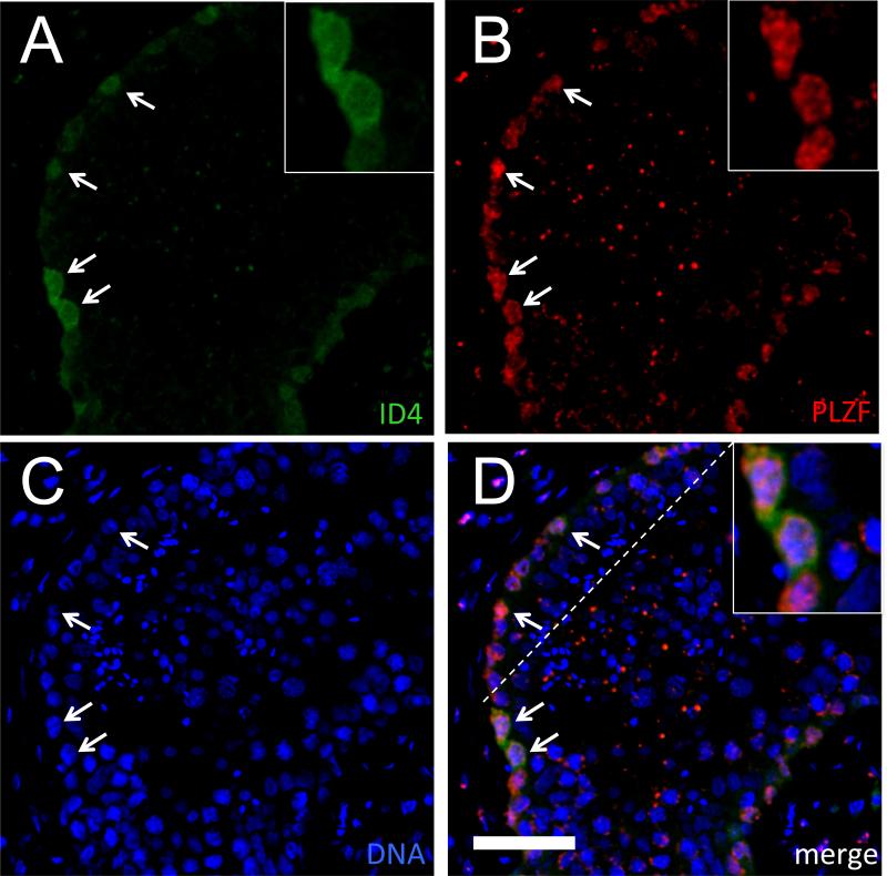
Figure 3.
Co-localization of anti-ID4 and anti-PLZF signals in the human seminiferous epithelium. Representative IF using rabbit monoclonal anti-ID4 (green; A) or mouse monoclonal anti-PLZF (red; B), DNA counterstain (blue; C), or merged image (D) from a 21 year old. Arrows denote ID4+/PLZF+ cells. Bar is 50 μm. Insets show selected ID4+/PLZF+ cells with both cytoplasmic and nuclear staining.

