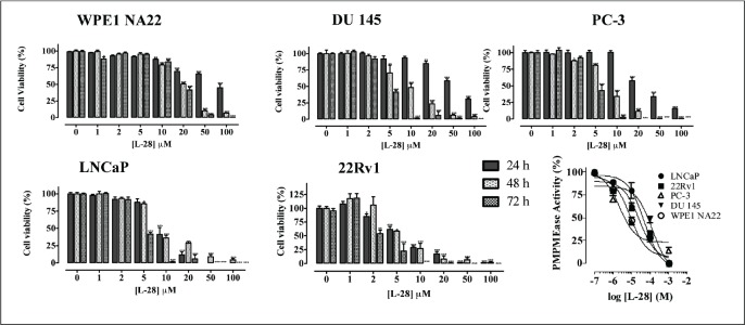
Figure 2. L-28 induces apoptosis in prostate cancer cell lines. Cells were plated in 96-wells at a density of 2×10⁴ as described in the methods. At 24, 48 and 72 h of treatment with varying concentrations of L-28, the cell viabilities were measured by fluorescence using the resazurin reduction assay. The bottom right panel shows the inhibition of PMPMEase activity in the different cell lysates by L-28. Each point represents the mean ± SEM of 4 determinations. *p < 0.01, **p < 0.001, and ***p < 0.0001 when compared to the normal prostate cells by paired t-test.

