Abstract
Leukocyte migration into tissues is characteristic of inflammation. It is usually measured in vitro as the average displacement of populations of cells towards a chemokine gradient, not acknowledging other patterns of cell migration. Here, we designed and validated a microfluidic migration platform to simultaneously analyze four qualitative migration patterns: chemo-attraction, -repulsion, -kinesis and -inhibition, using single-cell quantitative metrics of direction, speed, persistence, and fraction of cells responding. We find that established chemokines C5a and IL-8 induce chemoattraction and repulsion in equal proportions, resulting in the dispersal of cells. These migration signatures are characterized by high persistence and speed and are independent of the chemokine dose or receptor expression. Furthermore, we find that twice as many T-lymphocytes migrate away than towards SDF-1 and their directional migration patterns are not persistent. Overall, our platform characterizes migratory signature responses and uncovers an avenue for precise characterization of leukocyte migration and therapeutic modulators.
Introduction
The migration of leukocytes into sites of immune challenge is characteristic of inflammation 1, 2 and is tightly controlled by soluble 3-5 and immobilized chemical gradients 6. Migratory responses induced by chemical cues are traditionally classified into one of four different patterns that include directional migration towards a secreted protein gradient (chemoattraction) 4-6, migration in random directions (chemokinesis) 7, migration away from a source (chemorepulsion) 8, 9 and reduced migration in any direction (chemoinhibition). However, our ability to characterize these migration patterns is poor in standard migration assays. For example, Boyden chambers 10, Dunn and Zigmond chambers 11, 12, micropipette techniques 13, and most microfluidic-based assays 14-18 have significant limitations in quantifying the dynamic nature of the migration process, and typically only monitor a single averaged pattern. For instance, the Boyden chamber can be used to directly assess population-averaged migration in one direction (chemoattraction) but can only indirectly assess other patterns of migration (e.g. by comparing at least four measurements in a “checkerboard” assay10). It also lacks single cell resolution, and cannot identify heterogeneous migratory patterns within subsets of leukocytes. The Dunn and Zigmond chambers and micropipette techniques allow for single cell resolution 11-13, however, the ability to differentiate among different migration patterns is hampered by large variations in speed and directionality, which is typical for cells migrating on flat surfaces19. Until recently, microfluidic assays have focused on technology, such as controlling the shape and stability of chemical gradients17, competing gradients20, or on-chip neutrophil purification from whole blood samples 18, 21. However, the large variations in speed and directionality during leukocyte chemotaxis on flat surfaces in microfluidic devices limit precision for studies that require quantification of leukocyte migration 15-18. Newer microfluidic devices that confine moving leukocytes within small channels circumvent these problems and enable significantly higher precision measurements of leukocyte migration14, 22-24. Microfluidic devices enabled our group to define a normal range of human neutrophil velocity in healthy individuals14 and helped optimize a treatment that restores defective neutrophil directionality following burn injuries 22. However, like other chemotaxis assays, these devices were designed only to measure chemoattraction and ignored other cell migration patterns.
In this study, we design and validate a microfluidic device that employs two large-scale arrays of microchannels connected to facilitate the quantification of leukocyte migration both towards and away from chemical gradients. The device ensures that a precise number of cells are exposed to identical conditions at the initiation of the experiment and enables us to identify specific leukocyte migration signatures in response to well-established chemokines. Whereas the neutrophil chemoattractants fMet-Leu-Phe (fMLP) and Leukotriene B4 (LTB4) induce chemoattraction that is fast and persistent, interleukin-8 (IL-8) and complement component 5a (C5a) 25, 26 induce both chemoattraction and repulsion, in equal proportions, with high migratory persistence and speed. These effects are not dose-, receptor- or subset- dependent, but rather appear to be cell intrinsic. Similarly, stromal cell-derived factor 1 (SDF-1) acts on lymphocytes to induce both attraction and repulsion, although twice as many lymphocytes migrate in a chemorepulsive rather than in a chemoattractant manner and directional migration patterns are not persistent. Furthermore, we show that the neutrophil inhibitor Slit2 alters the size of migrating subpopulations of neutrophils without changing their overall migration patterns. The data presented in this report provides a quantitative analysis of leukocyte migration patterns not previously defined in vitro using traditional techniques. We suggest that multi-parameter assessment of migration has great potential for functional mechanistic analyses, and is critical for the optimal design of anti-inflammatory agents.
Results
Microfluidic device for evaluation of leukocyte migration
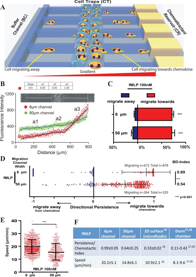
We designed a microfluidic device in which precise numbers of leukocytes are reproducibly loaded and uniformly distributed into a central main channel with cell traps. After loading, the cells leave the traps and migrate into side channels (6 × 6 μm or 50 × 6 μm for neutrophils and 10 × 6 μm for lymphocytes). Half of the channels lead into an array of chemokine reservoirs (on one side of the main channel) and half (on the opposite side) lead into a channel with buffer alone (Fig. 1A and Supplementary Fig. 1A). This design allows for the formation of two spatial chemokine gradients on each side of the central main channel, one with an increasing gradient towards the chemokine and the other with a decreasing concentration gradient towards the channel with buffer. We tested the stability of chemical gradients within the device channels, and found that they remained constant for ~ 12 hours in both 6 and 10 μm channels and for ~ 4 hours in 50 μm channels (Fig. 1B). The simultaneous presence of the two gradients enables quantitative measurements at single cell resolution of migration both towards and away from gradients, from the same initial condition for all cells in a given population.
Figure 1. Measuring attraction, repulsion, kinesis and inhibition during leukocyte responses to chemical gradients.
A) Cell migration in mechanically confined spaces can be characterized in relation to the direction of the chemical gradients and quantified for speed, directionality and persistence. Cells loaded in the Cell Traps (CT) can either migrate towards or away from the chemokine gradient. B) Characterization of the stable chemical gradients that can be established in the device. Fluorescein labeled dextran of 10kDa was used. After three hours a steep gradient is preserved in 6μm channels, while in 50μm channels a shallower gradient is preserved. a1 represents the migration channel from the buffer channel towards the cell loading chamber, a2 the cell loading chamber and a3 the migration channel from cell loading chamber towards the chemokine reservoir. C). Percentage of human neutrophils loaded in the migration channel that migrate towards or away from fMLP in 6 and 50μm wide migration channels. Neutrophils migrate in significantly greater numbers towards fMLP than away from it (p<0.001, Student's t test). Combined data from N=3 independent experiments. D) Scatterplot of migratory persistence of human neutrophils towards and away from a gradient of fMLP in 6 and 50μm migration channels. The Bi-Directional Index combines persistence and directionality in one parameter and is significantly higher in narrow (6μm) channels (p<0.001, Mann Whitney test). Bars represent mean ± SD. E) Migratory speed of neutrophils migrating towards fMLP in 6 and 50μm wide migration channels. Neutrophils migrate in significantly faster towards fMLP in 6 μm than in 50μm channels (p<0.001, Student's t test). Bars represent mean ± SD. F) Effect of migration channel width on migratory persistence and speed of cells migrating towards fMLP. Comparison between the Bi-Directional microfluidic device and established 2D microfluidic devices and Dunn chambers18, 27, 28. A total of 878 cells were analyzed in 6μm channels (672 migrated); 320 (264 migrated) in 50μm channels, N=3.
fMLP induces bidirectional migratory patterns in HL-60 cells
To validate the device, we first characterized the migratory patterns of a clonal population of HL-60 cells in response to fMLP gradients (maximum concentration, 100 nM). Using speed and Directional Persistence (DP – see methods section, Supplementary Fig. 1C), we divided individual cells into three groups 1) high persistence (DP [0.66-1]), 2) medium persistence (DP [0.33-0.66)), and 3) low persistence (DP [0-0.33)), see Supplementary Fig. 1 C-F. For HL-60 cells that respond to fMLP gradients, the largest cluster is comprised of cells that migrate with high persistence towards the chemoattractant (~67% of all migrating cells, DP [0.66-1], Supplementary Fig. 1E). The second largest cluster is that of cells that migrate towards the chemoattractant with low persistence, meaning they eventually reverse migrate back to their initial starting position (~ 13% cells, DP<0.33). A cluster of comparable size is comprised of cells that migrate in a random manner away from fMLP with low DP (~10% cells, −0.33<DP≤0) and a smaller cluster of HL-60 cells in the device were found to migrate away from fMLP with high DP (~ 4%, −1≤DP≤-0.66). Overall, these results highlight the known, dominant function of fMLP as a pro-migratory chemoattractant. Nevertheless our findings also demonstrate that significant numbers of clonal HL-60 cells migrate in a random and/or repulsed manner.
Higher migratory persistence in narrow migration channels
We compared the migratory response of human neutrophils to fMLP gradients in narrow 6 μm and wide 50 μm channels (Supplementary Movie 1 and 2). We found that the numbers and proportions of migrating neutrophils are comparable in narrow and wide channels. In 6 μm channels, a total of 71.5±3.8% neutrophils migrated towards fMLP and 5.4±1.7% migrated away from it, whereas in 50 μm channels, chemoattraction was noted in 71.2±3.4% and chemorepulsion in 9.7±2.8% of the neutrophils; a total of 23% cells (in 6 μm) and 20% cells (in 50 μm) did not migrate, respectively (Fig. 1C). Cell movement towards fMLP was significantly more persistent in 6μm channels, where 90% of moving neutrophils migrated with maximum directional persistence (DP = 1) and with a high Bi-Directional-Index (BD-Index = 0.89). In the wider 50μm channels, we found that cells failed to achieve maximal directional persistence and the BD-Index was significantly reduced (BD-Index = 0.54) vs. migration in 6μm channels (Fig. 1D, p<0.001). As illustrated in Fig 1E, migratory speed was also significantly faster in 6μm channels (20.2±5.1 μm min−1 vs. 14.8±6.1 μm min−1, p<0.001). The trend for reduced directional persistence and slower speed with decreasing mechanical confinement is further supported by the evaluation of neutrophil migration measurements in 2D surface microfluidic devices and traditional Dunn chambers (Fig 1F). In these assays, neutrophil migration takes place on flat surfaces, guided exclusively by chemical gradients and no mechanical restrictions, resulting in low migratory persistence (0.11-0.42 on flat surfaces vs. 0.99 in 6μm channels) and speed (8.1-10.9 μm min−1 vs. 20.2 μm min−1 in 6μm channels)18, 27, 28. Thus, confinement of cells within narrow channels which have cross sections comparable to extracellular matrix 29 is similar to that of leukocytes following paths of least resistance in tissues 30 and is associated with a fast and persistent migratory response.
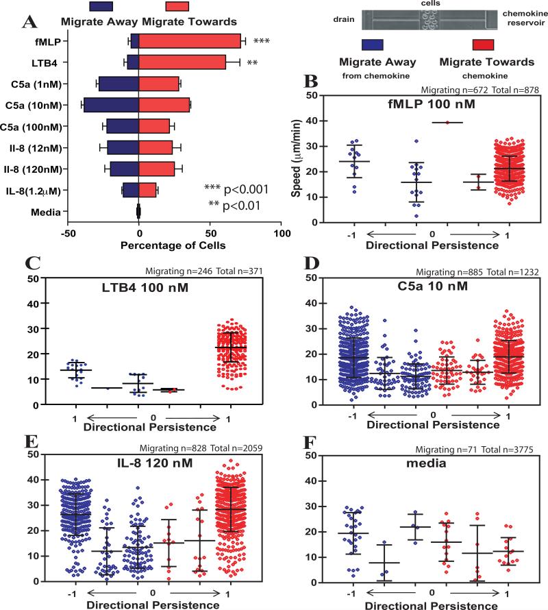
In separate experiments, we compared the response of human neutrophils to fMLP and LTB4. We observed that 76.9±3.9% and 68.9±9.7% of human neutrophils migrated to fMLP and LTB4, respectively. Of motile neutrophils, 93% and 89% migrated towards each chemoattractant with high directional persistence (DP > 0.66, Fig. 2A-C, p<0.01). We found that the average directional persistence towards LTB4 decreases from 0.99±0.09 to 0.74±0.19 (p<0.001) and the average BD-Index decreases from 0.79±0.56 to 0.70±0.28 (p<0.001) in wider vs. narrower channels (Supplementary Fig. 3). Moreover, we observed differences in the neutrophil migration patterns after neutrophils exit the wider vs. narrower migration channels and enter the chemokine reservoirs. While the neutrophils migrating through wide migratory channels continue to migrate inside the chemokine reservoirs, the neutrophils exiting narrow channels stop migrating quickly after entering. This phenomenon could be explained by the differences in the gradient slope inside the chemokine reservoirs. Whereas the flux of chemokine from the reservoir through the wider migration channels (300 μm2 cross section) can be almost an order of magnitude larger than in narrower migration channels (36 μm2), a larger gradient might form inside the reservoirs connected to wider vs. narrower channels and allow for ongoing migration within reservoirs.
Figure 2. Migration patterns of human neutrophils in response to fMLP, LTB4, C5a and IL-8.
A) Percentage of human neutrophils loaded in the cell-loading channel, which migrate towards or away from fMLP, LTB4, C5a and IL-8 at different concentrations. Human neutrophils migrate in significantly greater numbers towards fMLP and LTB4 than away (p<0.001 and p<0.01 respectively, Student's t test). Human neutrophils migrate towards and away from C5a and IL-8 in similar proportions. Directionality and speed of neutrophils in response to B) fMLP (100nM, N=4), C) LTB4 (100nM, N=3), D) C5a (10 nM, N=4), E) IL-8 (120 nM, N=4), and F) cell culture media alone (N=6). Bars represent mean ± SD. For all graphs red color represents migration towards a chemokine and blue represents migration away from a chemokine.
C5a and IL-8 induce random neutrophil migration patterns
We next compared the patterns of migration of human neutrophils to well-established chemoattractants IL-8 and C5a 25, 26, 31-33. While previous studies have defined average population based responses to these chemokines, we found that each chemokine elicits a distinct migration signature by the characteristic direction, speed and directional persistence of individual cells (Fig. 2, Table 1). We found that the response of human neutrophils to IL-8 (120nM) and C5a (10nM) is more heterogeneous, with two distinct subpopulations of comparable size moving with high persistence towards and away from the gradient (Supplementary Movie 3). A total of 35.8±1.2% of neutrophils migrated towards and 38.4±3.0% migrated away from the C5a gradient. Similarly, 25.4±5.2% of neutrophils migrated towards and 19.9±3.6% migrated away from the IL-8 gradient (Fig. 2A). Neutrophils that respond to C5a also include a significant fraction of cells (5-10%) that migrate with low persistence (DP < 0.33) in both directions (Fig 2D). Speed was found to be similar in the two populations responding to C5a at 17.9±6.6 μm min−1 (attracted subpopulation) and 16.6±7.8 μm min−1 (repelled subpopulation, Supplementary Fig. 2A). Furthermore, we found that neutrophils migrated with marked persistence towards IL-8, but had a lower persistence when migrating away from IL-8 (DP: 0.94 vs. −0.73 p<0.001). Migratory speed of the attracted population in response to IL-8 was higher (27.6±9.4 μm min−1) than that of the repelled population (22.0±10.4 μm min−1) (p<0.001). In control experiments using media only, a small number of human neutrophils migrated and their direction and DP was heterogeneous (Fig. 2F).
Table 1.
Summary of the quantitative analysis of cell migration patterns in this study. Traditional measures of chemotaxis, chemokinesis, chemoinhibition, and chemorepulsion are included for comparison.
| Cell type | Tested Molecules | Traditional Definition | Total cells moving (%) (mean ± SEM) | Direction | Directional Persistence | Speed | ||||
|---|---|---|---|---|---|---|---|---|---|---|
| Name | Combinations/Concentrations | Toward (%) (mean ± SEM) | Away (%) (mean ± SEM) | Toward (mean ± SD) | Away (mean ± SD) | Toward (μm min−1) (mean ± SD) | Away (μm min−1) (mean ± SD) | |||
| Human Neutrophils | fMLP | 100 nM | attractant | 76.9±3.9 | 71.5±3.8 | 5.4±1.7 | 0.99±0.05 | 0.54±0.48 | 20.2±5.1 | 16.5±7.8 |
| LTB4 | 100 nM | attractant | 68.9±9.7 | 61.1±10.3 | 7.8±2.6 | 0.99±0.09 | 0.60±0.46 | 22.4±6.0 | 11.3±4.1 | |
| C5a | 1 nM | attractant | 57.4±1.9 | 28.5±2.0 | 28.8±1.9 | 0.88±0.27 | 0.82±0.34 | 22.3±8.1 | 24.2±8.1 | |
| 10 nM | 73.8±2.9 | 35.8±1.0 | 38.0±2.5 | 0.83±0.32 | 0.76±0.36 | 17.9±6.6 | 16.6±7.8 | |||
| 100 nM | 44.0±6.4 | 21.9±3.1 | 22.1±3.3 | 0.84±0.30 | 0.76±0.35 | 12.8±5.6 | 12.0±6.3 | |||
| IL8 | 12 nM | attractant | 45.8±11.2 | 23.8±6.2 | 21.9±5.2 | 0.82±0.36 | 0.85±0.30 | 18.6±8.3 | 14.2±6.3 | |
| 120 nM | 45.3±8.8 | 25.4±5.2 | 19.9±3.6 | 0.94±0.17 | 0.73±0.38 | 27.6±9.4 | 22.0±10.4 | |||
| 1.2 μM | 22.7±2.6 | 12.1±1.5 | 10.5±1.4 | 0.89±0.24 | 0.62±0.38 | 21.5±11.3 | 12.3±9.1 | |||
| Slit2 | 5 μg mL−1 | inhibitor | 0.8±0.7 | 0.3±0.3 | 0.5±0.4 | 1.0±0.0 | 0.99±0.02 | 17.2±5.5 | 16.3±8.4 | |
| fMLP + Slit2 | 25.8±6.9 | 23.1±5.8 | 2.6±1.2 | 0.99±0.06 | 0.56±0.46 | 21.5±5.9 | 21.4±7.8 | |||
| Media | --- | --- | 1.7±0.8 | 0.9±0.4 | 0.8±0.4 | 0.53±0.4 | 0.83±0.32 | 13.7±7.6 | 18.5±8.3 | |
| HL-60 | fMLP | 100 nM | attractant | 49.1±9.1 | 39.8±7.6 | 9.3±1.8 | 0.88±0.27 | 0.34±0.37 | 20.7±7.1 | 8.3±8.1 |
| Slit2 | 1.5 μg mL−1 | inhibitor | 12.1±3.4 | 4.5±2.6 | 7.6±4.4 | 0.56±0.36 | 0.47±0.39 | 12.8±6.2 | 8.8±7.4 | |
| fMLP+Slit2 (0.6 μg mL−1) | 23.5±2.9 | 19.3±1.3 | 4.2±2.0 | 0.88±0.26 | 0.54±0.41 | 17.9±5.4 | 9.7±8.3 | |||
| fMLP+ Slit2 (1.5 μg mL−1) | 16.8±5.7 | 10.6±2.1 | 6.2±3.8 | 0.82±0.30 | 0.45±0.34 | 12.8±5.9 | 7.0±6.6 | |||
| Media | --- | --- | 5.8±2.6 | 3.4±2.0 | 2.3±1.4 | 0.62±0.38 | 0.48±0.39 | 10.3±4.8 | 5.3±5.6 | |
| Human CD3 T-lymphocytes | SDF-1 | 10 ngmL−1 | attractant | 3.8±0.6 | 2.1±0.1 | 1.7±0.6 | 0.56±0.45 | 0.58±0.42 | 5.9±4.7 | 12.1±5.1 |
| SDF-1 | 10 μg mL−1 | repellent | 39.0±4.5 | 13.3±2.0 | 25.7±3.1 | 0.15±0.28 | 0.27±0.36 | 7.7±3.4 | 9.9±5.0 | |
| Media | --- | --- | 4.8±1.2 | 1.4±0.3 | 3.5±1.2 | 0.41±0.43 | 0.64±0.36 | 5.5±3.6 | 5.1±6.3 | |
| Human CD4+ T-lymphocytes | SDF-1 | 10 μg mL−1 | repellent | 39.8±5.8 | 16.4±2.5 | 24.4±2.5 | 0.18±0.26 | 0.36±0.37 | 7.0±3.2 | 7.9±3.8 |
| Media | --- | --- | 13.1±1.2 | 6.2±0.8 | 7.0±0.5 | 0.53±0.37 | 0.56±0.40 | 7.1±3.7 | 8.1±3.8 | |
Effect of chemokine concentration on the migratory signatures
We next investigated concentration-dependent changes in migration patterns in response to IL-8 and C5a. As illustrated in Figure 2A, we found that changes in chemokine concentrations primarily affect the size of the migrating population and had no effect on directionality. Increasing IL-8 concentrations from 120nM to 1.2 μM resulted in a 50% decrease in the total fraction of migrating cells, while increasing C5a concentrations from 10 to 100 nM resulted in a 30% reduction of responding cells. We also found that the highest concentrations of IL-8 (1.2 μM) and C5a (100nM) reduced migratory speed in both the attracted and repelled subpopulations (as compared to 120nM IL-8 and 10nM C5a (Table 1)). Reducing the concentration of C5a to 1 nM also decreased the percent of migrating cells, an effect not seen with lower IL-8 concentration (12nM, Fig. 2A). Interestingly, the migratory speed and directional persistence were both significantly higher in response to the lowest concentration of C5a (1 nM vs. 10 and 100 nM, p<0.001 and p<0.05, respectively).
Donor variability of neutrophil migration patterns
We found a large variability in the response rates of neutrophils to IL-8 in different volunteer blood donors, and this difference was not observed with C5a and fMLP. A variety index, defined as the ratio between sample standard deviation and sample mean, was ~12 fold higher for IL-8 than for C5a, and ~4 fold higher for IL-8 than for fMLP (Supplementary Fig. 2B). We speculated that this variability may be related to the heterogeneity of IL-8 receptor expression and/or IL-8 signaling responses within leukocytes, or it may be related to wide differences in the circulating levels of IL-8 in healthy donors 34. Nevertheless, we did not find any difference in bi-directional migration signatures of neutrophils in response to IL8 between individual blood donors.
Effect cell confinement on bi-directional migration patterns
As shown in Supplementary Fig. 3A, we found that bi-directional migratory patterns are not affected by migratory channel width. Neutrophils migrate in approximate equal proportions both towards and away from C5a and IL-8 in both narrow 6μm wide and in 50μm wide channels (Supplementary Movie 3 and 4). However, the migratory persistence and speed of migrating neutrophils was significantly lower in the 50μm wide channels compared to the 6μm channels (p<0.001, Fig. 1F, Supplementary Fig. 3B-C). Directional persistence also decreases significantly (p<0.001) both towards and away from both IL-8 and C5a in 50μm wide channels. As shown in Supplementary Fig. 3C, the overall migration pattern (BD-Index) in wider channels does not change for C5a but the pattern becomes more random (lower BD-Index) in response to IL-8 (BD-Index from 0.20±0.88 to −0.02±0.37, p<0.001).
Effect of receptor expression on bi-directional migration
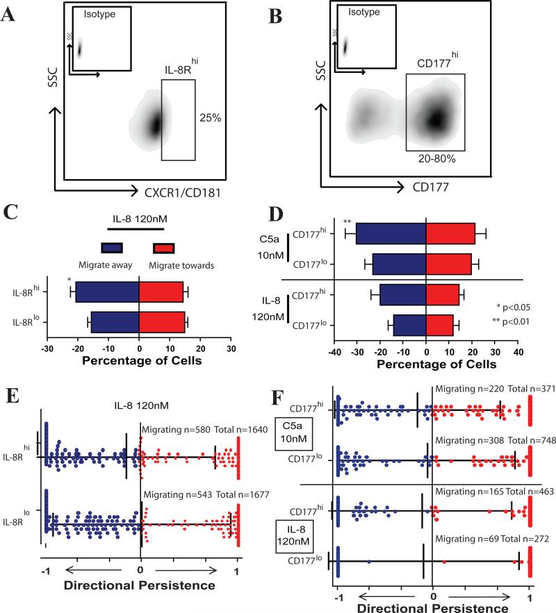
We also wished to determine if the chemoattraction and chemorepulsion response(s) of neutrophils in IL-8 and C5a gradients are associated with cellular phenotype. We FACS-sorted IL-8Rhi-expressing (CXCR1/CD181) (highest 25%ile) and IL-8Rlo (lowest 25%ile) cells and evaluated their respective migratory response to IL-8 (Fig. 3, Table 3). We found that both groups responded with a similar pattern and displayed bi-directional migratory behavior. Similar numbers of IL-8hi and IL-8Rlo cells responded to IL-8 (~35% vs. ~31% respectively). Nevertheless, the IL-8Rhi neutrophils showed a significant bias for migrating away from IL-8 (20.8±2.1% away vs. 14.5±1.6% towards, p<0.05). IL-8Rlo neutrophils migrated in equal proportions in both directions (15.1±1.1% towards vs. 16.2±1.1% away from IL-8). We did not find significant differences in directional persistence and speed between the two groups (Supplementary Fig. 4A, B).
Figure 3. Migratory patterns of neutrophil subpopulations.
A) Human neutrophils were fluorescence-activated cell sorted in two groups: highest and lowest quartile expressing IL-8R (CXCR1, CD181). B) Subpopulations of neutrophils expressing high levels of CD177 (CD177hi) were sorted and compared to CD177lo cells. The percentage of CD177hi cells ranged from 20-80% of total neutrophils. C) IL-8Rhi and IL-8Rlo expressing neutrophils show bi-directional migration patterns, but greater numbers of IL-8Rhi cells migrate away than towards IL-8 (p<0.05, Student's t test). D) Both CD177hi and CD177lo migrate in a bi-directional manner in response to C5a or IL-8. Greater numbers of CD177hi cells migrate in response to C5a vs. CD177lo cells (ns p=0.06). Higher numbers of CD177hi cells migrate away vs. towards C5a (p<0.01, Student's t test). E) Migratory patterns of IL-8Rhi and IL-8Rlo neutrophils in an IL-8 gradient; no difference was found in directional persistence. Bars represent mean and standard deviation of Bi-Directional Index. F) Migratory persistence of neutrophil subpopulations towards and away from a gradient of IL-8 and C5a. There is no difference in migration patterns of the subpopulations. Bars represent mean ± SD. Data from N=3 independent experiments. Number of cells analyzed: IL-8Rhi 580 migrating cells; IL-8Rlo 543 cells; CD177hi C5a 220 cells; CD177lo C5a 308 cells; CD177hi IL-8 165 cells; CD177lo IL-8 69 cells.
Table 3.
Summary of the quantitative analysis of cell migration patterns of different human neutrophil subpopulations
| Neutrophil subpopulations | Tested Molecules | Total cells migrating (%) (mean ± SEM) | Direction | Directional Persistence | Speed | |||||
|---|---|---|---|---|---|---|---|---|---|---|
| Name | Concentration | Toward (%) (mean ± SEM) | Away (%) (mean ± SEM) | Toward (mean ± SD) | Away (mean ± SD) | Toward (μm min−1) (mean ± SD) | Away (μm min−1) (mean ± SD) | |||
| IL-8R/CXCR1 | high | IL8 | 120 nM | 35.4±3.4 | 14.5±1.6 | 20.8±2.1 | 0.92±0.24 | 0.89±0.26 | 24.2±9.0 | 23.0±7.4 |
| low | 31.3±1.7 | 15.1±1.1 | 16.2±1.1 | 0.92±0.22 | 0.85±0.27 | 22.4±6.9 | 19.7±9.9 | |||
| CD177 | high | C5a | 10 nM | 54.9±8.5 | 21.6±4.6 | 30.3±4.9 | 0.79±0.32 | 0.84±0.28 | 21.8±8.5 | 21.4±8.4 |
| low | 42.9±5.6 | 19.7±3.3 | 23.1±3.4 | 0.88±0.23 | 0.88±0.25 | 23.4±8.4 | 20.4±7.5 | |||
| CD177 | high | IL8 | 120 nM | 34.5±6.0 | 14.6±2.1 | 19.9±4.0 | 0.94±0.21 | 0.90±0.23 | 26.4±10.5 | 24.5±9.3 |
| low | 26.7±3.3 | 12.2±0.3 | 14.5±3.0 | 0.95±0.18 | 0.99±0.07 | 25.2±8.5 | 28.5±5.9 | |||
We also compared the migration response(s) of the CD177 (NB1)35 expressing neutrophil subpopulation to C5a and IL-8. While the bi-directional migration pattern was present in both CD177hi and CD177lo populations, we found that a greater number of CD177hi cells migrate away than towards C5a (30.3±4.9% away vs. 21.6±4.6% towards, p<0.01, Fig. 3D). CD177hi cells did not show a statistical significant migratory bias towards or away from IL-8. In contrast, CD177lo cells migrated in equal proportions both towards and away from either C5a or IL-8. Furthermore, we did not find differences in speed and directional persistence between CD177hi and CD177lo populations (Fig 3F, Supplementary Fig. 4 C-F, Table 3). Overall, our measurements indicate that regardless of chemokine receptor expression and subpopulation, neutrophils respond in a bi-directional migration pattern to C5a and IL-8.
Migration of T lymphocytes in response to SDF-1
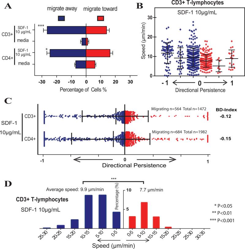
We next examined the patterns of CD3+ T lymphocyte migration in response to SDF-1. In the presence of 10 μg mL−1 SDF-1, 39.0±4.5% of the T cells migrated, with twice as many cells migrating away than towards the SDF-1 gradient (25.7±3.1% vs. 13.3±2.0%; p<0.001; Fig. 4A). We observed that the migratory patterns of T-lymphocytes are predominantly non-persistent, and ~90% of the cells migrating towards and ~70% of the cells migrating away from SDF-1 change their direction at least once during each migration experiments. Only 1.4 % of the CD3+ T cells migrate with high persistence towards and 4.7% migrate with high persistence away from SDF-1 (Fig. 4B). However, the cells migrating away from SDF-1 move faster than cells migrating towards SDF-1 (Fig. 4D, p<0.001). We did not observe any dominant chemoattractant effect of lower concentrations of SDF-1 (100ng mL−1). These results clearly demonstrate that SDF-1 has overall dispersive and chemorepulsive effects on a heterogeneous population of T lymphocytes.
Figure 4. Migration of human T lymphocytes in response to SDF-1.
A). Percentage of human T lymphocytes migrating towards or away from SDF-1 (10 μg mL−1) and media (control) Significantly greater numbers of T cells (both pooled CD3+ and CD4+ cells) migrate away from SDF-1 than towards (p<0.05, Student's t test). B). Distribution of speed and bi-directional index of human CD3+ T lymphocytes in response to SDF-1 (10 μg mL−1). Average persistence of the cells migrating towards and away from SDF-1 are 0.15 and 0.27, respectively. Only ~1.2% of the lymphocytes migrated persistently towards SDF-1, while ~4.7 % of the lymphocytes in the device migrated away from SDF-1 with high persistence. C) Distribution of Bi-Directional Index of T-cell subpopulations. Persistence is very low in all subgroups. D) Histogram of speed of human CD3+ T lymphocytes in response to 10 μg mL−1 SDF-1 in both directions (total 564 cells analyzed: 368 repelled cells and 196 attracted cells). Bars represent mean ± SD. Graphs represent combined data of N=3 independent experiments.
Migration of CD4+ T cell subpopulations in response to SDF-1
We also evaluated the chemoattractive or chemorepulsive migratory signature patterns within CD4+ T-cells. As shown in Fig. 4A we found that CD4+ T-cells are also repelled by SDF-1 in greater numbers (24.4±2.5%) vs. those responding by chemoattraction (16.4±2.5% p<0.05; Supplementary Movie 5). The overall migratory characteristics of pooled CD3+ and CD4+ T cells were very similar (CD3 BD-Index −0.12, CD4 BD-Index −0.15, Figure 4C, Table 1). Collectively, our results indicate that low persistent attraction and repulsive migratory signatures may be a general feature of T-cell motility in response to SDF-1.
Slit2 inhibits neutrophil chemotaxis to fMLP
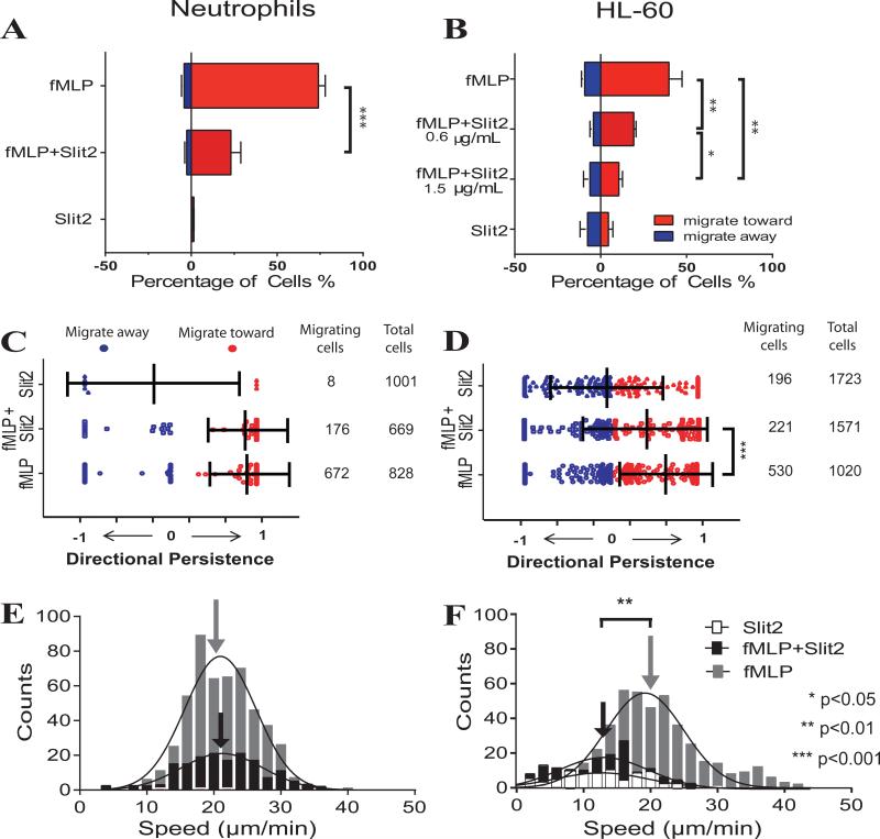
In a final series of experiments, we wished to validate our microfluidic device for the analysis of anti-inflammatory agents. In these assays, we used Slit2 as a prototype inhibitor of neutrophil migratory responses36-38. As illustrated in Fig. 5A, Slit2 (5 μg mL−1) reduced the fraction of human neutrophils migrating towards fMLP (100 nM) by ~3 fold (from 74% of cells migrating to 23%, p<0.001). Similarly, we evaluated the effect of Slit2 on the migration of the clonal HL-60 cell line and found that its inhibitory effect was concentration-dependent (Fig.5). Lower numbers of cells migrated at higher concentrations of Slit2 (p<0.05). Slit2 (1.5 μg mL−1) reduced the number of HL-60 cells that migrate towards fMLP (100 nM) by ~3 fold (from 40% to 11%, p<0.01, Fig. 5B). Slit2 did not reduce the persistence of migrating neutrophils, but it slightly decreased the bi-directional index of migrating HL-60 cells from 0.63±0.57 to 0.39±0.68 (p<0.001, Fig. 5 C, D). In control experiments, where Slit-2 was present in media alone, a migratory response was evident only in 0.8±0.7 % of human neutrophils and 12.1±3.4% of HL-60 cells.
Figure 5. Inhibitory effect of Slit2 on fMLP-induced human neutrophil and HL-60 migration.
Percentage of A) human neutrophils and B) HL-60 cells that migrate towards or away from fMLP with or without the presence of Slit2 (N=4 independent experiments). For both cell types Slit2 significantly inhibits fMLP-induced migration (p<0.01 and p<0.001 respectively, Student's t test). C) Migratory persistence of human neutrophils is not affected by Slit2 (fMLP BD-Index = 0.89, fMLP+Slit2 BD-Index = 0.84). Directional persistence of migrating HL-60 cells is decreased by Slit2 (fMLP BD-Index = 0.63, fMLP+Slit2 BD-Index=0.39; p<0.001, Mann Whitney test). Bars represent mean and standard deviation of Bi-Directional Index. Speed of E) neutrophils and F) HL-60 migrating towards fMLP with or without Slit2 (arrows indicate the mean speed. Slit2 decreases migratory speed of HL-60 cells from 20.7±7.1μm min−1 to 12.8±5.9μm min−1 (p<0.01, Student's t test). Colors are: grey, fMLP alone; black, fMLP and Slit2; white, Slit2 alone. Bars represent mean ± SD. Combined data of N=4 (neutrophils) and N=3 (HL-60) independent experiments is shown. For neutrophils and fMLP, a total of 672 migrating out of 828 total cells were analyzed, fMLP+Slit (176 migrating/669 total), HL-60 (fMLP 530/1020), fMLP+Slit (221/1571).
We also found that only ~20% of all human neutrophils respond to fMLP within the first 30 minutes. On the other hand ~65% of HL-60 cells started to migrate within the first 30 minutes after exposure to fMLP. We observed that Slit2 did not delay the initiation of the neutrophil and HL-60 migratory response to fMLP (Supplementary Fig. 5 A, B). While Slit2 did not alter the migratory speed of neutrophils (Fig. 5E & Supplementary Fig. 5C), it reduced the migratory speed of HL-60 cells from 20.7±7.1 μm min−1 to an average of 12.8±5.9 μm min−1 (p<0.01) (Fig. 5F & Supplementary Fig. 5D). Overall, these data demonstrate that Slit2 decreases the size of the chemotactic population of human neutrophils, but it does not decrease migratory speed, persistence or the initiation of the migratory response. This signature effect of Slit2 may have important implications for its development as an anti-inflammatory therapeutic.
Discussion
In this study, we define a new paradigm, whereby individual chemokines elicit distinct signature migratory responses in different leukocyte populations. We show that typical leukocyte migratory responses are the summation of qualitative migratory patterns including attraction, repulsion, kinesis and inhibition. We quantify these patterns using four metrics: a) percentage of cells migrating in response to a chemokine, b) directionality, c) speed, and d) persistence of migration and use these metrics to quantify migration signatures with higher precision. By enabling precise, quantitative studies of cell migration, our microfluidic platform creates opportunities to design novel anti-inflammatory agents that target specific migration patterns.
Key features in the design of our device enable high precision characterization of cell migration towards and away from chemical gradients. Cells migrate into side channels from pre-defined locations and at predefined numbers. The gradient characteristic is defined by the design of the device, and thus is highly reproducible. While it is well accepted that leukocyte migratory behavior may be dependent on the context of the local microvascular and tissue environment 30, 39, this important pathophysiological issue is not acknowledged in data obtained from traditional assays such as Dunn and Zigmond chambers 11, 12, micropipette techniques 13 as well as the majority of microfluidic cell migration platforms17, 19, where cells migrate on flat surfaces. In our device, leukocytes migrate in conditions of mechanical confinement inside narrow side microchannels. Narrow channels recapitulate environmental cues that neutrophils and lymphocytes encounter in vivo within the extracellular matrix, where leukocytes move between cells within tissues 29, 40-45. Confining the cells to microchannels has the added advantage of allowing for more precise measurements of migratory speed and directional persistence14, 22-24. The patterns of cell migration through these microchannels are distinct from the more “noisy” patterns of cells on 2D flat surfaces encountered in traditional cell migration assays11-13. Microfabricated channels are also more precise and better suited for precise measurements of cell migration than 3D gels which have heterogeneous pore size, tortuous neutrophils paths, and low orientation bias46. Furthermore, advantages of confining cells in microchannels are demonstrated by the results of our experiments using channels of different sizes. We found that cells within 6 × 6 μm channels display faster migratory speed and higher directional persistence that 50 × 6 μm, while still able to reverse their direction inside the smaller channels in the case of low persistence migration patterns. The higher directional persistence and speed through narrower channels is likely a result of the mechanical confinement. Similar differences in speed and persistence have been reported for epithelial cells in channels of various sizes 47. Differences in the chemokine gradients in channels of different sizes are unlikely to contribute to the observed disparity since we found cell migration characteristics through narrow channels to be similar in the presence of various chemokine concentrations and various gradients corresponding to these.
A key finding enabled by our devices is that C5a and IL-8, two well-established chemoattractants 25, 31, 32, 48-50, elicit simultaneous neutrophil migration both towards and away from the chemokine gradient. This finding challenges the current views of neutrophil attraction by C5a 26 and provides a more nuanced view for the responses of neutrophils during inflammation. It suggests that complement activation and production of C5a not only serve as chemoattractants, but also activate and disperse neutrophils at the site of inflammation. While C5a is also capable of activating neutrophils to release enzymes and oxidants 51, the bi-directional migration patterns induced by localized release of C5a could explain the more extensive tissue damage in rapidly evolving disease states32. In addition, while the chemoattractive and chemorepulsive actions of IL-8 have been previously reported8, they appeared to be specific for conditions of high gradient steepness and high average chemokine concentrations. What we find is that bi-directional migration patterns are preserved even when IL-8 concentrations change. The differences between our findings and previously reported studies may be due to differences in the cell microenvironment corresponding to differences in experimental settings, such as mechanical confinement vs. flat surfaces, no-flow vs. shear stress, etc. This possibility is supported by the dissimilarities in the neutrophil migration speed, 2-7μm min−1 on flat surfaces vs. 18.6-21.5 μm min−1 in channels8, and other differences in responses in the two experimental systems.
Another possibility for the bi-directional migratory responses to C5a and IL-8 is that cells respond to chemokine stimulation in a more stochastic manner than previously appreciated. This possibility is supported by the finding that sorted populations of IL-8Rhi and IL-8Rlo populations show equivalent bi-directional migratory responses to IL-8. Also suggesting that homogenous levels of individual chemokine receptor in neutrophil subpopulations52, 53 do not necessarily result in a homogenous migratory response are our results using sorted CD177hi and CD177lo subpopulations, when we found that both subpopulations showed bi-directional migratory patterns in response to gradients of either C5a or IL-8. The bi-directional effect is not specific to cell confinement, since neutrophils migrate in equal proportions towards and away from gradients in channels wider than the cells, when cells get less physical guidance. These results suggest that bi-directional response(s) are more common than previously thought and warrant further investigation. We also suggest that bi-directional migratory behavior may be relevant to the different strategies that neutrophils employ in the course of encountering pathogens in vivo. For example, chemoattractants, such as fMLP or LTB4, may guide neutrophils over large distances towards sites of inflammation (e.g. neutrophils swarming enhancement by LTB433), whereas C5a and IL-8 activate neutrophils in a manner that serves to enhance motility and thus allow for dispersal of subsets of cells at the site of inflammation. Future studies to better understand this bi-directional migration paradigm could include technologies that transfer individual cells from the microfluidic devices to surface cytometry systems after the migration patterns have been measured. Unfortunately, this approach requires significant technological advances that are not available today.
Similarly, we found that high concentrations of SDF-1 also elicit simultaneous lymphocyte migration both towards and away from the chemokine in human CD3+ and CD4+ T-lymphocytes. Again, previous reports recognized the ability of SDF-1 to repel lymphocytes 9, 43, 54, but our new device reveals that this occurs simultaneously with a notable chemoattractant effect. Furthermore, we show that CD4+ T-lymphocytes have a migratory signature that is similar to pooled populations of CD3+ T cells, suggesting that the attracted and repulsed cells are not different CD3+ T-cell populations. One interesting finding is that few lymphocytes migrate persistently toward or away from SDF-1, while the majority of the cells switch directions rather frequently. It is thus important to take into account that when measuring leukocyte migration there is sufficient time and space for the cells to reverse their direction. For example, this pattern of bidirectional migration would be difficult to capture in Boyden chamber or transwell assays, where cells initially migrating through the membrane are not able to reverse their migration. In this manner, experimental data could be interpreted as persistent attraction of the cells due to the rectifying effect of the membrane and only corrected in checker-board assays. Overall, according to the results of our study, the patterns of migration induced by SDF-1, as well as C5a and IL-8, may be best classified as “dispersive” and appropriate for “patrolling” a tissue; they do not function as molecules that simply direct chemical migration towards a precise target.
Our observation that Slit2 inhibits the migration of human neutrophils and HL-60 cells is consistent with previous reports38, 55, 56. In addition, measurements using our platform suggest that Slit2 reduces the fraction of cells responding to the chemoattractant, without altering migratory speed or migratory persistence and without delaying the migratory response. This signature effect of Slit2, modulating the numbers of responsive cells but not their migration phenotype, may have important anti-inflammatory implications.
In summary, our characterization of leukocyte migration signatures in microfluidic devices reveals migration signatures that are complex and specific to each leukocyte-chemokine combination. These signatures are not fully appreciated using traditional leukocyte chemotaxis tools and assays, which focus mostly on the analysis of chemoattraction. Other patterns of migration that coexist with chemoattraction are usually ignored but may hold valuable cues for the activity of the chemokines in conditions of health and disease. We suggest that the analysis of migration signatures based on the direction, speed, persistence and percentage of moving cells, could have broad implications for advancing our understanding of the actions of chemokine-driven inflammatory responses, and could accelerate the screening for new therapeutics to modulate acute and chronic inflammation.
Methods
Microfluidic devices
The microfluidic devices were manufactured using standard microfabrication techniques. Briefly, three layers of negative photoresist (SU8, Microchem, Newton, MA), the first 3 μm thin, the second 6 μm thick, and the third 50 μm thick were patterned on a silicon wafer by sequentially employing three photolithography masks and processing cycles according to the instructions from the manufacturer. The wafer with patterned photoresist was used as a mold to produce PDMS (Polydimethylsiloxane, Fisher Scientific, Fair Lawn, NJ) parts, which were then bonded irreversibly to standard glass slides (75×25 mm, Fisher Scientific).
Neutrophil and T-lymphocyte isolation
For neutrophil isolation, human peripheral blood samples from healthy volunteers, aged 18yrs and older, were purchased from Research Blood Components, LLC. Peripheral blood was drawn in 10mL tubes containing a final concentration of 5 mM EDTA (Vacutainer; Becton Dickinson). Nucleated cells were isolated using a HetaSep gradient, followed by the EasySep™ Human Neutrophil Enrichment Kit (STEMCELL Technologies, Vancouver, Canada) according to the manufacturer's protocol. T-lymphocytes were isolated from human blood samples also obtained from healthy volunteers consented in accordance with IRB approval by Children's Hospital Boston. PBMC were harvested from peripheral blood by gradient centrifugation (2200 rpm for 30 minutes) using Ficoll (GE Healthcare, Waukesha, WI). CD3+ T cells were positively isolated from PBMC using magnetic beads (Dynabeads FlowComp Human CD3 isolation kit, Invitrogen, Carlsbad, CA) according to the manufacturer's protocol, yielding bead free cells. The purity of isolated CD3+ cells was determined using flow cytometry and found to be better than 98%. CD4+ T-cells were isolated by negative selection from PBMC using magnetic beads (RosetteSep™ Human CD4+ T Cell Enrichment Cocktail, STEMCELL Technologies, Vancouver, Canada) according to the manufacturer's protocol.
Cell culture and migration buffers
HL-60 cells (CCL-240, ATCC, Manassas, VA) were cultured at 37°C in 5% CO2 in Iscove's Modified Dulbecco's Medium (IMDM, ATCC, Manassas, VA) containing 20% FBS (Sigma-Aldrich, St. Louis, MO) and differentiated to neutrophil-like cells by DMSO-induced differentiation for a period of 5 days. Cells were sub-cultured every third day to a density of 1×10⁶ cells mL−1. During migration assays, neutrophils were suspended in a buffer of 0.2% human serum albumin (HSA, Sigma-Aldrich) in Hanks Buffered Salt Solution (HBSS, ATCC, Manassas,VA). T-lymphocytes were suspended in RPMI 1640 media (Cambrex, Charles City, IA) containing 10% fetal bovine serum, 2 mM L-glutamine, 100 U mL−1 penicillin/streptomycin (Gibco-Invitrogen), 1% sodium bicarbonate and 1% sodium pyruvate (Cambrex).
Chemotaxis measurements
The microfluidic device was primed 15 minutes prior to cell loading with chemokine and the combinations. fMLP (Sigma-Aldrich, St. Louis, MO); leukotriene B4 (LTB4, Cayman Chemicals, Ann Arbor, MI) IL-8 & C5a (R&D Systems Inc, Minneapolis, MN), and SDF-1 (Peprotech Inc, Rocky Hill, NY) were diluted in desired complete media. Slit2 (Abcam, Cambridge, MA) was employed alone or in combination with other chemokines. A blunt needle connected to the inlet of the device served as a loading reservoir. 20 μL of the solution of desired chemokines mixed with 100 nM human fibronectin (0.1%, Sigma-Aldrich) was added to the needle reservoir. A 1 mL syringe (Hamilton Syringes, Reno, NV) was then applied to the needle. By applying pressure through the syringe, the solution was instilled into the device. Time was allowed for the air initially trapped in dead-end chambers to diffuse out through the PDMS, under the pressure from the syringe while the outlet port was blocked. After 15 minutes, another 1 mL syringe filled with 100 μL cell media solution was applied to a new needle connected to the inlet, and used to wash the chemokine solution from the cell loading chamber and migration channels. After careful washing, 10 μL of cell solution containing 2 × 10⁵ cells was added to the needle reservoir and then gently delivered into the cell-loading chamber. The dead-end reservoirs were primed at the beginning of each experiment and served as the source of chemoattractant. The reservoirs are connected to the main channel through channels that have a much higher resistance than the rest of the device, such that immediately after the washing step, a chemokine free environment is created in the main channel and the buffer channel. The passive diffusion of chemoattractant from the reservoirs into the main channel creates a gradient where the highest concentration is in the reservoir and the lowest concentration is in the main channel as reported14. A second gradient forms between the main channel and the buffer channel by diffusion, with the higher concentration in the main channel. Cell migration was recorded using time-lapse imaging on a fully automated Nikon TiE microscope (10× magnification) with biochamber heated to 37 °C with 5% carbon dioxide gas, for 3 (HL-60 and Neutrophils) or 8 hours (T lymphocytes). Cell displacement was tracked manually using Image J (NIH). We tracked and defined a cell as a migrating cell when it entered a migration channel by at least one cell length. For neutrophils entering the narrow channels, this is approximately 20 μm, for lymphocytes, this length is approximately 10 μm. Separate experiments to characterize the formation of gradients inside the device were performed in the absence of cells, by replacing the chemoattractant with fluorescein-dextran solution (Sigma) of comparable molecular weight, and by subsequently analyzing the distribution and changes in fluorescence intensity from time-lapse imaging. The migratory response of human neutrophils was studied in narrow 6 μm and wide 50 μm channels. HL-60 cells were studied in narrow 6 μm channels and T lymphocytes were studied in 10 μm channels. All channels were 6 μm tall. Wider channels optimize T lymphocytes migration, because T cells have a round and less deformable nucleus compared to neutrophils, which have a polymorph nucleus that is highly deformable, enabling them to migrate through pores of smaller sizes29. For these reasons, 10 μm and 6 μm wide channels were used for lymphocyte and neutrophil migration assays respectively.
Analysis of cell migration
Cell migration was characterized along four parameters: 1) Percentage of cells migrating, 2) Direction of migration, 3) Speed of migration and 4) Directional Persistence. The percentage of cells migrating is calculated from the number of cells migrating in each direction divided by the total amount of cells loaded in the cell loading reservoir. The direction of migration can be either toward or away from the chemokine. Speed and Directional Persistence are determined by analyzing the trajectories of leukocytes moving along the channels for the following quantitative metrics: 1) relative displacement (the difference between the initial and the final cell position within the migration channel) and 2) the total distance traveled (the sum of the distances traveled in successive images) and 3) the duration of migration. Speed is determined by total distance traveled divided by the duration of migration. Directional Persistence (DP) measures the persistence of migration of individual cells relative to the direction of the chemokine gradient and is calculated as relative displacement divided by total traveled distance. For cells migrating through the side channels in the direction of the chemokine reservoir DP falls between 0 and +1; in contrast, for cells migrating away from the chemokine reservoirs DP falls between 0 and −1. DP is equal to “1” for cells migrating persistently toward the chemokine reservoir without changing direction and was equal to “−1” for cells migrating persistently away from the chemokine reservoir, without any change in direction. DP changes for cells that make U-turns and change direction, and is equal to “0” for cells that migrate back and forth through the channels and ultimately return to their initial starting position by the end of the experiment. The higher the DP, the more persistently the cells migrate in one direction. We divided individual cells into three groups 1) high persistence (DP [0.66-1] ), 2) medium persistence (DP [0.33-0.66) ), and 3) low persistence (DP [0-0.33) ).
The Bi-Directional Index (BD-Index) is the population based average migratory pattern that combines the directional persistence and the direction of all cells. It is calculated as the sum of DP of all cells towards the chemokine plus the sum of DP of all cells migrating away from the chemokine, divided by the number of migrating cells. The measures of DP, BD-Index, migration speed, and percentage of migrating cells were used to describe cell migratory behaviors.
Cell sorting
Human neutrophils were stained with FITC conjugated anti-CD177, anti-IL-8R (CD181, CXCR1, Biolegend, San Diego, CA), and either IgG1 or IgG2b (BD Pharmingen San Jose, CA) as a control for 30 min at 4°C and sorted on a FACSAria (BD Biosciences). Data was analyzed using FlowJo software (Tree Star, Ashland, OR).
Statistical analysis
For most comparisons of two experimental conditions, statistical significance was determined using the student t test for normally distributed data and the Mann Whitney test for data that did not have normal distribution. To compare multiple populations we employed one-way ANOVA with Tukey's Post Hoc test. Differences were considered statistically significant when p values were less than 0.05.
Supplementary Material
Table 2.
Summary of the quantitative analysis of cell migration patterns of human neutrophils in 50μm wide migration channels.
| Cell type | Tested Molecules | Traditional Definition | Total cells migrating (mean ± SEM) | Direction | Directional Persistence | Speed | ||||
|---|---|---|---|---|---|---|---|---|---|---|
| Name | Concentration | Toward (%) (mean ± SEM) | Away (%) (mean ± SEM) | Toward (mean ± SD) | Away (mean ± SD) | Toward (μm min−1) (mean ± SD) | Away (μm min−1) (mean ± SD) | |||
| Human Neutrophils | fMLP | 100 nM | attractant | 80.9±5.0 | 71.2±3.4 | 9.7±2.8 | 0.64±0.25 | 0.19±0.23 | 14.8±6.1 | 13.0±5.7 |
| LTB4 | 100 nM | attractant | 85.8±4.9 | 81.1±4.5 | 4.7±1.5 | 0.74±0.19 | 0.14±0.24 | 19.5±5.0 | 13.6±9.1 | |
| C5a | 10nM | attractant | 54.4±10.5 | 26.1±5.3 | 28.3±5.4 | 0.31±0.27 | 0.34±0.24 | 9.6±4.6 | 8.1±3.0 | |
| IL8 | 120nM | attractant | 40.0±6.2 | 19.9±2.7 | 20.1±3.8 | 0.24±0.26 | 0.29±0.23 | 10.9±5.1 | 9.2±4.0 | |
| Media | --- | --- | 0.6±0.3 | 0.3±0.1 | 0.4±0.1 | 0.04±0.03 | 0.07±0.06 | 2.5±0.6 | 2.4±0.5 | |
Acknowledgements
We thank Bashar Hamza, M.S. for help with the HL-60 cells and microfabrication. All microfabrication was performed at the NIBIB funded, BioMEMS Resource Center (EB002503). We thank Matthew Hilbert, Namrata Jain, Nora Kochupurakkal for technical support with the purification of leukocyte subpopulations. Flow cytometry was performed in the IDDRC Stem Cell Core Facility at Boston Children's Hospital that is supported by National Institutes of Health award NIH-P30-HD18655. This projected was funded by National Institutes of Health grants GM092804, Shriners Hospitals for Children research grant to DI and AI04675 and AI092305 to DMB. Leo Boneschansker was partially funded by a grant from Groningen International Program of Science in Medicine (GIPS-M) at the University of Groningen, The Netherlands and by NIH grants T32DK07726 and T32AI007529.
Footnotes
Contributions
L.B., J.Y. and D.I. developed the microfluidic device. The experiments were designed by L.B., J.Y., D.M.B. and D.I. Experiments were performed by L.B. and J.Y. Data was analyzed by L.B., J.Y. and E.W. The manuscript was written by L.B., J.Y., D.M.B. and D.I. All authors discussed the results and implications and commented on the manuscript at all stages.
Conflicts of interest:
DMB has received grant funding from Pfizer and Astellas for unrelated studies. No other conflicts of interest are declared.
References
- 1.Ridley AJ, et al. Cell migration: integrating signals from front to back. Science. 2003;302:1704–1709. doi: 10.1126/science.1092053. [DOI] [PubMed] [Google Scholar]
- 2.Robbins SLC, Ramzi S. Pathologic basis of disease. Saunders; Philadelphia (USA): 1979. [Google Scholar]
- 3.Blanchet X, Langer M, Weber C, Koenen RR, von Hundelshausen P. Touch of chemokines. Front Immunol. 2012;3:175. doi: 10.3389/fimmu.2012.00175. [DOI] [PMC free article] [PubMed] [Google Scholar]
- 4.Migeotte I, Communi D, Parmentier M. Formyl peptide receptors: a promiscuous subfamily of G protein-coupled receptors controlling immune responses. Cytokine Growth Factor Rev. 2006;17:501–519. doi: 10.1016/j.cytogfr.2006.09.009. [DOI] [PubMed] [Google Scholar]
- 5.Nataf S, Davoust N, Ames RS, Barnum SR. Human T cells express the C5a receptor and are chemoattracted to C5a. J Immunol. 1999;162:4018–4023. [PubMed] [Google Scholar]
- 6.Weber M, et al. Interstitial dendritic cell guidance by haptotactic chemokine gradients. Science. 2013;339:328–332. doi: 10.1126/science.1228456. [DOI] [PubMed] [Google Scholar]
- 7.Keller HU. A proposal for the definition of terms related to locomotion of leukocytes and other cells. Bull World Health Organ. 1980;58:505–509. [PMC free article] [PubMed] [Google Scholar]
- 8.Tharp WG, et al. Neutrophil chemorepulsion in defined interleukin-8 gradients in vitro and in vivo. J Leukoc Biol. 2006;79:539–554. doi: 10.1189/jlb.0905516. [DOI] [PubMed] [Google Scholar]
- 9.Poznansky MC, et al. Active movement of T cells away from a chemokine. Nature medicine. 2000;6:543–548. doi: 10.1038/75022. [DOI] [PubMed] [Google Scholar]
- 10.Boyden S. The chemotactic effect of mixtures of antibody and antigen on polymorphonuclear leucocytes. J Exp Med. 1962;115:453–466. doi: 10.1084/jem.115.3.453. [DOI] [PMC free article] [PubMed] [Google Scholar]
- 11.Zicha D, Dunn GA, Brown AF. A new direct-viewing chemotaxis chamber. J Cell Sci. 1991;99(Pt 4):769–775. doi: 10.1242/jcs.99.4.769. [DOI] [PubMed] [Google Scholar]
- 12.Zigmond SH. Ability of polymorphonuclear leukocytes to orient in gradients of chemotactic factors. J Cell Biol. 1977;75:606–616. doi: 10.1083/jcb.75.2.606. [DOI] [PMC free article] [PubMed] [Google Scholar]
- 13.Gerisch G, Keller HU. Chemotactic reorientation of granulocytes stimulated with micropipettes containing fMet-Leu-Phe. J Cell Sci. 1981;52:1–10. doi: 10.1242/jcs.52.1.1. [DOI] [PubMed] [Google Scholar]
- 14.Butler KL, et al. Burn injury reduces neutrophil directional migration speed in microfluidic devices. PLoS One. 2010;5:e11921. doi: 10.1371/journal.pone.0011921. [DOI] [PMC free article] [PubMed] [Google Scholar]
- 15.Irimia D, et al. Microfluidic system for measuring neutrophil migratory responses to fast switches of chemical gradients. Lab Chip. 2006;6:191–198. doi: 10.1039/b511877h. [DOI] [PMC free article] [PubMed] [Google Scholar]
- 16.Kim BJ, Wu M. Microfluidics for mammalian cell chemotaxis. Ann Biomed Eng. 2012;40:1316–1327. doi: 10.1007/s10439-011-0489-9. [DOI] [PMC free article] [PubMed] [Google Scholar]
- 17.Li Jeon N, et al. Neutrophil chemotaxis in linear and complex gradients of interleukin-8 formed in a microfabricated device. Nat Biotechnol. 2002;20:826–830. doi: 10.1038/nbt712. [DOI] [PubMed] [Google Scholar]
- 18.Sackmann EK, et al. Microfluidic kit-on-a-lid: a versatile platform for neutrophil chemotaxis assays. Blood. 2012;120:e45–53. doi: 10.1182/blood-2012-03-416453. [DOI] [PMC free article] [PubMed] [Google Scholar]
- 19.Hartman RS, Lau K, Chou W, Coates TD. The fundamental motor of the human neutrophil is not random: evidence for local non-Markov movement in neutrophils. Biophys J. 1994;67:2535–2545. doi: 10.1016/S0006-3495(94)80743-X. [DOI] [PMC free article] [PubMed] [Google Scholar]
- 20.Lin F, et al. Neutrophil migration in opposing chemoattractant gradients using microfluidic chemotaxis devices. Ann Biomed Eng. 2005;33:475–482. doi: 10.1007/s10439-005-2503-6. [DOI] [PubMed] [Google Scholar]
- 21.Agrawal N, Toner M, Irimia D. Neutrophil migration assay from a drop of blood. Lab Chip. 2008;8:2054–2061. doi: 10.1039/b813588f. [DOI] [PMC free article] [PubMed] [Google Scholar]
- 22.Kurihara T, et al. Resolvin D2 Restores Neutrophil Directionality and Improves Survival after Burns. FASEB. 2013 doi: 10.1096/fj.12-219519. in press. [DOI] [PMC free article] [PubMed] [Google Scholar]
- 23.Ambravaneswaran V, Wong IY, Aranyosi AJ, Toner M, Irimia D. Directional decisions during neutrophil chemotaxis inside bifurcating channels. Integr Biol (Camb) 2010;2:639–647. doi: 10.1039/c0ib00011f. [DOI] [PMC free article] [PubMed] [Google Scholar]
- 24.Irimia D, Charras G, Agrawal N, Mitchison T, Toner M. Polar stimulation and constrained cell migration in microfluidic channels. Lab Chip. 2007;7:1783–1790. doi: 10.1039/b710524j. [DOI] [PMC free article] [PubMed] [Google Scholar]
- 25.Baggiolini M, Clark-Lewis I. Interleukin-8, a chemotactic and inflammatory cytokine. FEBS Lett. 1992;307:97–101. doi: 10.1016/0014-5793(92)80909-z. [DOI] [PubMed] [Google Scholar]
- 26.Marder SR, Chenoweth DE, Goldstein IM, Perez HD. Chemotactic responses of human peripheral blood monocytes to the complement-derived peptides C5a and C5a des Arg. J Immunol. 1985;134:3325–3331. [PubMed] [Google Scholar]
- 27.Ferguson GJ, et al. PI(3)Kgamma has an important context-dependent role in neutrophil chemokinesis. Nat Cell Biol. 2007;9:86–91. doi: 10.1038/ncb1517. [DOI] [PubMed] [Google Scholar]
- 28.Sitrin RG, Sassanella TM, Landers JJ, Petty HR. Migrating human neutrophils exhibit dynamic spatiotemporal variation in membrane lipid organization. Am J Respir Cell Mol Biol. 2010;43:498–506. doi: 10.1165/rcmb.2009-0286OC. [DOI] [PMC free article] [PubMed] [Google Scholar]
- 29.Wolf K, et al. Physical limits of cell migration: control by ECM space and nuclear deformation and tuning by proteolysis and traction force. J Cell Biol. 2013;201:1069–1084. doi: 10.1083/jcb.201210152. [DOI] [PMC free article] [PubMed] [Google Scholar]
- 30.Lammermann T, Germain RN. The multiple faces of leukocyte interstitial migration. Semin Immunopathol. 2014;36:227–251. doi: 10.1007/s00281-014-0418-8. [DOI] [PMC free article] [PubMed] [Google Scholar]
- 31.Baggiolini M, Dewald B, Moser B. Interleukin-8 and related chemotactic cytokines--CXC and CC chemokines. Adv Immunol. 1994;55:97–179. [PubMed] [Google Scholar]
- 32.Ward PA. The dark side of C5a in sepsis. Nat Rev Immunol. 2004;4:133–142. doi: 10.1038/nri1269. [DOI] [PubMed] [Google Scholar]
- 33.Lammermann T, et al. Neutrophil swarms require LTB4 and integrins at sites of cell death in vivo. Nature. 2013;498:371–375. doi: 10.1038/nature12175. [DOI] [PMC free article] [PubMed] [Google Scholar]
- 34.Vasaturo A, et al. A novel chemotaxis assay in 3-D collagen gels by time-lapse microscopy. PloS one. 2012;7:e52251. doi: 10.1371/journal.pone.0052251. [DOI] [PMC free article] [PubMed] [Google Scholar]
- 35.Matsuo K, Lin A, Procter JL, Clement L, Stroncek D. Variations in the expression of granulocyte antigen NB1. Transfusion. 2000;40:654–662. doi: 10.1046/j.1537-2995.2000.40060654.x. [DOI] [PubMed] [Google Scholar]
- 36.Li HS, et al. Vertebrate slit, a secreted ligand for the transmembrane protein roundabout, is a repellent for olfactory bulb axons. Cell. 1999;96:807–818. doi: 10.1016/s0092-8674(00)80591-7. [DOI] [PubMed] [Google Scholar]
- 37.Wong K, Park HT, Wu JY, Rao Y. Slit proteins: molecular guidance cues for cells ranging from neurons to leukocytes. Curr Opin Genet Dev. 2002;12:583–591. doi: 10.1016/s0959-437x(02)00343-x. [DOI] [PubMed] [Google Scholar]
- 38.Wu JY, et al. The neuronal repellent Slit inhibits leukocyte chemotaxis induced by chemotactic factors. Nature. 2001;410:948–952. doi: 10.1038/35073616. [DOI] [PMC free article] [PubMed] [Google Scholar]
- 39.Lammermann T, et al. Rapid leukocyte migration by integrin-independent flowing and squeezing. Nature. 2008;453:51–55. doi: 10.1038/nature06887. [DOI] [PubMed] [Google Scholar]
- 40.McDonald B, et al. Intravascular danger signals guide neutrophils to sites of sterile inflammation. Science. 2010;330:362–366. doi: 10.1126/science.1195491. [DOI] [PubMed] [Google Scholar]
- 41.Jacobelli J, et al. Confinement-optimized three-dimensional T cell amoeboid motility is modulated via myosin IIA-regulated adhesions. Nature immunology. 2010;11:953–961. doi: 10.1038/ni.1936. [DOI] [PMC free article] [PubMed] [Google Scholar]
- 42.Kreisel D, et al. In vivo two-photon imaging reveals monocyte-dependent neutrophil extravasation during pulmonary inflammation. Proc Natl Acad Sci U S A. 2010;107:18073–18078. doi: 10.1073/pnas.1008737107. [DOI] [PMC free article] [PubMed] [Google Scholar]
- 43.Miller MJ, Wei SH, Parker I, Cahalan MD. Two-photon imaging of lymphocyte motility and antigen response in intact lymph node. Science. 2002;296:1869–1873. doi: 10.1126/science.1070051. [DOI] [PubMed] [Google Scholar]
- 44.Sumen C, Mempel TR, Mazo IB, von Andrian UH. Intravital microscopy: visualizing immunity in context. Immunity. 2004;21:315–329. doi: 10.1016/j.immuni.2004.08.006. [DOI] [PubMed] [Google Scholar]
- 45.Weninger W, Biro M, Jain R. Leukocyte migration in the interstitial space of non-lymphoid organs. Nat Rev Immunol. 2014;14:232–246. doi: 10.1038/nri3641. [DOI] [PubMed] [Google Scholar]
- 46.Moghe PV, Nelson RD, Tranquillo RT. Cytokine-stimulated chemotaxis of human neutrophils in a 3-D conjoined fibrin gel assay. J Immunol Methods. 1995;180:193–211. doi: 10.1016/0022-1759(94)00314-m. [DOI] [PubMed] [Google Scholar]
- 47.Irimia D, Toner M. Spontaneous migration of cancer cells under conditions of mechanical confinement. Integr Biol (Camb) 2009;1:506–512. doi: 10.1039/b908595e. [DOI] [PMC free article] [PubMed] [Google Scholar]
- 48.Baggiolini M, Walz A, Kunkel SL. Neutrophil-activating peptide-1/interleukin 8, a novel cytokine that activates neutrophils. J Clin Invest. 1989;84:1045–1049. doi: 10.1172/JCI114265. [DOI] [PMC free article] [PubMed] [Google Scholar]
- 49.Rot A. Neutrophil attractant/activation protein-1 (interleukin-8) induces in vitro neutrophil migration by haptotactic mechanism. Eur J Immunol. 1993;23:303–306. doi: 10.1002/eji.1830230150. [DOI] [PubMed] [Google Scholar]
- 50.Yoshimura T, et al. Purification of a human monocyte-derived neutrophil chemotactic factor that has peptide sequence similarity to other host defense cytokines. Proc Natl Acad Sci U S A. 1987;84:9233–9237. doi: 10.1073/pnas.84.24.9233. [DOI] [PMC free article] [PubMed] [Google Scholar]
- 51.Guo RF, Ward PA. Role of C5a in inflammatory responses. Annu Rev Immunol. 2005;23:821–852. doi: 10.1146/annurev.immunol.23.021704.115835. [DOI] [PubMed] [Google Scholar]
- 52.Gohring K, et al. Neutrophil CD177 (NB1 gp, HNA-2a) expression is increased in severe bacterial infections and polycythaemia vera. Br J Haematol. 2004;126:252–254. doi: 10.1111/j.1365-2141.2004.05027.x. [DOI] [PubMed] [Google Scholar]
- 53.Beyrau M, Bodkin JV, Nourshargh S. Neutrophil heterogeneity in health and disease: a revitalized avenue in inflammation and immunity. Open Biol. 2012;2:120134. doi: 10.1098/rsob.120134. [DOI] [PMC free article] [PubMed] [Google Scholar]
- 54.Poznansky MC, et al. Thymocyte emigration is mediated by active movement away from stroma-derived factors. J Clin Invest. 2002;109:1101–1110. doi: 10.1172/JCI13853. [DOI] [PMC free article] [PubMed] [Google Scholar]
- 55.Chaturvedi S, et al. Slit2 prevents neutrophil recruitment and renal ischemia-reperfusion injury. Journal of the American Society of Nephrology : JASN. 2013;24:1274–1287. doi: 10.1681/ASN.2012090890. [DOI] [PMC free article] [PubMed] [Google Scholar]
- 56.Tole S, et al. The axonal repellent, Slit2, inhibits directional migration of circulating neutrophils. J Leukoc Biol. 2009;86:1403–1415. doi: 10.1189/jlb.0609391. [DOI] [PubMed] [Google Scholar]
Associated Data
This section collects any data citations, data availability statements, or supplementary materials included in this article.


