Abstract
In the United States, 250,000 people tear their anterior cruciate ligament (ACL) annually with females at higher risk of ACL failure then males. By predicting muscle forces during low impact maneuvers we may be able to estimate possible muscle imbalances that could lead to ACL failure during highly dynamic maneuvers. The purpose of this initial study was to predict muscle forces in males and females similar in size and activity level, during squat and lunge maneuvers. We hypothesized that during basic low impact maneuvers (a) distribution of quadriceps forces are different in males and females and (b) females exhibit quadriceps dominance when compared to males. Two males and three females performed squatting and lunging maneuvers while electromyography (EMG) data, motion capture data, and ground reaction forces were collected. Nine individual muscle forces for muscles that cross the knee were estimated using an EMG-driven model. Results suggest that males activate more their rectus femoris muscle than females, who in turn activate more their vastus lateralis muscle at their maximum flexion angle, and more their vastus medialis muscle when ascending from a squat. During the lunge maneuver, males used greater biceps femoris force than females, throughout the lunge, and females exhibited higher semitendinosus force. Quadriceps dominance was evident in both males and females during the prescribed tasks, and there was no statistical difference between genders. Understanding individual muscle force distributions in males and females during low impact maneuvers may provide insights regarding failure mechanisms during highly dynamic maneuvers, when ACL injuries are more prevalent.
Keywords: EMG-Driven Model, Muscle Force, Quadriceps Dominance, Squatting, Lunging, Lower Extremity
Introduction
An anterior cruciate ligament (ACL) tear is a rapidly growing injury that in the United States, 250,000 people suffer from annually [1]. When performing cutting and landing maneuvers, females are four to six times more likely to injure their anterior cruciate ligament (ACL) than males [2]. In the past 30 years, frequency of ACL injury in female high school athletes has increased to ten times that of high school males and five times that of female collegiate athletes [3], [4]. Due to this substantial increase in female ACL injury, numerous studies [5], [6], [7], [8], [9], [10], [11], and [12] have been conducted to determine possible causes of ACL failure. It has been recognized that females tend to land from a jump with lower angles of knee flexion and higher knee adduction than males [12], [13], [14], and [15]. Lower knee flexion angles in females may be caused by different muscle activation patterns between knee flexors and extensors when compared to males [16]. For example, it has been demonstrated that females often exhibit quadriceps dominance during drop jumps, cutting, and single leg squatting maneuvers [6], [12], [14], [16], and [17]. Quadriceps dominance refers to greater activation of quadriceps muscles than hamstring muscles to achieve knee stabilization. Higher quadriceps activation results in unequal flexion/extension loading on the knee, specifically when decelerating during landing, which contributes to ACL strain and potential injury [6], [7], [13], [14], and [16]. However, results from Rozzi et al. [11] demonstrated females increase hamstring activation to achieve joint stability, instead of increased quadriceps activation.
EMG-driven models have been developed as one method for estimating muscle activation and have been used to understand the impact of muscle forces on joint loading [18], [19], [20], [21], [22], [23], [24] and [25]. EMG-driven models derive muscle force from muscle activation through predictive optimization algorithms [26, 27]. By using an EMG-driven model, the impact of muscle force distribution in females can be assessed by initially exploring individual muscle forces in fundamental low impact maneuvers [28]. In this study, Buchanan’s EMG-driven model [27] was used to predict muscle forces for males and females, who are similar in size and level of activity, during squat and lunge maneuvers. Based on literature, it has been hypothesized that during fundamental low impact maneuvers (a) distributions of quadriceps and hamstring force are different in males and females and (b) females exhibit quadriceps dominance when compared to males.
Methods
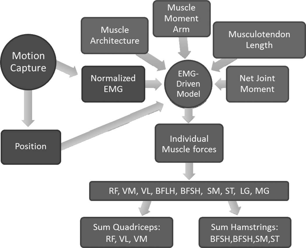
A motion capture system, force plates, and wireless electromyogram (EMG) were used to collect subject kinematics and muscle activation, which were inputs for an EMG-driven model. A musculoskeletal model was also developed in OpenSIM™ to generate time-varying musculotendon lengths, moment arms, and net joint moments as well as initial input muscle parameters for the EMG-driven model (Figure 1).
Figure 1.
Schematic of research for each subject and each trial. Circles represent activities, rectangles represent data, and the arrows represent both what is produced from an activity and what is an input for the activity. Motion analysis is the first activity, which produces both body positions during motion and EMG activation of muscles. The EMG-driven model has five inputs and produces individual muscle forces. The EMG-driven model outputs 9 muscles: rectus femoris (RF), vastus laterals (VL), vastus medialis (VM), semitendinosus (ST), semimembranosus (SM), bicep femoris-short head (BFSH), bicep femoris-long head (BFLH), medial gastrocnemius (MG), and lateral gastrocnemius (LG). Total quadriceps force is the sum of the RF, VL, VM. Total hamstring force is the sum of the BFLH, BFSH, SM and ST.
Subject data collection
Two healthy athletic males (ages 21, 24) and three healthy athletic females (age 18, 19, 21) who participate in advanced aerobic activates five to six times a week. Subjects have no history of knee injury performed ten squatting and ten lunging trials. A VICON™ (Vicon, Oxford, UK) motion capture system tracked 37 markers positioned on each subject. Ground reaction forces were collected at 1kHz using two Bertec™ force plates (Bertec Corporation, Colombus, USA). A Trigno™ wireless EMG system (Delsys Inc., Boston, USA) was used to measure voltage differentials at 1kHz on seven right leg muscles. Skin was cleaned and shaved, and surface electrodes were placed on the muscle belly parallel to the muscle fibers of the subject’s extensor muscles—the vastus lateralis (VL), vastus medialis (VM), and rectus femoris (RF)—and flexor muscles—the medial hamstrings (MH), lateral hamstrings (LH), medial gastrocnemius (MG), and lateral gastrocnemius (LG).
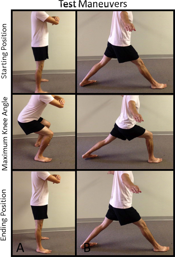
Subjects performed two motions: squatting and lunging (Figure 2). For squatting motion, subjects stood feet shoulder-width apart, descended into a squat until reaching their maximum knee flexion angle while not exceeding 90°, and then rose back into standing position. Subjects were instructed to keep their torso straight with lower lumbar tucked in and arms crossed in front of their body. Each subject performed 10 five-second squats with 15 seconds rest between squats. After squatting trials, subjects performed 10 five-second lunging trials with 15 seconds rest between lunges. Lunging motions started with subject’s right foot anterior to the body and knee in full extension. The left foot was extended posteriorly in full extension. Subjects descended until reaching their maximum knee flexion angle while not exceeding 90°. The right ankle joint remained flexed through the motion and the knee joint did not pass anteriorly to the ankle joint.
Figure 2.
Picture of subject performing test maneuvers A) squat and B) lunge. The top row displays the subject’s starting position before descending. The second row is the maximum knee flexion angle of the subject and the third row is the subject ascending back to the start position.
Each subject performed three maximum voluntary contraction (MVC) trials. Trials consisted of 10 tuck jumps. Subjects were instructed to squat then jump as high as possible and touch their knees to their chest before landing. Subjects were given 15 seconds of rest between jumps and one minute of rest between trials. Raw EMG data from each subject’s motion and MVC trials were filtered using a 40Hz Butterworth high pass filter, rectified, and filtered using a 4Hz Butterworth low pass filter. Filtered motion EMG data was then normalized to the filtered peak MVC data.
Musculoskeletal modeling
An EMG-driven model [27] was used to predict individual muscle forces for each subject during each of the 10 trials in both squatting and lunging trials. Four main components define the EMG-driven model: the anatomical model, the muscle activation dynamics model, the muscle contraction model, and the calibration process [27]. For each subject, an anatomically scaled model was created using a lower body SIMM model developed by Delp et al. [29]. 3D coordinate files were created in Visual 3D and then scaled by height and weight in OpenSIM™ 2.4.1. Inverse kinematics was performed on marker position data. The OpenSIM™ model was used to produce time-varying musculotendon lengths and moment arms as well as parameters for muscle architectures of VL, VM, RF, semimembranosus (SM), semitendinosus (ST), bicep femoris long head (BFLH), bicep femoris short head (BFSH), MG, and LG. Muscle architecture in this model includes pennation angle, maximum force, optimal fiber length, tendon slack length, and physiological cross sectional area (PCSA). Initial input parameters can be seen in Table 1 and were taken from Delp et al. [30].
Table 1.
Initial muscle parameters of the EMG- Driven model.
| Initial EMG -Driven Model Muscle Start Parameters | |||||||||
|---|---|---|---|---|---|---|---|---|---|
| BFLH | BFSH | LG | MG | RF | SM | ST | VL | VM | |
| pennation_angle (deg) | 0 | 0.401 | 0.140 | 0.297 | 0.087 | 0.262 | 0.087 | 0.087 | 0.087 |
| max_force (N) | 896 | 804 | 683 | 1558 | 1169 | 1288 | 410 | 1871 | 1294 |
| C1 | 0 | 0 | 0 | 0 | 0 | 0 | 0 | 0 | 0 |
| C2 | 0 | 0 | 0 | 0 | 0 | 0 | 0 | 0 | 0 |
| delay | 50 | 50 | 50 | 50 | 50 | 50 | 50 | 50 | 50 |
| shape_factor (m) | 0.01 | 0.01 | 0.01 | 0.01 | 0.01 | 0.01 | 0.01 | 0.01 | 0.01 |
| optimal_fiber_length (m) | 0.109 | 0.173 | 0.064 | 0.063 | 0.114 | 0.08 | 0.201 | 0.084 | 0.089 |
| tendon_slack_length (m) | 0.326 | 0.089 | 0.38 | 0.39 | 0.31 | 0.359 | 0.256 | 0.157 | 0.126 |
| PCSA (cm2) | 6.6 | 6.6 | 8 | 18.3 | 12.8 | 16 | 5.4 | 30.7 | 21.2 |
The muscle activation dynamic model computed neural activation using normalized EMG through a second-order differential equation:
| [1] |
where d was the electromechanical delay and α, C1, and C2, are the coefficients that define the second-order dynamics. These parameters (d, α, C1, and C2) map the EMG values, e(t), to neural activation values u(t) [27].
Normalized EMG data from the medial hamstring was used as input for the ST and SM, while the normalized EMG data from the lateral hamstring was input for the BFLH and BFSH. EMG-force relationships, which include C1, C2, shape factor (used to represent the muscle nonlinear properties), and muscle parameters, were optimized within their constraints (Table 2), and neural activation was converted to muscle activation.
Table 2.
EMG-Driven model muscle parameter constraints.
| EMG Driven model Parameter Constraints | |
|---|---|
| pennation_angle (deg) | - |
| max_force(N) | - |
| C1 | ±0.9 |
| C2 | ±0.9 |
| delay | 0.0 – 100 |
| shape_factor | 0.1 |
| optimal_fiber_length (m) | ±0.05 |
| tendon_slack_length (m) | ±0.1 |
| timescale | 0.1 |
| gain | 0.5–2.0 |
| Lambda | 0 – 0.25 |
| alpha | 0.012 – 0.01 |
Neural command u(t) was then used as input to muscle activation a(t) through the sub-model called Muscle Activation Dynamics. Once a(t) was determined, activations and muscle tendon lengths were input into a modified Hill model to estimate muscle force [19]. During the third sub-model, Muscle Contractile Dynamics the optimal fiber length, tendon slack length, total muscle-tendon unit length, and the pennation angle were all used as input in this sub-model. Parameters were collected from the scaled OpenSIM models. The following equation was used to calculate muscle forces:
| [2] |
Where F was the muscle-tendon force comprised of active and passive force components. Time varying moment arms, collected from the musculoskeletal model were multiplied to the corresponding muscle force producing joint moment. Further details regarding the EMG-driven model can be found in previous literature [18, 19, 20, 25, 26, 27, 31].
Inverse dynamics (ID) was performed in OpenSIM™ and used to calculate net joint flexion-extension moment and filtered using a 1Hz Butterworth lowpass filter. Error between the ID net joint moment and estimated forward dynamics (FD) net joint moment was minimized during the optimization process. Final individual muscle parameters, individual muscle forces, and ID versus FD net joint moments were first averaged per motion and then averaged by gender for each subject. Data from one male subject was disregarded; thus the following results are based on two male and three female subjects. Total ID versus FD optimization error was calculated.
Data Analysis
BFSH and BFLH were summed to generate one biceps femoris (BIC) force. The RF, VL,VM, BIC, ST, and SM forces were averaged by subject and then gender at 30° knee flexion, maximum joint angle (93° ± 4° male and 80° ± 19° female), and 30° knee extension during squatting and lunging maneuvers. BIC was the sum of the BFSH and BFLH. Average RF, VL, and VM force was summed by gender to generate total quadriceps force. Average BIC, ST, and SM force was summed by gender to generate total hamstring force. Distribution of quadriceps muscles in males and females at 30° knee flexion, maximum joint angle, and 30° knee extension were compared during the squat and lunge maneuvers. Similar analysis was performed on the hamstring muscles.
Individual subject quadriceps and hamstring forces were determined per trial. Quadriceps and hamstring forces were summed to generate total force and quadriceps force was taken as a percentage of total force for each trial. Maximum quadriceps percent of total force was compared at 30° knee flexion, maximum joint angle, and 30° knee extension during squatting and lunging maneuvers. Peak percent of quadriceps force was compared between males and females at the designated joint angles during squatting and lunging. A two-way ANOVA followed by an independent t-test was performed on all trials which were separated by gender *P < 0.05 and **P <0 .01.
Results
Muscle Force Estimation
Squatting
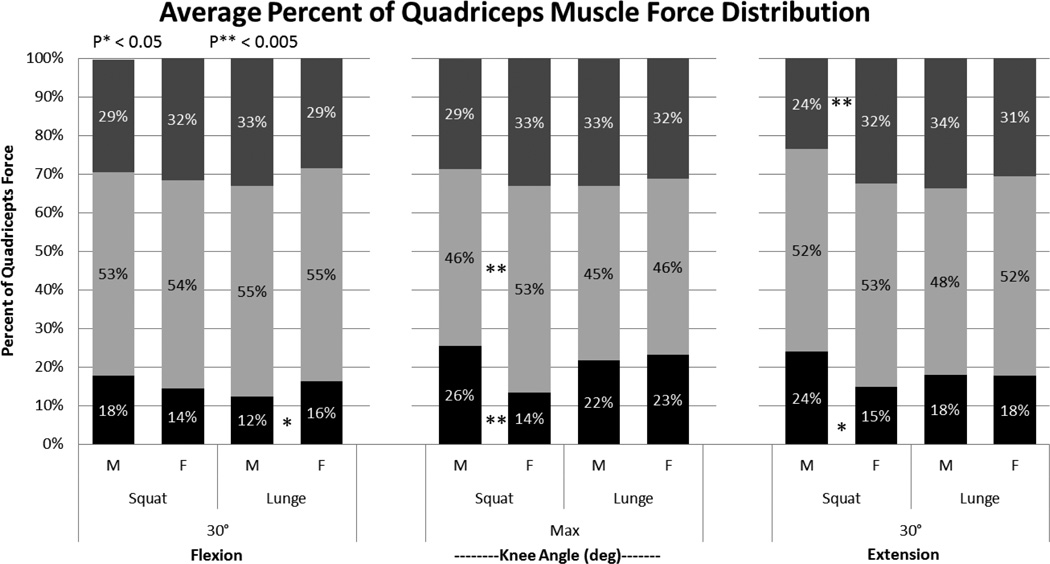
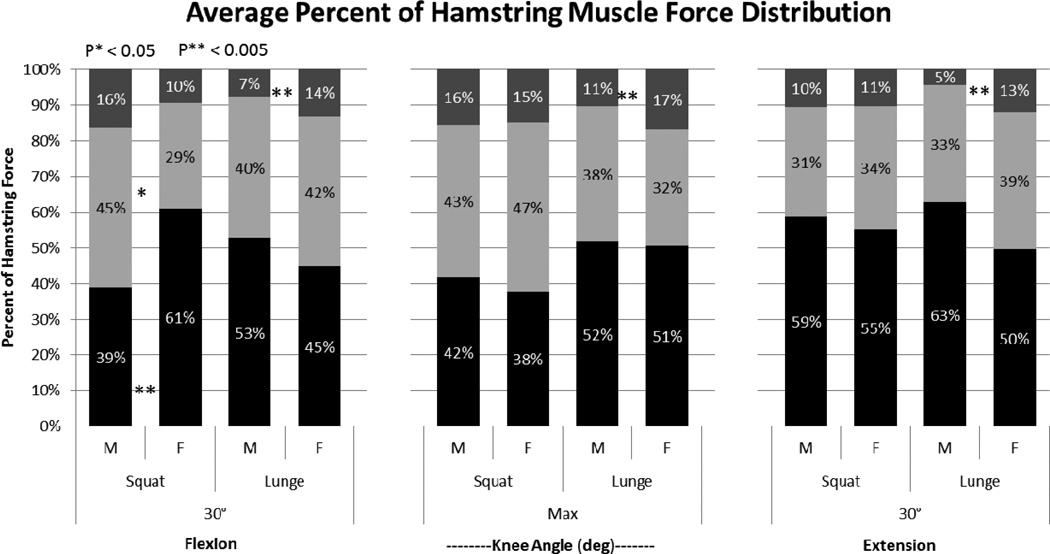
Average predicted male VM and RF forces are approximately 400N during maximum flexion angle and 30° knee extension, accounting for 50% of the quadriceps force respectively (Figure 3). Predicted female VM force was 100N greater than predicted female RF force and accounts for 32% of the total quadriceps force. Females exhibit lower RF force and percent of quadriceps force then males at maximum knee flexion and 30° knee extension. During maximum knee flexion, females average 53% of quadriceps force through their VL, while males only generate 46%. Average predicted flexor forces during a squat are similar in trend between males and females. At 30° knee flexion, average BIC percent of hamstring force was 61% in females and 39% in males (Figure 4). Females also exhibit a higher percent of force through their SM than males at 30° knee flexion. Distribution of muscle forces for the remaining range of motion was similar between males and females. Maximum muscle force values for males and females are scalable, as males generate forces approximately 33% higher than females, which corresponds to males’ 33% larger mass.
Figure 3.
Average quadriceps muscle force distribution between rectus femoris (Black), vastus lateralis (Light Gray), vastus medialis (Dark Gray) in males and females during squat and lunge task. Data represents average of all subject trials at 30° knee flexion, maximum knee joint angle, and 30° knee extension. Average maximum knee joint angle was 93° ± 4° for the male squat, 80° ± 19° female squat, 97° ± 7° male lunge, and 93° ± 6° for the female lunge. A two way ANOVA and independent t-test was performed on male vs. female average muscle force data at each joint angle.
Figure 4.
Average hamstring muscle force distribution between biceps femoris (Black), semimembranosus (Light Gray), and semitendinosus (Dark Gray) in males and females during squat and lunge task. Data represents average of all subject trials at 30° knee flexion, maximum knee joint angle, and 30° knee extension. Average maximum knee joint angle was 90° ± 4° for the male squat, 80° ± 19° female squat, 97° ± 7° male lunge, and 93° ± 6° for the female lunge. A two way ANOVA and independent t-test was performed on male vs. female average muscle force data at each joint angle. Biceps femoris is the sum of the biceps femoris long and short head.
Lunging
Average percent of quadriceps force through the RF was higher in females than males at 30° knee flexion (Figure 4). For the remainder of the motion, males and females displayed similar distribution of hamstring muscle force and muscle force trends. Estimated maximum muscle force in males was 900N for the VL, 600N for the VM, and 400N for the RF. In females, maximum estimated force was 600N for the VL, 400N for the VM, and 250N for the RF. Female flexor muscles are estimated with an average force of 300N per muscle. Estimated extensor muscle forces in males are 300N for the SM, GL and GM, whereas the BFLH and BFDH generated a maximum force of 1200N. During the range of motion, females consistently generate a higher percent of ST force (Figure 5). Between males and females, BIC and SM are similar in trend and distribution.
Figure 5.
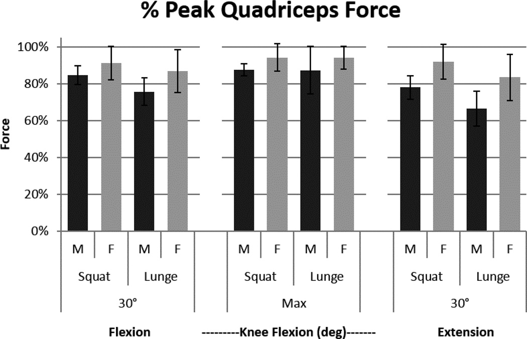
Data represents percent of peak quadriceps force of total force and one standard deviation at 30° knee flexion, maximum knee joint angle, and 30° knee extension between males(dark gray) and females(light gray) during squatting and lunging. Average maximum knee joint angle was 93° ± 4° for the male squat, 80° ± 19° female squat, 97° ± 7° male lunge, and 93° ± 6° for the female lunge. Total force was calculated by summing quadriceps and hamstring muscles at each joint angle. A two way ANOVA and independent t-test was performed on male vs. female peak quadriceps force of total force at each joint angle. Quadriceps dominance in males when compared to quadriceps dominance in females during a squat and lunge maneuver were not statistically significant.
EMG-Driven Optimization Parameters
When comparing male squatting and lunging optimized muscle parameters, individual muscles’ optimal fiber length, shape factor, and tendon slack length are within 2cm of each other (Table 3, Table 4). Larger differences were seen when comparing male squatting C1 and C2 values with lunging C1 and C2 values. Changes in delay and C1 and C2 values show the model applying a time delay. EMG-driven model optimization error for males during squatting was 2.2N/m ± 1.9 N/m and 3.8 N/m ± 3.0 N/m during lunging.
Table 3.
Averaged male and female final optimized muscle parameters from EMG-Driven model during a squat.
| Squat Average EMG Driven Optimized Parameters | ||||||||||||||||||
|---|---|---|---|---|---|---|---|---|---|---|---|---|---|---|---|---|---|---|
| Flexors | Biceps Femoris Long-Head | Biceps Femoris Short-Head | Semimembranosus | Semitendinosus | ||||||||||||||
| Male | Female | Male | Female | Male | Female | Male | Female | |||||||||||
| Avg | Std | Avg | Std | Avg | Std | Avg | Avg | Avg | Std | Avg | Std | Avg | Std | Avg | Std | |||
| pennation_angle | 0 | (0) | 0 | (0) | 0.401 | (0) | 0.387 | (0) | 0.262 | (0) | 0.262 | (0) | 0.087 | (0) | 0.087 | (0) | ||
| max_force | 896 | (0) | 896 | (0) | 804.000 | (0) | 807.407 | (0) | 1288.000 | (0) | 1288.000 | (0) | 410.000 | (0) | 410.000 | (0) | ||
| C1 | −0.263 | (0.61) | 0.186 | (0.194) | −0.299 | (0.51) | 0.029 | (0.505) | 0.305 | (0.53) | 0.068 | (0.746) | 0.183 | (0.21) | −0.072 | (0.543) | ||
| C2 | −0.250 | (0.639) | 0.120 | (0.24) | −0.298 | (0.512) | −0.087 | (0.447) | 0.306 | (0.532) | 0.010 | (0.773) | 0.178 | (0.218) | 0.213 | (0.488) | ||
| delay | 68.188 | (30.261) | 44.588 | (20.043) | 72.229 | (22.397) | 48.336 | (26.683) | 30.238 | (27.88) | 44.762 | (35.563) | 40.803 | (5.642) | 43.949 | (27.769) | ||
| shape_factor | 0.028 | (0.016) | 0.050 | (0.019) | 0.024 | (0.019) | 0.094 | (0.024) | 0.012 | (0.005) | 0.028 | (0.031) | 0.015 | (0.011) | 0.033 | (0.017) | ||
| optimal_fiber_length | 0.113 | (0) | 0.111 | (0.003) | 0.178 | (0.005) | 0.176 | (0.003) | 0.083 | (0.003) | 0.081 | (0.001) | 0.208 | (0.004) | 0.208 | (0.004) | ||
| tendon_slack_length | 0.353 | (0.001) | 0.332 | (0.01) | 0.094 | (0.005) | 0.104 | (0.017) | 0.387 | (0.005) | 0.345 | (0.027) | 0.273 | (0.012) | 0.266 | (0.01) | ||
| Extensors | Rectus Femoris | Vastus Lateralis | Vastus Medialis | |||||||||||||||
| Male | Female | Male | Female | Male | Female | Male | Female | |||||||||||
| Avg | Std | Avg | Std | Avg | Std | Avg | Std | Avg | Std | Avg | Std | Avg | Std | Avg | Std | |||
| pennation_angle | 0.087 | (0) | 0.087 | (0) | 0.087 | (0) | 0.087 | (0) | 0.087 | (0) | 0.087 | (0) | timescale | 0.100 | (0) | 0.1 | (0) | |
| max_force | 1169.000 | (0) | 1169.000 | (0) | 1871.000 | (0) | 1871.000 | (0) | 1294.000 | (0) | 1294.000 | (0) | damping | 0.100 | (0) | 0.1 | (0) | |
| C1 | −0.198 | (0.624) | 0.224 | (0.289) | −0.692 | (0.158) | −0.136 | (0.15) | −0.439 | (0.723) | 0.111 | (0.11) | gain_coefficient1 | 0.619 | (0.145) | 0.937 | (0.353) | |
| C2 | −0.164 | (0.62) | 0.238 | (0.257) | −0.759 | (0.299) | −0.144 | (0.14) | −0.450 | (0.699) | 0.125 | (0.097) | gain_coefficient2 | 0.731 | (0.129) | 1.055 | (0.204) | |
| delay | 45.371 | (53.765) | 36.387 | (15.714) | 70.054 | (25.137) | 36.516 | (19.333) | 60.640 | (14.824) | 37.513 | (22.289) | percent_change | 0.250 | (0.067) | 0.153 | (0.04) | |
| shape_factor | 0.063 | (0.009) | 0.074 | (0.012) | 0.080 | (0.003) | 0.100 | (0.018) | 0.075 | (0.007) | 0.097 | (0.036) | ||||||
| optimal_fiber_length | 0.113 | (0.004) | 0.114 | (0.002) | 0.088 | (0) | 0.084 | (0.001) | 0.092 | (0.004) | 0.089 | (0.002) | ||||||
| tendon_slack_length | 0.293 | (0.006) | 0.309 | (0.011) | 0.169 | (0.002) | 0.148 | (0.003) | 0.134 | (0.008) | 0.115 | (0.002) | ||||||
Table 4.
Averaged male and female final optimized muscle parameters from EMG-Driven model during a lunge.
| Lunge Average EMG-Driven Optimized Parameters | ||||||||||||||||||
|---|---|---|---|---|---|---|---|---|---|---|---|---|---|---|---|---|---|---|
| Flexors | Biceps Femoris Long -Head | Bicepts Femoris Short-Head | Semimembranosus | Semitendinosus | ||||||||||||||
| Male | Female | Male | Female | Male | Female | Male | Female | |||||||||||
| Avg | Std | Avg | Std | Avg | Std | Avg | Std | Avg | Std | Avg | Std | Avg | Std | Avg | Std | |||
| pennation_angle | 0 | (0) | 0 | (0) | 0.401 | (0) | 0.401 | (0) | 0.262 | (0) | 0.256 | (0) | 0.087 | (0) | 0.093 | (0) | ||
| max_force | 896 | (0) | 896 | (0) | 804 | (0) | 804 | (0) | 1288 | (0) | 1258.733 | (0) | 410 | (0) | 436.34 | (0) | ||
| C1 | −0.457 | (0.124) | −0.278 | (0.354) | −0.214 | (0.423) | −0.230 | (0.194) | 0.477 | (0.108) | −0.067 | (0.126) | 0.663 | (0.318) | −0.140 | (0.514) | ||
| C2 | −0.457 | (0.125) | −0.273 | (0.346) | −0.214 | (0.423) | −0.163 | (0.137) | −0.480 | (0.109) | −0.001 | (0.03) | 0.663 | (0.318) | −0.080 | (0.449) | ||
| delay | 83.872 | (15.054) | 83.261 | (18.337) | 71.371 | (16.991) | 67.389 | (9.788) | 21.129 | (19.407) | 51.901 | (12.473) | 15.988 | (15.454) | 64.414 | (5.973) | ||
| shape_factor | 0.056 | (0.002) | 0.056 | (0.02) | 0.026 | (0.011) | 0.073 | (0.007) | 0.055 | (0.015) | 0.078 | (0.016) | 0.060 | (0.049) | 0.053 | (0.034) | ||
| optimal fiber length | 0.109 | (0.003) | 0.108 | (0.001) | 0.176 | (0.001) | 0.176 | (0.004) | 0.083 | (0.002) | 0.084 | (0.007) | 0.209 | (0.005) | 0.202 | (0.009) | ||
| tendon_slack_length | 0.339 | (0.008) | 0.337 | (0.008) | 0.092 | (0.003) | 0.092 | (0.007) | 0.376 | (0.012) | 0.358 | (0.01) | 0.275 | (0.012) | 0.271 | (0.013) | ||
| Extensors | Rectus Femoris | Vastus Lateralis | Vastus Medialis | |||||||||||||||
| Male | Female | Male | Female | Male | Female | Male | Female | |||||||||||
| Avg | Std | Avg | Std | Avg | Std | Avg | Std | Avg | Std | Avg | Std | Avg | Std | Avg | Std | |||
| pennation_angle | 0.087 | (0) | 0.087 | (0) | 0.087 | (0) | 0.087 | (0) | 0.115 | (0) | 0.087 | (0) | timescale | 0.1 | (0) | 0.1 | (0) | |
| max_force | 1169 | (0) | 1169 | (0) | 1871 | (0) | 1871 | (0) | 1294 | (0) | 1294 | (0) | damping | 0.1 | (0) | 0.1 | (0) | |
| C1 | −0.464 | (0.142) | −0.343 | (0.346) | −0.619 | (0.409) | −0.260 | (0.212) | 0.018 | (0.409) | −0.374 | (0.274) | gain_coefficient1 | 0.841 | (0.18) | 0.975 | (0.102) | |
| C2 | −0.424 | (0.141) | −0.354 | (0.329) | −0.580 | (0.268) | −0.118 | (0.16) | 0.019 | (0.268) | −0.355 | (0.303) | gain_coefficient2 | 0.611 | (0.449) | 1.367 | (0.396) | |
| delay | 62.519 | (5.56) | 56.272 | (10.896) | 63.529 | (0.514) | 38.669 | (6.852) | 39.272 | (0.514) | 58.474 | (12.337) | percent_change | 0.242 | (0.02) | 0.154 | (0.107) | |
| shape_factor | 0.043 | (0.059) | 0.099 | (0.004) | 0.058 | (0.003) | 0.085 | (0.027) | 0.082 | (0.003) | 0.086 | (0.019) | ||||||
| optimal_fiber_length | 0.115 | (0.003) | 0.111 | (0.001) | 0.088 | (0) | 0.085 | (0.002) | 0.099 | (0) | 0.089 | (0.002) | ||||||
| tendon_slack_length | 0.305 | (0.004) | 0.276 | (0.01) | 0.170 | (0) | 0.151 | (0.003) | 0.182 | (0) | 0.115 | (0.003) | ||||||
When comparing female optimized muscle parameters during a squat and a lunge, results show many trends similar to male optimized muscle parameters. However, female individual muscle C1 and C2, shape factor, and delay coefficients were not similar between motions. Female results produced consistently higher individual muscle delay factors during a lunging motion (Table 4) than during a squatting motion (Table 3). EMG-driven optimization error for females during squatting was 2.1 N/m ± 1.8 N/m and 2.1 N/m ± 0.6 N/m during lunging. Female average gain coefficients were consistently higher for both maneuvers then males. Average gain coefficients were approximately 30% higher than males during the squat (Table 3). Female lunge average gain coefficient were larger than males approximately 13% for the hamstring muscles and 75% for the quadriceps muscles (Table 4).
Discussion
The purpose of this preliminary study was to estimate individual muscle forces for males and females, who are similar in size and activity level, during a squat and lunge maneuver. It was hypothesized that during fundamental low impact maneuvers (a) distribution of quadriceps forces are different in males and females and (b) females exhibit quadriceps dominance when compared to males. Results from this study suggest (a) statistical significance between the percent of quadriceps muscle distribution in males and females (Figure 3) as well as percent of and hamstring muscle distribution (Figure 4) during squatting and lunging. (b) When compared to males, females do not exhibit quadriceps dominance; however, males and females exhibited quadriceps dominance in both maneuvers (Figure 5). The results of this study suggest that males and females recruit different individual muscles within the same group i.e. quadricepts, to generate the same total force at the knee. Understanding the contribution of individual muscles in males and females during low impact maneuvers may shed insight into which individual muscles are generating the peak force during highly dynamic maneuvers where ACL failure is more prevalent.
Predicted Muscle Forces
Optimized muscle force estimation is one method for muscle activation interpretation. Though muscle force optimization is often used, there are few studies regarding muscle forces of healthy patients and, none which address muscle forces during squat and lunge maneuvers. Due to limited predicted muscle forces available in literature, muscle forces found in this study were compared to muscle forces reported in studies dealing with low impact planar motions such as gait and sit-to-stand. Estimated peak muscle force of the VM and VL were 800N during squatting and 1000N during lunging. Similar predictive muscle force studies found the VM and VL to range from 500N to 1500N [30] and [31]. Conversely, estimated maximum BFLH and BFSH force was 1400N, while the estimated forces from similar studies range from 250N to 750N [32], [33], and [34]. Higher force values could be due to: higher maximum forces, high delay parameters taken from Corcos et al. [35], and/or increased normalized EMG data. Male and female averaged squatting parameters revealed optimized optimal fiber lengths for the BFLH, BFSH, SM, ST, VL, and VM comparable to previously recorded lengths [36].
Hypothesis A
Hewett et al. [14] established that higher knee adduction moments during box jump landings are indicators of potential ACL injury. In this study, the squat maneuver was chosen to simulate a low impact maneuver comparable to the box jump landing. It is believed that higher knee adduction is caused by weak hamstring and gluteus muscles. Data from this study demonstrates that, during squatting, females exhibit higher VL muscle force and less RF force than males during maximum knee flexion and 30° knee extension. Since the maneuvers are slow and controlled, it was hypothesized that females are able to focus on keeping their knees parallel and generate higher VL force to resist knee adduction. Males generate higher RF force, which strictly extends the knee rather than resisting knee adduction. During squat descent, females generate higher percent BIC force and lower SM force than males. Females struggled to maintain a straight torso when descending into the squat (Figure 2). High BIC force could result from the females attempting to maintain hip extension at 30° knee flexion. Lunge quadriceps and hamstring distributions were similar in males and females. During descent, females generate slightly higher percent of RF force than males, and through the entire range of motion, females generated higher percent of ST force. ST force creates medial rotation of the knee, which also creates a higher knee adduction moment. When compared to males, female results show muscle distribution patterns in females either resist knee adduction during the squat or contribute to knee adduction in lunges.
Hypothesis B
Literature suggests that females are quadriceps dominant, which results in higher risk of ACL failure [6], [12], [15], [16], and [17]. Results from this study suggest that during fundamentally low impact maneuvers both males and females are quadriceps dominant. Quadriceps force was over 60% of the total maximum exerted force in males and females (Figure 4). These findings are consistent with previous literature [6], [7], [14], [15], and [16]. Though both genders were quadriceps dominant, the individual muscle that produced the maximum force within the quadriceps were different: males RF and females VL and VM. By looking at individual muscle forces in low impact motions we can better understand the what may cause higher risk of ACL failure in females. Further studies should be conducted to determine the impact of quadriceps dominance during highly dynamic maneuvers where ACL strain and injury are more prevalent.
Limitations
Five subjects were used in this preliminary study; a larger sample size would be needed to show population and gender based statistical analysis. Surface EMG was used to collect muscle activations and the net joint knee moment was calculated from the force of eight muscles. We expect that muscle force distribution patters would alter if all lower extremity muscles were included. The EMG-driven model was not calibrated to each subject [19] and [31] and initial muscle parameters were the same for males and females, based in the OpenSIMTK lower extremity muscle model [30]. The model is planar and optimizes in the flexion/extension plane of the knee. Net hip and ankle moments were not considered when estimating muscle forces. An upright torso during squatting could affect the predicted forces in the biarticulate muscles [38]. The RF, SM, ST, and BHLH are biarticulate muscles that contribute to rotations of the hip and knee. Optimizing muscle forces in six degrees-of-freedom and across the hip joint may alter the results of this study [31, 37, 38].
Summary
Results from this preliminary study suggest that during low impact maneuvers such as squatting and lunging males and females are both quadriceps dominant. Though both use more quadriceps then hamstrings they used different muscles within the quadriceps to produce the same motion. During squat ascent, females produce a higher VL force to resist knee adduction while males produces a larger RF force. Further studies focusing on individual muscle force estimation should: use larger sample selections, measure muscle parameters using Magnetic Resonance Imaging [39], perform a sensitivity analysis on the constraints of the EMG-driven model, perform subject specific calibration according to Lloyd and Besier [19], and be multi-joint model. Studies should also be conducted to test whether or not females continue to generate a higher percent of VL force than males or have an increase in knee adduction during highly dynamic motions. Results are consistent with highly dynamic motion studies, implying that even when performing low impact maneuvers, the ACL could be overloaded due to individual muscle forces within the quadriceps. We believe that injuries may be caused from individual muscle imbalances not merely imbalances within groups of muscles. This study that illuminates the need to look individual muscle and the effect they have on ACL failure.
Acknowledgements
This research was supported, in part, by NSF (BES 0966398) and NIH (R01AR046389-0851).
Footnotes
Publisher's Disclaimer: This is a PDF file of an unedited manuscript that has been accepted for publication. As a service to our customers we are providing this early version of the manuscript. The manuscript will undergo copyediting, typesetting, and review of the resulting proof before it is published in its final citable form. Please note that during the production process errors may be discovered which could affect the content, and all legal disclaimers that apply to the journal pertain.
Conflict of Interest:
There is no conflict of interest
References
- 1.Macleod TD, Snyder-Mackler L, Buchanan TS. Differences in neuromuscular control and quadriceps morphology between potential copers and noncopers following anterior cruciate ligament injury. J. Orthop. Sports Phys. Ther. 2014;44:76–84. doi: 10.2519/jospt.2014.4876. [DOI] [PMC free article] [PubMed] [Google Scholar]
- 2.Arendt EA, Agel J, Dick R. Anterior Cruciate Ligament Injury Patterns among collegiate men and women. J Athl Train. 1999;34:86–92. [PMC free article] [PubMed] [Google Scholar]
- 3.NCAA. NCAA injury surveillance system summary. Indianapolis: National Collegiate Athletic Association; 2002. [Google Scholar]
- 4.NFHS NFoSHSA. 2008–09 High School Athletics Participation Survey. Kansas City, Missouri: National Federation of State High School Associations; 2009. [Google Scholar]
- 5.Chappell JD, Yu B, Kirkendall DT, Garrett WE. A comparison of knee kinetics between male and female recreational athletes in stop-jump tasks. Am. J. Sports Med. 2002;30:261–267. doi: 10.1177/03635465020300021901. [DOI] [PubMed] [Google Scholar]
- 6.Myer GD, Ford KR, Hewett TE. The effects of gender on quadriceps muscle activation strategies during a maneuver that mimics a high ACL injury risk position. J. Electromyogr. Kinesiol. 2005;15:181–189. doi: 10.1016/j.jelekin.2004.08.006. [DOI] [PubMed] [Google Scholar]
- 7.Myer GD, Ford KR, Khoury J, Succop P, Hewett TE. Clinical correlates to laboratory measures for use in non-contact anterior cruciate ligament injury risk prediction algorithm. Clin. Biomech. (Bristol, Avon) 2010;25:693–699. doi: 10.1016/j.clinbiomech.2010.04.016. [DOI] [PMC free article] [PubMed] [Google Scholar]
- 8.Myer G, Brent J. Real-time assessment and neuromuscular training feedback techniques to prevent ACL injury in female athletes. Strength Cond. 2011;33:21–35. doi: 10.1519/SSC.0b013e318213afa8. [DOI] [PMC free article] [PubMed] [Google Scholar]
- 9.Paterno MV, Schmitt LC, Ford KR, Rauh MJ, Myer GD, Huang B, et al. Biomechanical measures during landing and postural stability predict second anterior cruciate ligament injury after anterior cruciate ligament reconstruction and return to sport. Am. J. Sports Med. 2010;38:1968–1978. doi: 10.1177/0363546510376053. [DOI] [PMC free article] [PubMed] [Google Scholar]
- 10.Norcross MF, Blackburn JT, Goerger BM, a Padua D. The association between lower extremity energy absorption and biomechanical factors related to anterior cruciate ligament injury. Clin. Biomech. (Bristol, Avon) 2010;25:1031–1036. doi: 10.1016/j.clinbiomech.2010.07.013. [DOI] [PubMed] [Google Scholar]
- 11.Rozzi SL, Lephart SM, Gear WS, Fu FH. Knee joint laxity and neuromuscular characteristics of male and female soccer and basketball players. Am. J. Sports Med. 1999;27:312–319. doi: 10.1177/03635465990270030801. [DOI] [PubMed] [Google Scholar]
- 12.Malinzak RA, Colby SM, Kirkendall DT, Yu B, Garrett WE. A comparison of knee joint motion patterns between men and women in selected athletic tasks. Clin. Biomech. (Bristol, Avon) 2001;16:438–445. doi: 10.1016/s0268-0033(01)00019-5. [DOI] [PubMed] [Google Scholar]
- 13.Hewett TE, Ford KR, Myer GD. Anterior cruciate ligament injuries in female athletes: Part 2, a meta-analysis of neuromuscular interventions aimed at injury prevention. Am. J. Sports Med. 2006;34:490–498. doi: 10.1177/0363546505282619. [DOI] [PubMed] [Google Scholar]
- 14.Hewett TE, Ford KR, Hoogenboom BJ, Myer GD. UNDERSTANDING AND PREVENTING ACL INJURIES : CONSIDERATIONS - UPDATE 2010. NAJSPT. 2010;5:234–251. [PMC free article] [PubMed] [Google Scholar]
- 15.Urabe Y, Kobayashi R, Sumida S, Tanaka K, Yoshida N, Nishiwaki GA, et al. Electromyographic analysis of the knee during jump landing in male and female athletes. Knee. 2005;12:129–134. doi: 10.1016/j.knee.2004.05.002. [DOI] [PubMed] [Google Scholar]
- 16.Youdas JW, Hollman JH, Hitchcock JR, Hoyme GJ, Johnsen JJ. Comparison of hamstring and quadriceps femoris electromyographic activity between men and women during a single -limb squat on both a stable and labile surface. 2007;21:105–111. doi: 10.1519/00124278-200702000-00020. [DOI] [PubMed] [Google Scholar]
- 17.Sigward SM, Powers CM. The influence of gender on knee kinematics, kinetics and muscle activation patterns during side-step cutting. Clin. Biomech. (Bristol, Avon) 2006;21:41–48. doi: 10.1016/j.clinbiomech.2005.08.001. [DOI] [PubMed] [Google Scholar]
- 18.Shao Q, Bassett DN, Manal K, Buchanan TS. An EMG-driven model to estimate muscle forces and joint moments in stroke patients. Comput. Biol. Med. 2009;39:1083–1088. doi: 10.1016/j.compbiomed.2009.09.002. [DOI] [PMC free article] [PubMed] [Google Scholar]
- 19.Lloyd D, Besier TF. An EMG-driven musculoskeletal model to estimate muscle forces and knee joint moments in vivo. J. Biomech. 2003;36:765–776. doi: 10.1016/s0021-9290(03)00010-1. [DOI] [PubMed] [Google Scholar]
- 20.Erdemir A, McLean S, Herzog W, van den Bogert AJ. Model-based estimation of muscle forces exerted during movements. Clin. Biomech. (Bristol, Avon) 2007;22:131–154. doi: 10.1016/j.clinbiomech.2006.09.005. [DOI] [PubMed] [Google Scholar]
- 21.Kumar D, Rudolph KS, Manal KT. EMG-driven modeling approach to muscle force and joint load estimations: case study in knee osteoarthritis. J. Orthop. Res. 2012;30:377–383. doi: 10.1002/jor.21544. [DOI] [PMC free article] [PubMed] [Google Scholar]
- 22.Sartori M, Reggiani M, Farina D, Lloyd DG. EMG-driven forward-dynamic estimation of muscle force and joint moment about multiple degrees of freedom in the human lower extremity. PLoS One. 2012;7:e52618. doi: 10.1371/journal.pone.0052618. [DOI] [PMC free article] [PubMed] [Google Scholar]
- 23.de Oliveira LF, Menegaldo LL. Individual-specific muscle maximum force estimation using ultrasound for ankle joint torque prediction using an EMG-driven Hill-type model. J. Biomech. 2010;43:2816–2821. doi: 10.1016/j.jbiomech.2010.05.035. [DOI] [PubMed] [Google Scholar]
- 24.McGowan CP, Kram R, Neptune RR. Modulation of leg muscle function in response to altered demand for body support and forward propulsion during walking. J. Biomech. 2009;42:850–856. doi: 10.1016/j.jbiomech.2009.01.025. [DOI] [PMC free article] [PubMed] [Google Scholar]
- 25.Manal K, Buchanan TS. An electromyogram-driven musculoskeletal model of the knee to predict in vivo joint contact forces during normal and novel gait patterns. J. Biomech. Eng. 2013;135 doi: 10.1115/1.4023457. 021014. [DOI] [PMC free article] [PubMed] [Google Scholar]
- 26.Besier TF, Fredericson M, Gold GE, Beaupré GS, Delp SL. Knee muscle forces during walking and running in patellofemoral pain patients and pain-free controls. J. Biomech. 2009;42:898–905. doi: 10.1016/j.jbiomech.2009.01.032. [DOI] [PMC free article] [PubMed] [Google Scholar]
- 27.Buchanan TS, Lloyd DG, Manal K, Besier TF. Neuromusculoskeletal modeling: estimation of muscle forces and joint moments and movements from measurements of neural command. J. Appl. Biomech. 2004;20:367–395. doi: 10.1123/jab.20.4.367. [DOI] [PMC free article] [PubMed] [Google Scholar]
- 28.Escamilla RF. Knee biomechnics of the squat exercise. Med. Sci. Sport. Exerc. 2001;33:127–141. doi: 10.1097/00005768-200101000-00020. [DOI] [PubMed] [Google Scholar]
- 29.Delp SL, Anderson FC, Arnold AS, Loan P, Habib A, John CT, et al. OpenSim: open-source software to create and analyze dynamic simulations of movement. IEEE Trans. Biomed. Eng. 2007;54:1940–1950. doi: 10.1109/TBME.2007.901024. [DOI] [PubMed] [Google Scholar]
- 30.Delp SL, Loan P, Hoy MG, Zajac FE, Topp EL, Rosen JM. An interactive graphics-based model of the lower extremity to study orthopedic surgical procedures. IEEE Trans. Biomed. Eng. 1990;37:757–767. doi: 10.1109/10.102791. [DOI] [PubMed] [Google Scholar]
- 31.Hamnett JS. Estimated muscle forces during running and cutting for ACL deficient, ACL reconstructed, and healthy subjects using a two-joint EMG-driven model. (Master of Science in Mechanical Engineering) The University of Delaware. 2011 [Google Scholar]
- 32.Sasaki K, Neptune RR. Individual muscle contributions to the axial knee joint contact force during normal walking. J. Biomech. 2010;43:2780–2784. doi: 10.1016/j.jbiomech.2010.06.011. [DOI] [PMC free article] [PubMed] [Google Scholar]
- 33.Yoshioka S, Nagano A, Hay DC, Fukashiro S. The minimum required muscle force for a sit-to-stand task. J. Biomech. 2012;45:699–705. doi: 10.1016/j.jbiomech.2011.11.054. [DOI] [PubMed] [Google Scholar]
- 34.Lin Y-C, Walter JP, Banks SA, Pandy MG, Fregly BJ. Simultaneous prediction of muscle and contact forces in the knee during gait. J. Biomech. 2010;43:945–952. doi: 10.1016/j.jbiomech.2009.10.048. [DOI] [PubMed] [Google Scholar]
- 35.Corcos DM, Gottlieb GL, Latash ML, Almeida GL, Agarwal GC. Electromechanical delay: An experimental artifact. J. Electromyogr. Kinesiol. 1992;2:59–68. doi: 10.1016/1050-6411(92)90017-D. [DOI] [PubMed] [Google Scholar]
- 36.Arnold AS, Delp SL. Rotational moment arms of the medial hamstrings and adductors vary with femoral geometry and limb position: implications for the treatment of internally rotated gait. J. Biomech. 2001;34:437–447. doi: 10.1016/s0021-9290(00)00232-3. [DOI] [PubMed] [Google Scholar]
- 37.Powers CM. The influence of abnormal hip mechanics on knee injury: a biomechanical perspective. J. Orthop. Sports Phys. Ther. 2010;40:42–51. doi: 10.2519/jospt.2010.3337. [DOI] [PubMed] [Google Scholar]
- 38.Tsai L-C, Powers CM. Increased hip and knee flexion during landing decreases tibiofemoral compressive forces in women who have undergone anterior cruciate ligament reconstruction. Am. J. Sports Med. 2013;41:423–429. doi: 10.1177/0363546512471184. [DOI] [PubMed] [Google Scholar]
- 39.Tsai L-C, Colletti PM, Powers CM. Magnetic resonance imaging-measured muscle parameters improved knee moment prediction of an EMG-driven model. Med. Sci. Sports Exerc. 2012;44:305–312. doi: 10.1249/MSS.0b013e31822dfdb3. [DOI] [PubMed] [Google Scholar]


