Abstract
The gene encoding the mu-opioid receptor (MOR) generates a remarkable diversity of subtypes, the functional significance of which remains largely unknown. The structure of MOR could be a critical determinant of MOR functionality and its adaptations to chronic morphine exposure. Since MOR antinociception has sexually dimorphic dimensions, we determined the influence of sex, stage of estrus cycle and chronic systemic morphine on levels of MOR splice variant mRNA in rat spinal cord. Chronic systemic morphine influenced the spinal expression of mRNA encoding rMOR-1B2 and rMOR-1C1 in a profoundly sex-dependent fashion. In males, chronic morphine resulted in a 2-fold increase in expression levels of rMOR-1B2 and rMOR-1C1 mRNA. This effect of chronic morphine was completely absent in females. Increased density of MOR protein in spinal cord of males accompanied the chronic morphine-induced increase in MOR variant mRNA, suggesting that it reflected an increase in corresponding receptor protein. These results suggest that tolerance/dependence results, at least in part, from different adaptational strategies in males and females. The signaling consequences of the unique composition of the C-terminus tip of rMOR-1C1 and rMOR-1B2 could point the way to defining the molecular components of sex-dependent tolerance and withdrawal mechanisms.
Keywords: mu-opioid receptor, splice variants, sexual dimorphism, opioid tolerance
Introduction
OPRM1, the only identified gene encoding the mu-opioid receptor (MOR), (Chen et al. 1993) generates a remarkable diversity of MOR subtypes (Pasternak 2001). Most are C-terminal variants that are generated through alternative splicing between exon 3 and multiple downstream exons (Pasternak & Pan 2013). The discovery of these and other splice variants (e.g., exon 11 variants with extensive and 5’ splicing, exon 1 variants containing a single transmembrane domain) provide a molecular biology framework for the concept of multiple mu-opioid receptors inferred from much earlier pharmacological studies (Wolozin & Pasternak 1981).
Some functional differences have been identified among MOR splice variants. Many opioids show marked differences in efficacy and potency at C-terminal MOR variants (Pasternak et al. 2004, Pan et al. 2005b, Bolan et al. 2004, Pan et al. 2005a). Morphine-induced internalization also varies among the C-terminal variants, e.g., morphine given intracerebroventricularly internalizes mMOR-1C1 but not mMOR-1 (Abbadie & Pasternak 2001).
Differences among MOR splice variants also vary with respect to agonist selectivity. Knockout of exon 1 abolishes morphine analgesia, but not that of either morphine-6β-glucuronide or heroin (Schuller et al. 1999). Conversely, knockout of exon 11 greatly reduces analgesic responsiveness to morphine-6β-glucuronide and heroin, but not to morphine or methadone (Pan et al. 2009). MOR transcripts also vary in their expression patterns and levels of expression across brain regions (Abbadie et al. 2000b, Abbadie et al. 2000c), implying region-specific processing, and differing physiological functions. MOR splice variants have also been implicated to explain incomplete cross-tolerance among agonists and variability of agonist potency/effectiveness in patients.
Strikingly, MOR splice variants have not been studied with respect to the well-documented sexual dimorphism in MOR antinociception in laboratory animals (Barrett et al. 2002, Cicero et al. 1997, Cicero et al. 1996, Boyer et al. 1998, Krzanowska & Bodnar 1999, Cook et al. 2000, Peckham & Traynor 2006) and, in humans, (Sarton et al. 2000). This laboratory has recently reported two striking manifestations of sexually dimorphic properties of MOR in spinal cord: (1) in opioid naïve spinal tissue the propensity of MOR to heterodimerize with the kappa opioid receptor (KOR) is substantially greater in females than males, the magnitude of which is dependent on stage of estrus cycle, i.e., spinal levels of the MOR/KOR heterodimer during proestrus are significantly greater than during diestrus (Chakrabarti et al. 2010, Liu et al. 2011) and (2), chronic systemic morphine treatment augments MOR Gs coupling in the spinal cord of males but not females (Chakrabarti et al. 2012).
The structure of MOR could be a determinant of both the facility with which spinal MOR heterodimerizes with KOR as well as the G proteins with which MOR preferentially couples. Accordingly, we investigated the influence of stage of estrus cycle and chronic systemic morphine (in both males and females) on spinal cord levels of mRNA encoding MOR splice variants. We selected MOR variants representing each of the two main classes of splicing that have been identified in rodents and humans. For example, MOR-1C1 and MOR-1B2 represent variants resulting from extensive 3’ splicing at the end of the C-terminus but containing the seven transmembrane spanning domains and the N-terminus of MOR-1. MOR-1G1 represents variants resulting from extensive 5’ splicing containing six transmembrane spanning domains and an altered N-terminus.
Results reveal a striking sexual dimorphism in the ability of chronic systemic morphine to selectively influence the generation of spinal MOR variants. Implications for sex-based opioid tolerance mechanisms are discussed.
Materials and methods
Animals
Sprague-Dawley rats (Charles River, Kingston, NY; 225–300 g) were maintained in an approved controlled environment on a 12 h light/dark cycle. Food and water were available ad libitum. Experimental procedures were reviewed and approved by the Institutional Animal Care and Use Committee of SUNY Downstate Medical Center.
Determination of stage of estrous cycle
Stage of cycle was determined using histology of vaginal smears. Predominance of small leukocytes was indicative of diestrus; a predominance of large round nucleated cells was indicative of proestrus.
Systemic administration of morphine
Morphine was administered via the subcutaneous implantation of morphine base pellets (75 mg each) (Chakrabarti et al. 2012, Villar & Bhargava 1992). On day 7, animals were sacrificed by decapitation. Lumbar spinal tissue was expelled by injecting ice-cold saline into the rostral end of the spinal cord and either immediately submerged in RNA Later (Ambion) solution for RNA extraction or ice-cold 50 mM Tris buffer (pH 7.4) for radioligand binding studies.
Tissue preparation for RNA extraction
Transverse sections of whole lumbar spinal cord were homogenized in a Teflon glass homogenizer in Trizol reagent. Homogenates were spun (3,000g, 5 min), supernatants were mixed with 0.2 ml of chloroform (incubated for 3 min, room temperature), after which they were centrifuged at 12,000g for 15 minutes at 4°C. The aqueous phase was precipitated with 0.5 ml of isopropanol. Resulting pellets of total RNA were washed twice in 75% ethanol and either stored in 75% ethanol (−80°C) or resuspended in nuclease free water (130 ng/μl) for immediate use. Aliquots (260 ng; 2 μl) of each sample were used for RT-PCR. Every sample was analyzed in an Agilent Bioanalyzer to evaluate the quality of the RNA and quantify its concentration. Samples with RNA Integrity numbers less than seven were discarded.
RT-PCR
A Step One Plus apparatus (Applied Biosystems, Life technologies, Grand Island, NY) was programmed to run the following reverse transcriptase real time PCR (RT-PCR) scheme: 7 min at 42°C (retro transcription); 10 s at 95°C (denaturation). PCR amplification was achieved by 40 cycles of the following: 5 s at 95°C, 10 s at 58°C, 4 s, at 72°C. Amplicon dissociation was achieved by 15 s at 95°C, 1 min at 60°C, and 15 s at 95°C. Reactions were performed using TAKARA One Step SYBR® Ex Taq ™ qRT-PCR Kit (Takara Bio Inc., Madison, WI), containing SYBR RT-PCR mix (12.5µl), Taq polymerase 2.5U (0.5 µl), retrotranscriptase mix (0.5 µl), forward primer (0.2 µM, 0.5 µl), reverse primer (0.2 µM, 0.5 µl), RNAse free water (8.5 µl) and total RNA (260 ng; 2 µl).
All oligonucleotides used for quantitative RT-PCR produced amplicons ranging from 100 bp to 250 bp. Each was designed and tested against a rat database for possible unwanted products by using the Primer Blast online software: The following were the primers employed: rMOR-1/rMOR-1G1: f-GAACAGCAAAACTCCACTCGAG; r-GCAATGGAGCAGTTTCTGCCT; rMOR-1A: f-AGGGAACATCCCTCCACGGCT; r-GGACTTTGCCTCAAGTTACTGACCTT; rMOR-1B2: f-CCAACCTCGTCCACGATCGAACA; r-TGTCTCTACTGACTGAGGCTCCTGGT; rMOR-1C1: f-ACGTGGTCTTTGAAATCGCGTGC; r-ACTTCTCACTAGCACATCCCTGTGT; rMOR-1G1; f-TCCGCATTCCAAAAACTGGACAGG; r-ACTGGTCGCTAAGGCGTCTGC; rMOR-1R; f-AGGGAACATCCCTCCACGGCTAATA; r-ACCTACCATTGTGGGGGCCAAG. All RT-PCR reactions including negative controls were run in duplicates.
The number of cycles required for each amplicon to reach detection, via interaction with SYBR green, was defined as the threshold cycle (CT). The CT for each of the six MOR variants studied and glucose-6-phosphate dehydrogenase (G6PD) was concomitantly determined from spinal cord of opioid naïve, diestrus, proestrus female and male rats. G6PD was used as both a reference internal control gene to normalize the RT-PCRs for the amount of RNA added to the reactions and to calculate a relative mRNA value for the splice variants of interest, which was calculated as 2−ΔCT where (ΔCT) is the difference between the CT for each MOR variant and G6PD (Livak & Schmittgen 2001).
To assess the influence of chronic morphine on MOR variant expression, we first calculated the difference between variant-specific mRNA levels in spinal cord of opioid naïve and chronic morphine-treated rats (ΔCT), which was expressed relative to the ΔCT of the variant mRNA of the lowest abundance (rMOR-1R), (ΔΔCT). Expression levels of rMOR-1R were not influenced by chronic morphine. MOR variant mRNA doubles with each PCR cycle at the exponential phase of a PCR reaction, when the CT is recorded. Since CT is inversely proportional to mRNA levels, the relative expression of specific MOR variants in naïve vs. opioid tolerant spinal cord was calculated as 2−ΔΔCT (Livak & Schmittgen 2001).
Radioligand binding studies
Receptor number (Bmax) and KD estimates before and following chronic systemic morphine treatment were determined by saturation binding using [3H]DAMGO. Lumbar spinal tissue obtained from two placebo- or chronic morphine-treated rats were pooled, homogenized in 50 mM Tris buffer (pH 7.4) and centrifuged at 40,000g for 30 min at 4°C. Pellets were resuspended in Tris buffer, incubated (30 min, 25°C) and centrifuged again (40,000g, 30 min). Resulting pellets, were resuspended in 50 mM potassium phosphate buffer (pH 7.2) for duplicate determination of [3H]DAMGO binding (0.1 -10 nM; 25°C, 90 min; ≈100 µg protein/tube). Assays were terminated by adding ice-cold buffer and filtration over Whatman GF/B filters. Filter-bound radioactivity was detected using liquid scintillation spectrophotometry. Nonspecific binding was determined in the presence of 10 μM naltrexone.
Statistical analysis
One-way ANOVA followed by Tukey’s multiple comparison test were used to compare mRNA levels among groups. Nonlinear regression analysis was used to estimate Bmax and KD derived from saturation binding studies. All binding data were best fit by a one-site model.
Results
Expression levels of spinal MOR splice variants in spinal tissue obtained from naïve proestrus and diestrus female and male rats
The relative abundance of spinal cord mRNA (2−ΔCT ; see methods) encoding rMOR-1, rMOR-1A, rMOR-1B2, rMOR-1C1, rMOR-1G1 and rMOR-1R was assessed with RT-PCR using the mRNA encoding G6PD as the internal reference. One-way ANOVA followed by Tukey’s multiple comparison test were used to analyze data pertaining to each MOR variant. Table 1 illustrates that levels of mRNA encoding rMOR-1 (the most abundant variant mRNA), rMOR-1A, rMOR-1C1, and rMOR-1R did not differ among any of the groups. However, levels of mRNA encoding rMOR-1B2 and rMOR-1G1 was significantly (~3-fold) greater in the spinal cord of diestrus females vs. males (F2,15 =8.44 and F2,12=4.875, respectively, P<0.05 for both, Table 1). These differences notwithstanding, spinal cord levels of rMOR-1B2 and rMOR-1G1 did not significantly differ between diestrus and proestrus or proestrus vs. male rats.
Table 1.
Relative spinal content of mRNA encoding MOR variants in diestrus, proestrus female and male rats
| MOR Variants | Relative mRNA levels (2−ΔCT) | ||
|---|---|---|---|
| Diestrus Female | Proestrus Female | Male | |
| rMOR-1 | 1.671 ± 0.222 | 1.251 ± 0.255 | 1.591 ± 0.205 |
| rMOR-1A | 0.3171 ± 0.06 | 0.2498 ± 0.054 | 0.14912 ± 0.0258 |
| rMOR-1B2 | 0.00573 ± 0.001* | 0.00315 ± 0.0006 | 0.00177 ± 0.0002 |
| rMOR-1C1 | 0.000563 ± 0.0001 | 0.000772 ± 0.0003 | 0.000393 ± 0.00003 |
| rMOR-1G1 | 0.0821 ± 0.0194* | 0.03489 ± 0.015 | 0.02498 ± 0.0038 |
| rMOR-1R | 0.000617 ± 0.0001 | 0.000415 ± 0.00034 | 0.000136 ± 0.0001 |
The CT values, number of amplification cycles to reach threshold detection, were obtained for MOR variants and for the control gene G6PD using RT-PCR. Comparative mRNA levels of each MOR variant were calculated as 2−ΔCT where ΔCT is the difference between the CT for each MOR variant and that of G6PD (n=4-8).
p<0.05 in comparison to males.
Chronic morphine differentially influences the abundance of specific spinal MOR splice variants in a sex-dependent fashion
Since spinal cord of opioid naïve proestrus and diestrus female rats did not differ in the content of mRNA encoding those MOR splice variants under study and stage of estrus cycle is disrupted by chronic morphine (Craft et al. 1999), stage of estrus cycle was not considered as a separate factor in investigations of sex-dependent effects of chronic systemic morphine on spinal MOR variant mRNA expression.
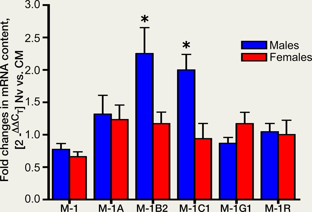
Chronic systemic morphine influenced the spinal expression of some MOR variants in a sex-dependent fashion (Fig 1). In the spinal cord of males, chronic morphine increased expression levels ≈2-fold of mRNA encoding rMOR-1B2 and rMOR-1C1 (ANOVA; F5,30=6.636, p<0.05 for both rMOR-1B2/rMOR-1C1; n=5–8) (Fig. 1). However, in striking contrast to males, in females, chronic morphine failed to alter spinal levels of mRNA encoding rMOR-1B2 and rMOR-1C1. Notably, spinal levels of mRNA encoding rMOR-1, rMOR-1A, rMOR-1G1 and rMOR-1R remained invariable in the spinal cord of females as well as males following chronic morphine.
Figure 1. Sex-dependent effect of chronic morphine on the mRNA expression of MOR variants.
Male and female rats were treated with escalating doses of morphine base pellets (see methods). Data are calculated as fold change in MOR mRNA content, where 2−ΔΔCT represents the change in spinal mRNA content in opioid naïve (Nv) vs. chronic morphine treated (CM) groups relative to that of rMOR-1R. Chronic morphine significantly upregulated two splice variants of MOR, rMOR-1B2 (M-1B2) and rMOR-1C1 (M-1C1) in only males (*= ~ 2 fold; p<0.05). n=5-8).
Chronic systemic morphine upregulates density of MOR in the spinal cord of male rats
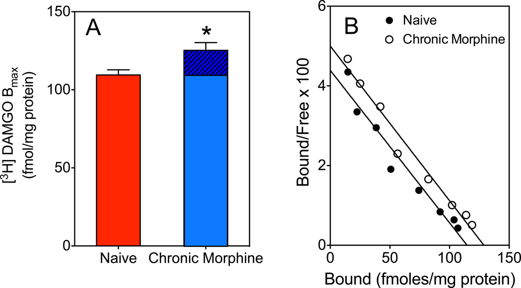
Radioligand binding studies were employed to determine the effect of chronic morphine on the MOR density and agonist affinity in spinal cord of male rats (Fig. 2). [3H]DAMGO Bmax±SEM and KD±SEM in lumbar spinal cord of naïve male rats were 109.5±3.0 fmol/mg protein and 1.49±0.04 nM, respectively. Following chronic systemic morphine there was a small but significant increase (13%; p<0.05) in [3H]DAMGO Bmax (125.3±4.5 fmol/mg protein) without a significant change in KD (1.60±0.03 nM).
Figure 2. Effect of chronic systemic morphine on MOR density in spinal cord of male rat.
Lumbar spinal tissue obtained from two placebo or chronic morphine-treated male rats were dissected and pooled for each saturation binding experiment using [3H]DAMGO. (A) Mean [3H]DAMGO Bmax±SEM was determined in spinal cord from naïve (109.5±3.0 fmol/mg protein) and chronic morphine treated (125.3±4.5 fmol/mg protein) groups (*p<0.05; n=6). Hatched portion indicates the increment in [3H]DAMGO Bmax following chronic-morphine treatment. (B) A representative scatchard plot is shown for naïve and chronic morphine-treated spinal cord. No significant difference was observed between [3H]DAMGO KDs in naïve (1.49±0.04 nM) and morphine-treated spinal cord (1.60±0.03 nM) group (p>0.05).
Discussion
This is the first report to suggest that chronic systemic treatment with morphine, sufficient to produce profound analgesic tolerance and dependence at the time of sacrifice (Villar & Bhargava 1992), not only alters the content of spinal cord mRNAs that encode specific MOR splice variants but also does so in a sex-dependent fashion. Sexually dimorphic alterations in the relative abundance of selective MOR variants, which could have unique signaling attributes, provides a molecular context for understanding sex-based accommodations to chronic morphine that have been reported previously (Chakrabarti et al. 2012).
In spinal cord of males, but not females, chronic morphine was found to augment mRNA encoding rMOR-1C1 and rMOR-1B2. Poor correlation between steady state isoform-specific mRNA and the protein it encodes has been reported (Mons & Cooper 1994). Nevertheless, it seems reasonable to conjecture that a selective increase in the mRNA encoding rMOR-1B2 and rMOR-1C1 likely suggests a parallel change in the content and activity of rMOR-1B2 and rMOR-1C1 receptor protein. This inference is supported by saturation binding analyses, which revealed that a small but significant increase in MOR receptor density accompanies the increment in rMOR-1B2/rMOR-1C1 mRNA following chronic systemic morphine.
Analgesic tolerance formation was not directly assessed in the current study. However, the regimen of chronic systemic morphine exposure is well established to produce profound analgesic tolerance and dependence at the time of animal sacrifice (Villar & Bhargava 1992). Importantly, sexual dimorphism in the magnitude of analgesic tolerance remains controversial in both rats (Craft et al. 1999, Holtman et al. 2004) and mice (Hosseini et al. 2010, Kest et al. 2000). Thus, it is highly unlikely that differential magnitudes of analgesic tolerance in males vs. females per se could explain the observed sex-dependent effects of chronic morphine on spinal rMOR-1B2/rMOR-1C1 mRNA. Moreover, since the magnitude of differences in MOR agonist efficacy and potency among splice variants is relatively small in comparison to the magnitude of tolerance that can develop, it also seems unlikely that presumptive tolerance-associated upregulation of rMOR-1C1 and rMOR-1B2 per se would significantly contribute to tolerance by altering the agonist response profile to mu agonists (Pasternak & Pan 2013).
C-terminal splice variants of MOR have been reported to differ in their endocytotic response to chronic morphine (Koch et al. 2001, Koch et al. 1998). However, given the well documented disconnects between the ability of opioid agonists to induce MOR endocytosis and/or G protein uncoupling vs. analgesic tolerance (Sternini et al. 1996, Madia et al. 2012), the differential intracellular trafficking of C-terminal splice variants, per se, is not likely to be a primary factor modulating morphine analgesic tolerance in vivo.
There are, however, unique structural characteristics of rMOR-1C1 and rMOR-1B2, which reside in the composition of the tip of the C-terminus that could influence their functionality. The C-terminal twelve amino acids (aa) of rMOR-1, LENLEAETAPLP, are replaced in rMOR-1C1 by 65 aa, PALAVSVAQIFTGYPSPTHGEKPCKSYRDRPRPCGRTWSLKSRAESNVEHFHCGAALI-YNNVNFI [encoded for by exon 7 (bp 44966586-4496614), exon 8 (bp 45027017-450027076), exon 9a (bp 45034092-45034145) and exon 9b (bp 45032373-45032473) of the OPRM gene located on chromosome 1]. In rMOR-1B2, the C-terminal twelve aa of rMOR-1 are replaced by seven aa, EPQSVET, (Zimprich et al. 1995, Pan 2005, Pan et al. 2005b), encoded by exon 5 [exon 5b (bp 44845374- 44847120) and 5a (bp 44847121-44847400) of the OPRM1 gene]. Importantly, the unique C-terminus of rMOR-1B2/rMOR-1C1 contains numerous consensus sequences for phosphorylation by a variety of kinases that are not present in rMOR-1. (e.g., protein kinase C, protein kinase A, casein kinase II, ribosomal factor S6 kinase, cyclin-dependent kinase 5 and cGMP dependent protein kinase, etc).
Phosphorylation patterns of G protein coupled receptors not only influence the nature of the particular G protein to which receptors couple (Lefkowitz et al. 2002, Zamah et al. 2002), but also influence biased agonism, agonist-specific stabilization of receptor conformations that preferentially activate specific downstream signaling pathways (Kahsai et al. 2011). Moreover, the induction by chronic morphine of newly synthesized MOR variants and the intracellular trafficking of newly synthesized MORs could themselves constitute signals that herald novel adaptations. These considerations suggest that the male-specific upregulation of spinal rMOR-1B2/rMOR-1C1 in response to chronic morphine could substantially influence the aggregate signaling consequences of mu opioids. In this regard, it is relevant to note that a derivative of the endogenous MOR ligand, endomorphin 2, has been shown to elicit analgesic tolerance and related biochemical sequelae (Muranyi et al. 2013). Thus, male-specific adaptations to chronic morphine described herein could be relevant to physiological adaptations to stimuli that result in sustained utilization/activation of the endogenous MOR analgesic system. This is particularly so since MOR-1C1 has been shown to be present in laminae I/II of rat and human spinal cord (Abbadie et al. 2000b, Abbadie et al. 2000a), areas rich in endomorphin 2 (Martin-Schild et al. 1999, Schreff et al. 1998).
Recently, we reported on the heterodimerization of MOR with KOR in spinal cord that was not only sex-dependent but also dependent on the stage of the estrus cycle (Chakrabarti et al. 2010, Liu et al. 2011), the bases for which remain unknown. We had speculated that sex and estrus cycle dependence of MOR/KOR heterodimerization resulted from sex- and estrus cycle-dependent expression of spinal MOR variants. However, this is unlikely in light of the current finding that spinal expression levels of mRNA encoding each of the six splice variants studied did not vary between diestrus and proestrus females.
Surprisingly, spinal levels of mRNA encoding rMOR-1B2 and rMOR-1G1 were significantly higher in the spinal cord of diestrus female vs. male rats. The physiological/pharmacological significance of this difference remains to be defined. However, it should be noted that differences in analgesic responsiveness among diestrus, proestrus and male rats have been reported (Boyer et al. 1998). Furthermore, since diestrus is the longest single stage of the estrus cycle, studies comparing males and (non-cycled) females most likely involve females in diestrus. Therefore, differences in spinal rMOR-1B2/rMOR-1G1 could undergird, at least in part, male-female differences in opioid antinociception that have been reported, definitive proof of which will require the development of MOR variant-selective agonists and antagonists.
In summary, the sexually dimorphic ability of chronic systemic morphine to upregulate spinal rMOR-1C1 and rMOR-1B2 mRNA could suggest that spinal MOR variants are differentially recruited in males vs. females to mediate sex-dependent adaptations to chronic morphine, e.g., (Chakrabarti et al. 2012). Knowledge of unique signaling properties of rMOR-1C1 and rMOR-1B2, which reside in the composition of the tip of the C-terminus, could point the way to defining the molecular determinants of sex-dependent tolerance and withdrawal mechanisms and treatments thereof.
Acknowledgments
Supported by NIDA grant DA027663 (to NJL and ARG).
Abbreviations
- MOR
Mu-opioid receptor
- aa
amino acids
- G6PD
Glucose-6-phosphate dehydrogenase
Footnotes
Competing interests
The authors declare that they have no competing interests.
References
- Abbadie C, Gultekin SH, Pasternak GW. Immunohistochemical localization of the carboxy terminus of the novel mu opioid receptor splice variant MOR-1C within the human spinal cord. Neuroreport. 2000a;11:1953–1957. doi: 10.1097/00001756-200006260-00029. [DOI] [PubMed] [Google Scholar]
- Abbadie C, Pan Y, Drake CT, Pasternak GW. Comparative immunohistochemical distributions of carboxy terminus epitopes from the mu-opioid receptor splice variants MOR-1D, MOR-1 and MOR-1C in the mouse and rat CNS. Neuroscience. 2000b;100:141–153. doi: 10.1016/s0306-4522(00)00248-7. [DOI] [PubMed] [Google Scholar]
- Abbadie C, Pan YX, Pasternak GW. Differential distribution in rat brain of mu opioid receptor carboxy terminal splice variants MOR-1C-like and MOR-1-like immunoreactivity: evidence for region-specific processing. J Comp Neurol. 2000c;419:244–256. doi: 10.1002/(sici)1096-9861(20000403)419:2<244::aid-cne8>3.0.co;2-r. [DOI] [PubMed] [Google Scholar]
- Abbadie C, Pasternak GW. Differential in vivo internalization of MOR-1 and MOR-1C by morphine. Neuroreport. 2001;12:3069–3072. doi: 10.1097/00001756-200110080-00017. [DOI] [PubMed] [Google Scholar]
- Barrett AC, Smith ES, Picker MJ. Sex-related differences in mechanical nociception and antinociception produced by mu- and kappa-opioid receptor agonists in rats. Eur J Pharmacol. 2002;452:163–173. doi: 10.1016/s0014-2999(02)02274-4. [DOI] [PubMed] [Google Scholar]
- Bolan EA, Pan YX, Pasternak GW. Functional analysis of MOR-1 splice variants of the mouse mu opioid receptor gene Oprm. Synapse. 2004;51:11–18. doi: 10.1002/syn.10277. [DOI] [PubMed] [Google Scholar]
- Boyer JS, Morgan MM, Craft RM. Microinjection of morphine into the rostral ventromedial medulla produces greater antinociception in male compared to female rats. Brain Res. 1998;796:315–318. doi: 10.1016/s0006-8993(98)00353-9. [DOI] [PubMed] [Google Scholar]
- Chakrabarti S, Liu NJ, Gintzler AR. Formation of {micro}-/{kappa}-opioid receptor heterodimer is sex-dependent and mediates female-specific opioid analgesia. Proc Natl Acad Sci U S A. 2010;107:20115–20119. doi: 10.1073/pnas.1009923107. [DOI] [PMC free article] [PubMed] [Google Scholar]
- Chakrabarti S, Liu NJ, Zadina JE, Sharma T, Gintzler A. Pleiotropic opioid regulation of spinal endomorphin 2 release and its adaptations to opioid withdrawal are sexually dimorphic. J Pharmacol Exp Ther. 2012;340:56–63. doi: 10.1124/jpet.111.186874. [DOI] [PMC free article] [PubMed] [Google Scholar]
- Chen Y, Mestek A, Liu J, Hurley JA, Yu L. Molecular cloning and functional expression of a mu-opioid receptor from rat brain. Mol Pharmacol. 1993;44:8–12. [PubMed] [Google Scholar]
- Cicero TJ, Nock B, Meyer ER. Gender-related differences in the antinociceptive properties of morphine. J Pharmacol Exp Ther. 1996;279:767–773. [PubMed] [Google Scholar]
- Cicero TJ, Nock B, Meyer ER. Sex-related differences in morphine's antinociceptive activity: relationship to serum and brain morphine concentrations. J Pharmacol Exp Ther. 1997;282:939–944. [PubMed] [Google Scholar]
- Cook CD, Barrett AC, Roach EL, Bowman JR, Picker MJ. Sex-related differences in the antinociceptive effects of opioids: importance of rat genotype, nociceptive stimulus intensity, and efficacy at the mu opioid receptor. Psychopharmacology (Berl) 2000;150:430–442. doi: 10.1007/s002130000453. [DOI] [PubMed] [Google Scholar]
- Craft RM, Stratmann JA, Bartok RE, Walpole TI, King SJ. Sex differences in development of morphine tolerance and dependence in the rat. Psychopharmacology (Berl) 1999;143:1–7. doi: 10.1007/s002130050911. [DOI] [PubMed] [Google Scholar]
- Holtman JR, Jr, Sloan JW, Wala EP. Morphine tolerance in male and female rats. Pharmacol Biochem Behav. 2004;77:517–523. doi: 10.1016/j.pbb.2003.12.020. [DOI] [PubMed] [Google Scholar]
- Hosseini M, Karami R, Khodabandeloo F, Khatami L. Gender dependent differences Iin analgesic effect and tolerance to morphine. Pharmacologyonline. 2010;3:92–101. [Google Scholar]
- Kahsai AW, Xiao K, Rajagopal S, Ahn S, Shukla AK, Sun J, Oas TG, Lefkowitz RJ. Multiple ligand-specific conformations of the beta2-adrenergic receptor. Nat Chem Biol. 2011;7:692–700. doi: 10.1038/nchembio.634. [DOI] [PMC free article] [PubMed] [Google Scholar]
- Kest B, Palmese C, Hopkins E. A comparison of morphine analgesic tolerance in male and female mice. Brain Res. 2000;879:17–22. doi: 10.1016/s0006-8993(00)02685-8. [DOI] [PubMed] [Google Scholar]
- Koch T, Schulz S, Pfeiffer M, Klutzny M, Schroder H, Kahl E, Hollt V. C-terminal splice variants of the mouse mu-opioid receptor differ in morphine-induced internalization and receptor resensitization. J Biol Chem. 2001;276:31408–31414. doi: 10.1074/jbc.M100305200. [DOI] [PubMed] [Google Scholar]
- Koch T, Schulz S, Schroder H, Wolf R, Raulf E, Hollt V. Carboxyl-terminal splicing of the rat mu opioid receptor modulates agonist-mediated internalization and receptor resensitization. J Biol Chem. 1998;273:13652–13657. doi: 10.1074/jbc.273.22.13652. [DOI] [PubMed] [Google Scholar]
- Krzanowska EK, Bodnar RJ. Morphine antinociception elicited from the ventrolateral periaqueductal gray is sensitive to sex and gonadectomy differences in rats. Brain Res. 1999;821:224–230. doi: 10.1016/s0006-8993(98)01364-x. [DOI] [PubMed] [Google Scholar]
- Lefkowitz RJ, Pierce KL, Luttrell LM. Dancing with different partners: protein kinase A phosphorylation of seven membrane-spanning receptors regulates their G protein-coupling specificity. Mol Pharmacol. 2002;62:971–974. doi: 10.1124/mol.62.5.971. [DOI] [PubMed] [Google Scholar]
- Liu NJ, Chakrabarti S, Schnell S, Wessendorf M, Gintzler AR. Spinal Synthesis of Estrogen and Concomitant Signaling by Membrane Estrogen Receptors Regulate Spinal {kappa}- and {micro}-Opioid Receptor Heterodimerization and Female-Specific Spinal Morphine Antinociception. J Neurosci. 2011;31:11836–11845. doi: 10.1523/JNEUROSCI.1901-11.2011. [DOI] [PMC free article] [PubMed] [Google Scholar]
- Livak KJ, Schmittgen TD. Analysis of relative gene expression data using real-time quantitative PCR and the 2(-Delta Delta C(T)) Method. Methods. 2001;25:402–408. doi: 10.1006/meth.2001.1262. [DOI] [PubMed] [Google Scholar]
- Madia PA, Navani DM, Yoburn BC. [(35)S]GTPgammaS binding and opioid tolerance and efficacy in mouse spinal cord. Pharmacol Biochem Behav. 2012;101:155–165. doi: 10.1016/j.pbb.2011.11.001. [DOI] [PubMed] [Google Scholar]
- Martin-Schild S, Gerall AA, Kastin AJ, Zadina JE. Differential distribution of endomorphin 1- and endomorphin 2-like immunoreactivities in the CNS of the rodent. J Comp Neurol. 1999;405:450–471. [PubMed] [Google Scholar]
- Mons N, Cooper DMF. Adenylyl cyclase mRNA expression does not reflect the predominant Ca2+/calmodulin-stimulated activity in the hypothalamus. J. Neuroendocrinol. 1994;6:665–671. doi: 10.1111/j.1365-2826.1994.tb00633.x. [DOI] [PubMed] [Google Scholar]
- Muranyi M, Cinar R, Kekesi O, et al. Ligand-specific regulation of the endogenous mu-opioid receptor by chronic treatment with mu-opioid peptide agonists. BioMed research international. 2013 doi: 10.1155/2013/501086. 501086. [DOI] [PMC free article] [PubMed] [Google Scholar]
- Pan L, Xu J, Yu R, Xu MM, Pan YX, Pasternak GW. Identification and characterization of six new alternatively spliced variants of the human mu opioid receptor gene, Oprm. Neuroscience. 2005a;133:209–220. doi: 10.1016/j.neuroscience.2004.12.033. [DOI] [PubMed] [Google Scholar]
- Pan YX. Diversity and complexity of the mu opioid receptor gene: alternative pre-mRNA splicing and promoters. DNA Cell Biol. 2005;24:736–750. doi: 10.1089/dna.2005.24.736. [DOI] [PubMed] [Google Scholar]
- Pan YX, Xu J, Bolan E, Moskowitz HS, Xu M, Pasternak GW. Identification of four novel exon 5 splice variants of the mouse mu-opioid receptor gene: functional consequences of C-terminal splicing. Mol Pharmacol. 2005b;68:866–875. doi: 10.1124/mol.105.011858. [DOI] [PubMed] [Google Scholar]
- Pan YX, Xu J, Xu M, Rossi GC, Matulonis JE, Pasternak GW. Involvement of exon 11-associated variants of the mu opioid receptor MOR-1 in heroin, but not morphine, actions. Proc Natl Acad Sci U S A. 2009;106:4917–4922. doi: 10.1073/pnas.0811586106. [DOI] [PMC free article] [PubMed] [Google Scholar]
- Pasternak DA, Pan L, Xu J, Yu R, Xu MM, Pasternak GW, Pan YX. Identification of three new alternatively spliced variants of the rat mu opioid receptor gene: dissociation of affinity and efficacy. J Neurochem. 2004;91:881–890. doi: 10.1111/j.1471-4159.2004.02767.x. [DOI] [PubMed] [Google Scholar]
- Pasternak GW. Incomplete cross tolerance and multiple mu opioid peptide receptors. Trends Pharmacol Sci. 2001;22:67–70. doi: 10.1016/s0165-6147(00)01616-3. [DOI] [PubMed] [Google Scholar]
- Pasternak W, Pan Y-X. Mu opioids and their receptors: Evolution of a concept. Pharmacological Reviews. 2013;65:1257–1317. doi: 10.1124/pr.112.007138. [DOI] [PMC free article] [PubMed] [Google Scholar]
- Peckham EM, Traynor JR. Comparison of the antinociceptive response to morphine and morphine-like compounds in male and female Sprague-Dawley rats. J Pharmacol Exp Ther. 2006;316:1195–1201. doi: 10.1124/jpet.105.094276. [DOI] [PubMed] [Google Scholar]
- Sarton E, Olofsen E, Romberg R, den Hartigh J, Kest B, Nieuwenhuijs D, Burm A, Teppema L, Dahan A. Sex differences in morphine analgesia: an experimental study in healthy volunteers. Anesthesiology. 2000;93:1245–1254. doi: 10.1097/00000542-200011000-00018. ; discussion 1246A. [DOI] [PubMed] [Google Scholar]
- Schreff M, Schulz S, Wiborny D, Hollt V. Immunofluorescent identification of endomorphin-2-containing nerve fibers and terminals in the rat brain and spinal cord. Neuroreport. 1998;9:1031–1034. doi: 10.1097/00001756-199804200-00014. [DOI] [PubMed] [Google Scholar]
- Schuller AG, King MA, Zhang J, et al. Retention of heroin and morphine-6 beta-glucuronide analgesia in a new line of mice lacking exon 1 of MOR-1. Nat Neurosci. 1999;2:151–156. doi: 10.1038/5706. [DOI] [PubMed] [Google Scholar]
- Sternini C, Spann M, Anton B, Keith DE, Bunnett NW, von Zastrow M, Evans CJ, Brecha NC. Agonist-selective endocytosis of m-opioid receptor by neurons in vivo. Proc. Natl. Acad. Sci. USA. 1996;93:9241–9246. doi: 10.1073/pnas.93.17.9241. [DOI] [PMC free article] [PubMed] [Google Scholar]
- Villar VM, Bhargava HN. Pharmacodynamics and kinetics of loss of tolerance and physical dependence on morphine induced by pellet implantation in the rat. Pharmacology. 1992;45:319–328. doi: 10.1159/000139017. [DOI] [PubMed] [Google Scholar]
- Wolozin BL, Pasternak GW. Classification of multiple morphine and enkephalin binding sites in the central nervous system. Proc Natl Acad Sci U S A. 1981;78:6181–6185. doi: 10.1073/pnas.78.10.6181. [DOI] [PMC free article] [PubMed] [Google Scholar]
- Zamah AM, Delahunty M, Luttrell LM, Lefkowitz RJ. Protein kinase A-mediated phosphorylation of the beta 2-adrenergic receptor regulates its coupling to Gs and Gi. Demonstration in a reconstituted system. J Biol Chem. 2002;277:31249–31256. doi: 10.1074/jbc.M202753200. [DOI] [PubMed] [Google Scholar]
- Zimprich A, Simon T, Hollt V. Cloning and expression of an isoform of the rat mu opioid receptor (rMOR1B) which differs in agonist induced desensitization from rMOR1. FEBS Lett. 1995;359:142–146. doi: 10.1016/0014-5793(95)00028-8. [DOI] [PubMed] [Google Scholar]


