Abstract
Post-transplant lymphoproliferative disorder (PTLD) is a heterogeneous group of lymphoid proliferations caused by immunosuppression after solid organ or bone marrow transplantation. PTLD is categorized by early lesion, polymorphic PTLD and monomorphic PTLD. Fluorine-18 fluorodeoxyglucose-positron emission tomography/computed tomography (F-18 FDG-PET/CT) scans have clinical significance in the evaluation of PTLD following renal transplantation. We report imaging findings of a monomorphic non-Hodgkin lymphoma, post renal transplant seen on FDG PET/CT in a 32-year-old lactating woman. Whole body FDG- ET/CT demonstrated uptake in right external iliac and inguinal lymph nodes.
Keywords: Asymmetric lactating breast uptake, fluorodeoxyglucose-positron emission tomography/computed tomography, monomorphic non-Hodgkin lymphoma, renal transplant
INTRODUCTION
Post-transplant lymphoproliferative disorder (PTLD) is a rare, but life-threatening disorder, characterized by an uncontrolled proliferation of lymphocytes, caused by immunosuppressive drug-induced diminished immune surveillance.[1] The majority of monomorphic PTLD cases are non-Hodgkin's lymphoma of B-cell origin; in contrast, T-cell lymphoma and Hodgkin's disease are rare.[2] We report imaging features of fluorodeoxyglucose-positron emission tomography/computed tomography (FDG-PET/CT) with monomorphic non-Hodgkin lymphoma PTLD in a post renal transplant.
CASE REPORT
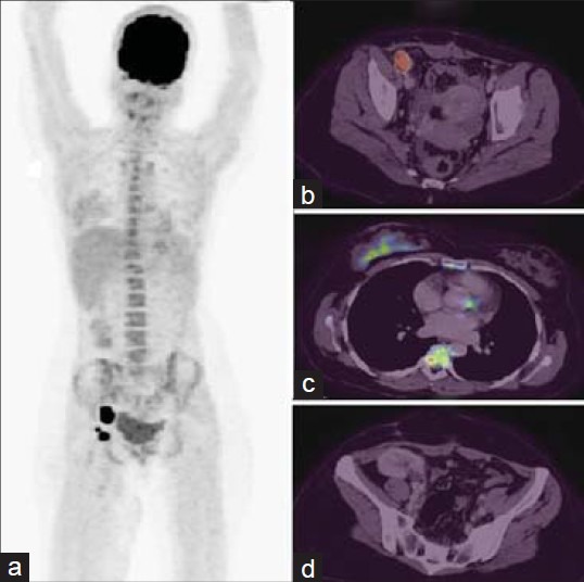
This was a case report of a 32-year-old female patient who had immunoglobulin A nephropathy underwent renal transplant 6 years before and was on immunosuppressants. She is postpartum 6 months and breast feeding, presented with swelling in the right inguinal region. She underwent an excision biopsy of the right inguinal node, which showed diffuse large B cell lymphoma. As a part of evaluation, she was subjected to whole body PET/CT, which showed increased uptake in multiple nodes in right external iliac and inguinal region [Figure 1a and b], also right breast parenchyma uptake [Figure 1c] and transplant kidney noted in right iliac fossa [Figure 1d]. As PET/CT scan confirmed involvement of multiple nodes, patient underwent chemotherapy and asked to stop breast feeding.
Figure 1.

Whole body fluorodeoxyglucose-positron emission tomography/computed tomography (PET/CT) maximum intensity projection image (a), axial fused PET/CT showed an intense uptake in the right external iliac lymphnodes (b), right breast parenchyma uptake due to lactation (c) and transplant kidney in right iliac fossa (d) noted
DISCUSSION
PTLD are a heterogeneous group of abnormal lymphoid proliferations that occur after solid organ transplant or hematopoietic transplantation. PTLDs consist of a disease spectrum ranging from hyperplasia to aggressive lymphomas with 60 to 70% being Epstein-Barr virus (EBV) positive.[1] The majority of cases are B-cell, although 10-15% is of T-cell origin or rarely Hodgkin lymphoma. In the initial stages of PTLD, proliferation is polyclonal. With mutation and selective growth, the lesion becomes oligoclonal and later, monoclonal. The incidence rate of monomorphic PTLD was 0.24% and the median time between the transplant and diagnosis of PTLD was 85.8 months.[2] The immunosuppression required to prevent graft rejection post transplantation impairs T-cell immunity, potentially allowing for uncontrolled proliferation of EBV-infected B-cells, which may result in a spectrum of B-cell proliferations that range from hyperplasia to true lymphoma.[3] The mechanisms that cause EBV-negative PTLD are not well-understood. It often presents with a later onset, monomorphic histology and a more aggressive course than EBV-positive PTLD, suggesting that it may have a different pathophysiology.[3]
Experience with the use of PET in PTLD has been limited to case reports and rather small single-center case series.[4,5,6] These reports reveal that PET scan may have a high sensitivity for the detection of PTLD lesions.[7,8] FDG uptake pattern have been useful in predicting the benign or malignant nature of nodes. In our case, as PET study revealed multiple nodal involvements, patient was subjected to chemotherapy. Our case thus illustrates that FDG PET/CT is an important imaging modality for the detection and accurate staging and restaging of PTLD and to decide upon appropriate treatment.
Footnotes
Source of Support: Nil
Conflict of Interest: None declared
REFERENCES
- 1.Elias C, Steven HS, Nancy LH, Stefano P, Harald S, Elaine SJ. The 2008 WHO classification of lymphoid neoplasms and beyond: Evolving concepts and practical applications Blood. 2011 May 12;117:5019–32. doi: 10.1182/blood-2011-01-293050. [DOI] [PMC free article] [PubMed] [Google Scholar]
- 2.Choi JH, Park BB, Suh C, Won JH, Lee WS, Shin HJ. Clinical characteristics of monomorphic post-transplant lymphoproliferative disorders. J Korean Med Sci. 2010;25:523–6. doi: 10.3346/jkms.2010.25.4.523. [DOI] [PMC free article] [PubMed] [Google Scholar]
- 3.Schubert S, Renner C, Hammer M, Abdul-Khaliq H, Lehmkuhl HB, Berger F, et al. Relationship of immunosuppression to Epstein-Barr viral load and lymphoproliferative disease in pediatric heart transplant patients. J Heart Lung Transplant. 2008;27:100–5. doi: 10.1016/j.healun.2007.09.027. [DOI] [PubMed] [Google Scholar]
- 4.O’Conner AR, Franc BL. FDG PET imaging in the evaluation of post-transplant lymphoproliferative disorder following renal transplantation. Nucl Med Commun. 2005;26:1107–11. doi: 10.1097/00006231-200512000-00010. [DOI] [PubMed] [Google Scholar]
- 5.von Falck C, Maecker B, Schirg E, Boerner AR, Knapp WH, Klein C, et al. Post transplant lymphoproliferative disease in pediatric solid organ transplant patients: A possible role for [18F]-FDG-PET(/CT) in initial staging and therapy monitoring. Eur J Radiol. 2007;63:427–35. doi: 10.1016/j.ejrad.2007.01.007. [DOI] [PubMed] [Google Scholar]
- 6.Bianchi E, Pascual M, Nicod M, Delaloye AB, Duchosal MA. Clinical usefulness of FDG-PET/CT scan imaging in the management of posttransplant lymphoproliferative disease. Transplantation. 2008;85:707–12. doi: 10.1097/TP.0b013e3181661676. [DOI] [PubMed] [Google Scholar]
- 7.Noraini AR, Gay E, Ferrara C, Ravelli E, Mancini V, Morra E, et al. PET-CT as an effective imaging modality in the staging and follow-up of post-transplant lymphoproliferative disorder following solid organ transplantation. Singapore Med J. 2009;50:1189–95. [PubMed] [Google Scholar]
- 8.Blaes AH, Cioc AM, Froelich JW, Peterson BA, Dunitz JM. Positron emission tomography scanning in the setting of post-transplant lymphoproliferative disorders. Clin Transplant. 2009;23:794–9. doi: 10.1111/j.1399-0012.2008.00938.x. [DOI] [PubMed] [Google Scholar]


