Abstract
Increase in glycolytic pathway, forms one of the major adaptations in various cancer types. This can be imaged using 18F-fluoro-deoxyglucose positron emission tomography/computed tomography (FDG PET). The intensity of FDG avidity is an indirect marker of the grade of the tumor. We present a case where FDG PET demonstrated a known chondrosarcoma and two other incidental lesions. The intensity of avidity in each of the lesions was grossly incongruent from the chondrosarcoma and further investigation proved the lesions to be two distinct primary malignancies, pathologically different from the known chondrosarcoma. We present the case to highlight the fact that the grade of FDG avidity is a clue to the pathological nature of the lesion and should always be considered while interpreting PET images.
Keywords: 18F-fluoro-deoxyglucose positron emission tomography, metabolic signature, pathological grade
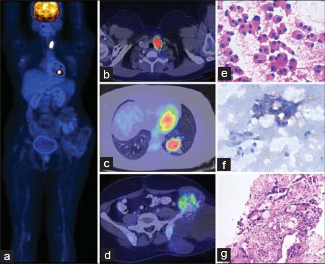
A 40-year-old female patient diagnosed to have Grade 1 chondrosarcoma of the left iliac bone presented with local re-growth of the tumor after initial excision done 3 years back. The whole body image of 18F-fluoro-deoxyglucose (FDG) PET scan, [Figure 1] shows very mild FDG avidity in the iliac mass (1d). Two additional lesions, one in the lower lobe of the left lung and one in the left lobe of the thyroid, were also identified. The mass in the lower lobe of the left lung showed intense FDG uptake with a standardized uptake value (SUVmax) of 17. The corresponding computed tomography images (1c) demonstrate speculated mass with central necrotic area. The lesion in the left lobe of the thyroid (1b) also showed intense hypermetabolism (SUVmax 30). The metabolic signature of the primary (SUVmax 3) is in congruence with its well differentiated nature (Grade 1 chondrosarcoma).[1,2] Both the lesion in the lung and the thyroid showed grossly incongruent metabolic signature from the primary. This prompted us to investigate both sites and the histopathology confirmed the lung mass to be primary adenocarcinoma (1g) while the thyroid lesion showed features of medullary carcinoma. Staining for calcitonin was also positive from the thyroid lesion (1f).
Figure 1.

Whole body image (a) showing different intensities of uptake of the three different lesion with the corresponding fused axial sections of the lesions (b-d). The pathology images of the thyroid lesion (e) along with special staining for calcitonin (f) and that of the lung lesion (g)
The current case attempts to highlight two interesting features. First being the incidental detection of three distinct primary lesions in a patient. The patient also had a family history of osteosarcoma in her son, which suggests the possibility of Li Fraumeni syndrome syndrome.[3,4] The second feature is an inherent property of FDG PET that makes it an indirect molecular marker to estimate the nature of underlying pathological process. The SUVmax values of the individual lesions in this case are quite different from each other. This clue helped make the correct diagnosis by not calling the lesions as metastatic and doing further evaluation. We present this case to highlight the fact that the metabolic activity of a lesion can be a potential clue to the pathological processes and this should be kept under consideration while interpreting PET images.
ACKNOWLEDGMENT
The authors would like to acknowledge Prof. Rodney J. Hicks, Peter Mac Callum Cancer Center, Melbourne, Australia, for the introducing the term “Metabolic Signature ” in PET imaging.
Footnotes
Source of Support: Nil
Conflict of Interest: None declared
REFERENCES
- 1.Tateishi U, Yamaguchi U, Seki K, Terauchi T, Arai Y, Hasegawa T. Glut-1 expression and enhanced glucose metabolism are associated with tumour grade in bone and soft tissue sarcomas: A prospective evaluation by 18F fluorodeoxyglucose positron emission tomography. Eur J Nucl Med Mol Imaging. 2006;33:683–91. doi: 10.1007/s00259-005-0044-8. [DOI] [PubMed] [Google Scholar]
- 2.Fuglø HM, Jørgensen SM, Loft A, Hovgaard D, Petersen MM. The diagnostic and prognostic value of 8F-FDG PET/CT in the initial assessment of high-grade bone and soft tissue sarcoma. A retrospective study of 89 patients. Eur J Nucl Med Mol Imaging. 2012;39:1416–24. doi: 10.1007/s00259-012-2159-z. [DOI] [PubMed] [Google Scholar]
- 3.Li FP, Fraumeni JF., Jr Soft-tissue sarcomas, breast cancer, and other neoplasms. A familial syndrome? Ann Intern Med. 1969;71:747–52. doi: 10.7326/0003-4819-71-4-747. [DOI] [PubMed] [Google Scholar]
- 4.Masciari S, Van den Abbeele AD, Diller LR, Rastarhuyeva I, Yap J, Schneider K, et al. F18-fluorodeoxyglucose-positron emission tomography/computed tomography screening in Li-Fraumeni syndrome. JAMA. 2008;299:1315–9. doi: 10.1001/jama.299.11.1315. [DOI] [PubMed] [Google Scholar]


