Abstract
Background
Risk of overuse injury among athletes is high due in part to repeated loading of the lower extremities. Compared to individuals with normal arch (NA) structure, those with high (HA) or low arch (LA) may be at increased risk of specific overuse injuries, including stress fractures. A high medial longitudinal arch may result in decreased shock absorbing properties due to increased rigidity in foot mechanics. While the effect of arch structure on dynamic function has been examined in straight line walking and running, the relationship between the two during multi-directional movements remains unstudied.
Objective
The purpose of this study was to determine if differences in plantar loading in football players occur during both walking and pivoting movements.
Method
Plantar loading was examined in 9 regions of the foot for 26 participants (16 NA, 10 HA).
Results
High arch athletes demonstrated increased maximum force in the lateral rear foot and medial forefoot, and force time integral in the medial forefoot while walking. HA athletes also demonstrated increased maximum force in the medial rear foot and medial and central forefoot during rapid pivoting.
Conclusions
The current findings demonstrate that loading patterns differ between football players with high and normal arch structure, which could possibly influence injury risk in this population.
Keywords: Plantar pressure, Overuse injury, Cutting task, Biomechanics
1. Introduction
Risk of lower extremity injury among young athletes is related to a variety of internal and external factors. It has been suggested that repeated impacts with the ground, coincident with the resultant ground reaction forces, can initiate numerous lower extremity injuries, including stress fractures [1], cartilage damage [2] and the development of osteoarthritis [3,4], amongst others. Anatomical differences in foot, including abnormal arch structure, increase the risk of lower extremity overuse injuries two-fold [5]. Athletes with both high and low arches may be at increased risk of lower extremity injury compared to athletes with normal arches [5–7]. A high medial longitudinal arch may increase rigid foot mechanics that result in decreased shock-absorbing properties when compared to athletes with normal arch structure [5,8,9]. Specifically, decreased motion (increased stiffness) between multiple segments within the foot has been shown among individuals with high arch structure [10]. An increased relative arch height is also linked to increased risk of multiple lower extremity injuries, including plantar fasciitis and lateral ankle sprains [9]. Furthermore, the biomechanical coupling of foot eversion-inversion with tibial internal-external rotation may also increase the risk of knee injury in athletes with a high arch [11].
Foot structure can be objectively assessed with a variety of clinical and research tools [12–15]. For example, the arch height index measurement system (AHIMS) is a non-invasive, easy to use device that is a reliable and valid method of measuring the foot structure [9,14,16,17]. Objective measurement could provide an effective tool for clinicians to identify an athlete's relative predisposition to injury [11].
Comparison of foot structure to dynamic function has been evaluated primarily during straight-line walking and running [7,10–12,18–23], with findings of changes in loading [12,20,22] and kinematic aspects of movement [11] among those with varying arch structures. The current literature indicates that further insight into injury mechanisms among athletes with abnormal arch height could result by evaluation of dynamic loading activities in shod conditions [10]. Specifically, differences in plantar loading between straight line tasks and multi-directional movements have been previously demonstrated in shod conditions. For example, when compared to running, individuals performing a side cut have demonstrated increased values on the medial aspect of the foot in peak pressure [23,24], pressure time integral [23] and percent of total load of the foot [24].
Shoe design for football may consist of different cleat patterns, cushioning characteristics and a vast variety of materials. The requirements for the sport and position often require different design considerations. For American football, cutting maneuvers are frequent and have been previously examined [25,26]. Ford et al. [26] compared a football cleat on multiple surfaces (synthetic and natural) and found significantly greater forefoot loading on the natural grass surface. Interestingly, the lateral aspect of the foot had greater peak pressure on a synthetic surface compared to natural surface when cutting with cleated footwear [26]. Oredurff et al. [25] identified similar pressure distribution patterns when wearing a similar cleat used in Ford et al. [26]. Several additional movements were examined and compared across different cleat patterns. The relationship between cushioning and support during cutting may be considered a challenging design feature in American football cleats. Additional questions are likely raised due to individual variation of foot structure, specifically arch height. Therefore, the purpose of the current study was to determine if loading patterns were different among high school football players with different arch structures. We hypothesized that individuals with a high arch would have increased plantar loading relative to individuals with a normal arch during barefoot walking and dynamic cutting activities while wearing sport-specific footwear.
2. Methods
2.1. Subjects
Twenty six participants underwent measurements of foot type and completed walking and slalom cutting protocols described below (Table 1). Informed consent, approved by the Institutional Review Board, was obtained for all participants or their legal guardian if under 18 years old. Participants completed both injury history and current athletic participation questionnaires. Inclusion criteria consisted of: (1) active member of a high school football team, (2) lower extremity injury history, including the absence of any current injury, and (3) current participation in an off-season conditioning program.
Table 1.
Demographic information for subjects with a normal arch compared to a high arch.
| Age (years) | Height (cm) | Mass (kg) | |
|---|---|---|---|
| Normal arch (n= 14) | 17.3 ± 0.4 | 179.2 ± 6.7 | 81.2 ± 10.8 |
| High arch (n = 8) | 17.6 ± 0.4 | 180.1 ± 5.0 | 88.5 ± 11.7 |
| p-Value | 0.170 | 0.723 | 0.119 |
Demographic data.
2.2. Static foot structure measurements
Foot measurements were made using the arch height index measurement system (AHIMS). While seated, dorsum height at 50% total foot length was divided by truncated foot length, measured from the heel to the first metatarsal head, to calculate arch height index (AHI) [9,17]. AHI classifications were determined based on 1.5 standard deviations from a mean value of 0.316±.027 from previously measured sample of 102 feet [9]. An AHI of 0.356 or greater was identified as high arch, while AHI of 0.275 or below was identified as low arch.
2.3. Dynamic foot function measurements
2.3.1. Barefoot walking
Novel emed-x system (Novel, St. Paul, MN) was embedded in a portable runway to measure the distribution of pressure under the foot during barefoot walking. Participants walked at a self-selected speed, while making contact with the plate with one foot per trial. Participants took a three step approach with the third step landing on the plate for a total of three trials per foot.
2.3.2. Shod cutting
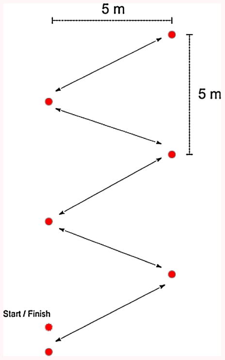
Participants were fitted with American football specific cleats designed for synthetic surfaces (Scorch Thrill FieldTurf, Adidas) and wore thin cotton socks. A slalom course was laid out on a synthetic playing surface (Fig. 1) on which participants performed a total of eight cutting steps (4 right, 4 left) per trial at maximum effort. A duplicate course was set up on the same field where participants were instructed the proper running and cutting technique and allowed to practice until proficient in testing methods prior to data collection.
Fig. 1.

Slalom course.
A Novel pedar-x system (Novel, St. Paul, MN) was used to measure in-shoe pressure data. Conforming insoles containing 99 sensors were placed inside each shoe beneath the plantar surface of the foot. Subject given a brief acclimation period and with an opportunity to adjust the footwear prior to the trials. A Velcro waistband was used to hold the pedar unit connected to the insoles, along with battery power and transmitter for wireless data collection. Each participant completed two trials through the slalom course.
3. Data analysis
Arch height and foot pressures from both feet were measured for each subject. Participants were classified as high, normal or low arch based on previous findings [9]. Data for both barefoot walking and shod cutting were sampled at 100 Hz with each measurement system. Prior to collection, conforming insoles were calibrated to a known load of 900 kPa. In each condition, force time integral (FTI), maximum force (MF) and peak pressure (PP) were calculated for seven defined regions of the foot: medial heel, lateral heel, medial midfoot, lateral midfoot, medial forefoot, central forefoot and lateral forefoot. Each region was defined as a percentage of the total foot, so regions were consistent across multiple insole sizes. Bare foot steps were defined as initial contact with the pressure plate until toe-off, and the specified regions of the foot were evaluated. Cutting steps were defined as initial contact with the ground until toe-off while accelerating towards the next sequential cone on the slalom course (Fig. 1). Each trial was synchronized with digital video to identify the frames when cutting steps occurred. Loading data were extracted only for the frames in which cutting steps were identified.
3.1. Statistical analysis
Mean and standard deviations were calculated for demographic information regarding each of the participants, along with peak pressure, force time integral and maximum force in each foot region. A repeated measures 2-way ANOVA was performed for both walking and cutting conditions (high vs normal arch) using PSW Statistics software (Version 17, SPSS Inc., Chicago, IL). Alpha level was defined as 0.05 to identify statistical significance between groups.
4. Results
4.1. AHI
Ten participants (38%) were identified as having high arch (HA) and 16 (62%) identified as normal arch (NA). Four participants classified as low arch were excluded from the analyses based on pre-determined exclusionary criteria. During in-shoe testing, two participants in each group were excluded due to insufficient data, resulting in 8 HA and 14 NA participants.
4.2. Barefoot gait
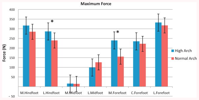
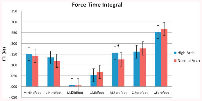
The HA group demonstrated greater maximum force in the lateral hindfoot (p = 0.008) and medial forefoot (p < 0.001) compared to the NA group (Fig. 2). The remaining foot regions were not statistically different in force between groups. Force Time Integral was greater for the HA athletes in the medial forefoot (p = 0.044), but not in any other foot region (Fig. 3). There were no differences in peak pressure between the two groups in all regions.
Fig. 2.

Maximum force during barefoot walking.
Fig. 3.

Force time integral during barefoot walking.
4.3. Shod cutting
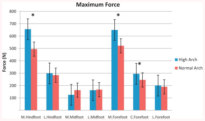
The HA athletes exhibited greater maximum force in the medial hindfoot (p = 0.031), medial forefoot (p = 0.007) and central forefoot (p = 0.025) compared to NA athletes (Fig. 4). No differences were found for any of the remaining foot regions, nor in any foot region for force time integral nor peak pressure (p > 0.05).
Fig. 4.

Maximum force during rapid cutting.
5. Discussion
The purpose of this study was to examine the difference in loading patterns of American football players with high and normal arch structures. The findings of the current study indicate that athletes with high arch height exhibit different loading patterns during barefoot walking and rapid cutting than those with normal arch structures. These differences are most evident in the lateral heel and medial forefoot. It has been suggested that a high arch structure results in a more rigid foot, less capable of dissipating forces related to contact with the ground. In the literature, a low arch foot has been described as a better shock absorber than a normal foot with high arch [8], and has shown decreased force and pressure compared to normal arch [11]. Further, those with a high arch have demonstrated stiffer foot mechanics, with less eversion at the ankle, rear/midfoot and mid/forefoot joints compared to low arch during dynamic loading [10]. These claims were supported in the current study, specifically regarding increased maximum force observed among those with high arch structure.
Not surprisingly, the movements performed in this study elicited different plantar loading patterns. However, in each condition, group differences in maximum force were shown in regions involved with the initial contact and toe off portion of the stance phase, where forces may be greatest. These regions of differential maximum force loading include the lateral rearfoot and medial forefoot in walking, and the medial rearfoot and forefoot while cutting.
Though loading measurements while walking and running have been studied, the specific evaluation of FTI (impulse) has rarely been reported. Representing the time over which a force is applied, impulse measurements offer valuable insight to evaluate pathomechanics associated with overuse injuries in specific foot regions [27]. In the present study, the highest impulse values of FTI occurred in the lateral and central forefoot, but were only increased in the medial forefoot among the high arch group during walking. Compared to other athletic tasks, impulse has been greatest in the medial forefoot during a side cut [23,27]. However, in the current study, no difference between arch conditions was observed for any foot region during pivoting, despite significant differences in maximum force.
A pivoting or cutting movement requires participants to land and push off from the medial aspect of the foot. When cutting, previous work has demonstrated increases in peak pressure [23,24,27], percent load [24], and FTI impulses [27] on medial aspects of the foot, regardless of arch classification. In the current study, the greatest maximum force occurred in the medial rear and forefoot while cutting. In particular, participants with a high arch increased maximum force in the medial heel and medial forefoot, which potentially increased the risk of stress-related injury in those regions associated with overuse. The physical demands of American football often require quick movements with rapid deceleration. While one prospective study reported more injuries to the lateral aspect of the foot among those with high arches [7], the present findings indicate that football players, in particular, with high arches may be at increased risk of overuse injuries to the medial aspect of the foot.
To our knowledge, the current study is the first to examine high arch compared to normal arch feet during athletic cutting movements. No athletes analyzed in the current study had low arches. Therefore, comparison of the effects of arch height on dynamic foot function should only be generalized to those with high and normal arch structures. It should also be noted that pivoting or cutting movements performed during running by athletes in the present study were evaluated on a synthetic playing surface. While the turf technology has improved to represent natural grass, differences in loading patterns between the two surfaces have been observed [26]. While performing similar cutting movements, athletes of similar age (16.9 ± 0.9 years) were shown to increase peak pressure in the central forefoot and lesser toes on a synthetic playing surface compared to natural grass. Conversely, relative load in the medial forefoot and lateral midfoot were greater on natural grass when compared to the synthetic turf. Results of the current study pertain only to movements on a synthetic surface, and should be interpreted as such.
Research into the influences of arch type on foot mechanics to injury risk has produced mixed results [28]. Further, barefoot and shod data should be interpreted independently, as differences between those with both low and high arches were reported under the two conditions [21]. For example, these findings contradict previous work of authors who suggested increased lateral loading in high arch and medial loading in low arch [7]. This may indicate that loading patterns differ between activities, and not primarily by arch type.
The clinical importance of the current findings could be significant in terms of identifying athletes that may be at increased risk of certain injuries. Arch structure classification may be incorporated in a pre-activity screening and the implementation of specific performance tools could potentially alter injury risk. For example, different footwear conditions have been shown to alter plantar loading when performing aggressive cutting movements similar to those outlined in the current study (Orendurff et al. [25]). When wearing a football boot with added midsole foam, peak plantar pressures decreased compared to a traditional football boot with no foam midsole.
Among athletes required to perform frequent and rapid pivoting/cutting movements, such as football players, those with a high arch may be at increased risk of overuse injury compared to normal arch counterparts as evidence by the increased relative and impulse loading. The results of the current study indicate that arch height index classification may be an effective tool for clinicians for the evaluation of the potential loading differences between those with varying foot structure within this specific athletic population.
Acknowledgments
The authors would like to acknowledge funding support from National Institutes of Health/NIAMS grants R01-AR049735, R01-AR055563, R01-AR056259, and R03-AR057551. The authors also acknowledge the Robert S. Heidt, Sr. – Wellington Foundation and adidas for the donation of the footwear.
References
- 1.Milgrom C, Giladi M, Kashtan H, Simkin A, Chisin R, Margulies J, et al. A prospective study of the effect of a shock-absorbing orthotic device on the incidence of stress fractures in military recruits. Foot & Ankle. 1985 Oct;6(2):101–4. doi: 10.1177/107110078500600209. [DOI] [PubMed] [Google Scholar]
- 2.Simon SR, Radin EL, Paul IL, Rose RM. The response of joints to impact loading. II. In vivo behavior of subchondral bone. Journal of Biomechanics. 1972 May;5(3):267–72. doi: 10.1016/0021-9290(72)90042-5. [DOI] [PubMed] [Google Scholar]
- 3.Radin EL, Paul IL, Rose RM. Role of mechanical factors in pathogenesis of primary osteoarthritis. Lancet. 1972 Mar;1(7749):519–22. doi: 10.1016/s0140-6736(72)90179-1. [DOI] [PubMed] [Google Scholar]
- 4.Reinker KA, Ozburne S. A comparison of male and female orthopaedic pathology in basic training. Military Medicine. 1979 Aug;144(8):532–6. [PubMed] [Google Scholar]
- 5.Kaufman KR, Brodine SK, Shaffer RA, Johnson CW, Cullison TR. The effect of foot structure and range of motion on musculoskeletal overuse injuries. The American Journal of Sports Medicine. 1999 Sep-Oct;27(5):585–93. doi: 10.1177/03635465990270050701. [DOI] [PubMed] [Google Scholar]
- 6.James SL, Bates BT, Osternig LR. Injuries to runners. The American Journal of Sports Medicine. 1978 Mar-Apr;6(2):40–50. doi: 10.1177/036354657800600202. [DOI] [PubMed] [Google Scholar]
- 7.Williams DS, 3rd, McClay IS, Hamill J. Arch structure and injury patterns in runners. Clinical Biomechanics (Bristol, Avon) 2001 May;16(4):341–7. doi: 10.1016/s0268-0033(01)00005-5. [DOI] [PubMed] [Google Scholar]
- 8.Simkin A, Leichter I, Giladi M, Stein M, Milgrom C. Combined effect of foot arch structure and an orthotic device on stress fractures. Foot & Ankle. 1989 Aug;10(1):25–9. doi: 10.1177/107110078901000105. [DOI] [PubMed] [Google Scholar]
- 9.Williams DS, McClay IS. Measurements used to characterize the foot and the medial longitudinal arch: reliability and validity. Physical Therapy. 2000 Sep;80(9):864–71. [PubMed] [Google Scholar]
- 10.Powell DW, Long B, Milner CE, Zhang S. Frontal plane multi-segment foot kinematics in high- and low-arched females during dynamic loading tasks. Human Movement Science. 2011 Feb;30(1):105–14. doi: 10.1016/j.humov.2010.08.015. [DOI] [PubMed] [Google Scholar]
- 11.Nigg BM, Cole GK, Nachbauer W. Effects of arch height of the foot on angular motion of the lower extremities in running. Journal of Biomechanics. 1993 Aug;26(8):909–16. doi: 10.1016/0021-9290(93)90053-h. [DOI] [PubMed] [Google Scholar]
- 12.Cavanagh PR, Morag E, Boulton AJ, Young MJ, Deffner KT, Pammer SE. The relationship of static foot structure to dynamic foot function. Journal of Biomechanics. 1997 Mar;30(3):243–50. doi: 10.1016/s0021-9290(96)00136-4. [DOI] [PubMed] [Google Scholar]
- 13.Cavanagh PR, Rodgers MM. The arch index: a useful measure from footprints. Journal of Biomechanics. 1987;20(5):547–51. doi: 10.1016/0021-9290(87)90255-7. [DOI] [PubMed] [Google Scholar]
- 14.Pohl MB, Farr L. A comparison of foot arch measurement reliability using both digital photography and calliper methods. Journal of Foot and Ankle Research. 2010;3:14. doi: 10.1186/1757-1146-3-14. [DOI] [PMC free article] [PubMed] [Google Scholar]
- 15.Yalcin N, Esen E, Kanatli U, Yetkin H. Evaluation of the medial longitudinal arch: a comparison between the dynamic plantar pressure measurement system and radiographic analysis. Acta Orthopaedica Traumatologica Turcica. 2010;44(3):241–5. doi: 10.3944/AOTT.2010.2233. [DOI] [PubMed] [Google Scholar]
- 16.Butler RJ, Hillstrom H, Song J, Richards CJ, Davis IS. Arch height index measurement system: establishment of reliability and normative values. Journal of the American Podiatric Medical Association. 2008 Mar-Apr;98(2):102–6. doi: 10.7547/0980102. [DOI] [PubMed] [Google Scholar]
- 17.Richards C, Card K, Song J, Hillstrom H, Butler R, Davis I, editors. A novel arch height index measurement (AHIMS); intra- and inter-rater reliability. Toledo, OH. The Annual Meeting of the American Society of Biomechanics; 2003. [Google Scholar]
- 18.Butler RJ, Davis IS, Hamill J. Interaction of arch type and footwear on running mechanics. The American Journal of Sports Medicine. 2006 Dec;34(12):1998–2005. doi: 10.1177/0363546506290401. [DOI] [PubMed] [Google Scholar]
- 19.Butler RJ, Hamill J, Davis I. Effect of footwear on high and low arched runners' mechanics during a prolonged run. Gait & Posture. 2007 Jul;26(2):219–25. doi: 10.1016/j.gaitpost.2006.09.015. [DOI] [PubMed] [Google Scholar]
- 20.Chuckpaiwong B, Nunley JA, Mall NA, Queen RM. The effect of foot type on in-shoe plantar pressure during walking and running. Gait & Posture. 2008 Oct;28(3):405–11. doi: 10.1016/j.gaitpost.2008.01.012. [DOI] [PubMed] [Google Scholar]
- 21.Molloy JM, Christie DS, Teyhen DS, Yeykal NS, Tragord BS, Neal MS, et al. Effect of running shoe type on the distribution and magnitude of plantar pressures in individuals with low- or high-arched feet. Journal of the American Podiatric Medical Association. 2009;99(4):330–8. doi: 10.7547/0980330. [DOI] [PubMed] [Google Scholar]
- 22.Teyhen DS, Stoltenberg BE, Collinsworth KM, Giesel CL, Williams DG, Kardouni CH, et al. Dynamic plantar pressure parameters associated with static arch height index during gait. Clinical Biomechanics (Bristol, Avon) 2009 May;24(4):391–6. doi: 10.1016/j.clinbiomech.2009.01.006. [DOI] [PubMed] [Google Scholar]
- 23.Wong PL, Chamari K, Mao de W, Wisloff U, Hong Y. Higher plantar pressure on the medial side in four soccer-related movements. British Journal of Sports Medicine. 2007 Feb;41(2):93–100. doi: 10.1136/bjsm.2006.030668. [DOI] [PMC free article] [PubMed] [Google Scholar]
- 24.Eils E, Streyl M, Linnenbecker S, Thorwesten L, Volker K, Rosenbaum D. Characteristic plantar pressure distribution patterns during soccer-specific movements. The American Journal of Sports Medicine. 2004 Jan-Feb;32(1):140–5. doi: 10.1177/0363546503258932. [DOI] [PubMed] [Google Scholar]
- 25.Orendurff MS, Rohr ES, Segal AD, Medley JW, Green JR, Kadel NJ. Regional foot pressure during running, cutting, jumping and landing. The American Journal of Sports Medicine. 2008;36(3):566–71. doi: 10.1177/0363546507309315. [DOI] [PubMed] [Google Scholar]
- 26.Ford KR, Manson NA, Evans BJ, Myer GD, Gwin RC, Heidt RS, Jr, et al. Comparison of in-shoe foot loading patterns on natural grass and synthetic turf. Journal of Science and Medicine in Sports. 2006 Dec;9(6):433–40. doi: 10.1016/j.jsams.2006.03.019. [DOI] [PubMed] [Google Scholar]
- 27.Queen RM, Haynes BB, Hardaker WM, Garrett WE., Jr Forefoot loading during 3 athletic tasks. The American Journal of Sports Medicine. 2007 Apr;35(4):630–6. doi: 10.1177/0363546506295938. [DOI] [PubMed] [Google Scholar]
- 28.Queen RM, Mall NA, Nunley JA, Chuckpaiwong B. Differences in plantar loading between flat and normal feet during different athletic tasks. Gait & Posture. 2009 Jun;29(4):582–6. doi: 10.1016/j.gaitpost.2008.12.010. [DOI] [PubMed] [Google Scholar]


