Abstract
Prior reports indicate that female athletes who demonstrate high knee abduction moments (KAMs) during landing are more responsive to neuromuscular training designed to reduce KAM. Identification of female athletes who demonstrate high KAM, which accurately identifies those at risk for noncontact anterior cruciate ligament (ACL) injury, may be ideal for targeted neuromuscular training. Specific neuromuscular training targeted to the underlying biomechanical components that increase KAM may provide the most efficient and effective training strategy to reduce noncontact ACL injury risk. The purpose of the current commentary is to provide an integrative approach to identify and target mechanistic underpinnings to increased ACL injury in female athletes. Specific neuromuscular training techniques will be presented that address individual algorithm components related to high knee load landing patterns. If these integrated techniques are employed on a widespread basis, prevention strategies for noncontact ACL injury among young female athletes may prove both more effective and efficient.
Keywords: prevention of anterior cruciate ligament injury, knee, drop vertical jump, landing mechanics, young athletes, biomechanics
Introduction
High knee abduction moments (KAMs) during landing predict noncontact anterior cruciate ligament (ACL) injury risk in young female athletes with high accuracy (4,17,25,43). Therefore, a field-based assessment algorithm was systematically developed and validated, which aimed to improve the potential to identify and target injury prevention training to female athletes with increased KAM (38,39). The validated field-based assessment algorithm delineated 5 biomechanical factors that combined to identify high KAM during landing with high accuracy (38,39).
The established links between lower limb mechanics and noncontact ACL injury risk led to the development of neuromuscular training interventions designed to prevent noncontact ACL injury by targeting deficits identified in specific populations (14,17,33–35,40,41). Injury prevention protocols have resulted in positive preventative and biomechanical changes in female athletic populations at high risk for knee injury (14,15,32,41). More specifically, pilot work indicates that female athletes categorized as high risk for noncontact ACL injury, based on previous coupled biomechanical and epidemiologic studies (17), may be more responsive to specially designed neuromuscular training (34). Neuromuscular training targeted to the specific biomechanical components that drive high KAM may provide the most efficient and effective training strategy to reduce non-contact ACL injury risk. Female athletes who demonstrate high KAM risk factor for noncontact ACL injury may be ideal for targeted neuromuscular training. The purpose of this commentary was to provide an integrative approach to identify and target mechanistic underpinnings to increased ACL injury in female athletes. Specific techniques will be presented that can be used for athletes for whom their high KAM is related to one or more components assessed in the ACL injury risk prediction algorithm.
Integration of Screening Techniques to Identify Specific Mechanisms That Underlie High Knee Abduction Moments and Methods to Target Risk Factors With Neuromuscular Training
The previously described methodology (36) demonstrates techniques to accurately capture and analyze measures of tibia length, knee valgus motion, knee flexion range of motion, body mass, and quadriceps to hamstrings ratio (QuadHam ratio), which are used in a high knee load prediction algorithm. Certain athletes are at a higher risk of noncontact ACL injury and may demonstrate one or more specific mechanisms that underlie predicted increased KAM. The following discussion will present representative athletes’ test measurements of specific landing mechanics or anthropometrics that result in the prediction of high KAM. Accordingly, specific neuromuscular training techniques will be presented that can be used to target each deficit that contributes to high KAM that is captured in the prediction algorithm.
Rapid Musculoskeletal Growth Contributes to High Knee Abduction Moments
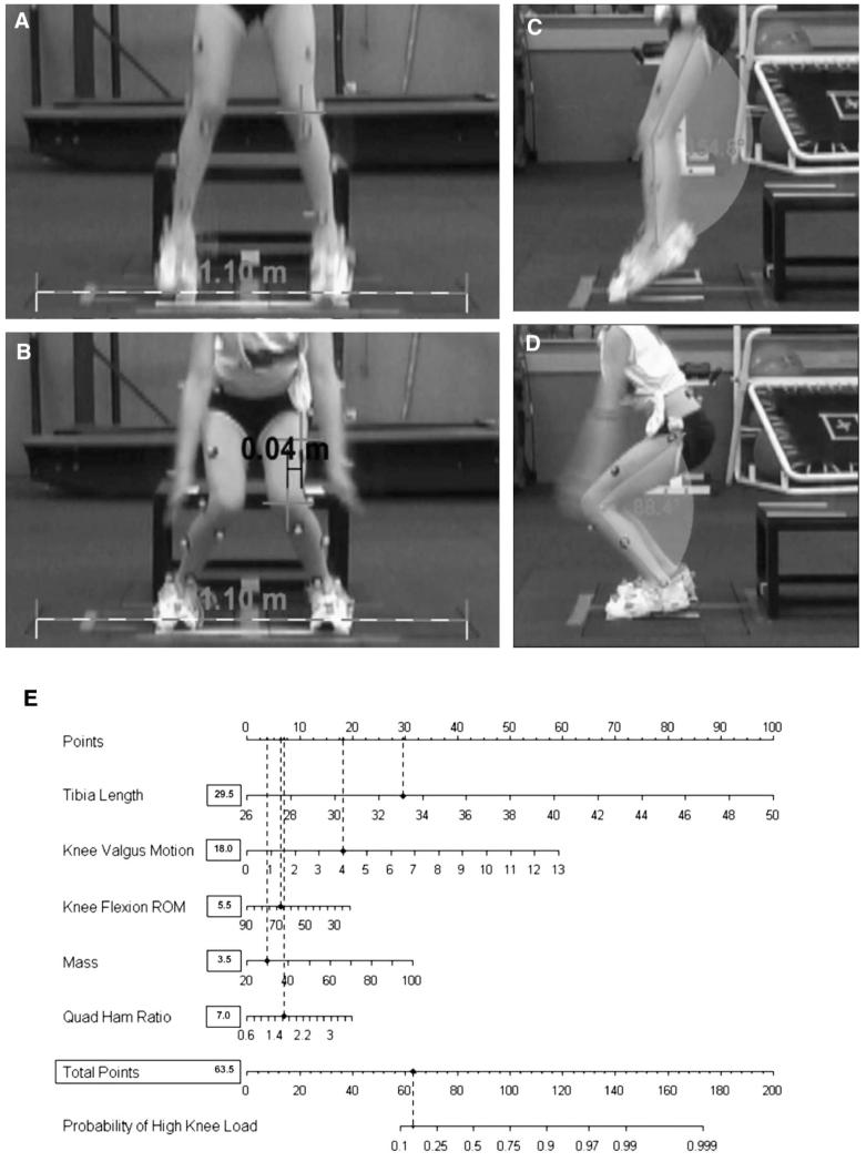
Rapid growth during maturation initiates increased stature and, in turn, an increased height of the center of mass. The increased musculoskeletal height, added to increased total body mass, may initiate greater joint forces that are more difficult to balance and dampen during high velocity maneuvers (13,16,18). Unlike adolescent male athletes who naturally increase hip abduction strength relative to body mass as they increase in age from 11 to 17 years, female athletes demonstrate no similar adaptation in hip abduction strength measures (5). The absence of adaptation-relative hip abduction strength to match the demands of growth and development in adolescent women as they mature may create decreased “core control.” The maturational invoked inertial imbalance (increased trunk load without adaptive hip and trunk control) may lead to abnormal joint alignments, increased KAM, and may be related to their increased risk of noncontact ACL injury compared with men following this developmental stage. Figure 1 presents an example of a representative subject whose combination of increased tibia length and mass, associated with her rapid growth, contributed to increased inertial demands on her lower extremity and increased risk to demonstrate high KAM landing mechanics when using the field-based ACL injury risk prediction algorithm. The completed algorithm for the representative subject (tibia length: 47 cm; knee valgus motion: 3.0 cm; knee flexion range of motion (ROM): 55.9°; mass: 71 kg; QuadHam ratio: 1.19) predicted that she would have a 96% (126.5 points) probability of high KAM during the drop vertical jump. Her actual KAM measure for the presented drop vertical jump quantified simultaneously with three-dimensional (3D) motion analysis was 48.5 N m of knee abduction load. Conversely, Figure 2 presents a representative subject whose combinations of decreased tibia length and mass before her rapid growth spurt diminishes her risk to demonstrate high KAM landing mechanics. As demonstrated in her completed algorithm, the representative subject (tibia length: 33.0 cm; knee valgus motion: 4.0 cm; knee flexion ROM: 66.4°; mass: 30.7 kg; QuadHam ratio: 1.64) would have a 14% (63.5 points) chance to demonstrate high KAM during the drop vertical jump. Her actual KAM measure for the presented drop vertical jump that was quantified simultaneously with 3D motion analysis was 13.4 N m of knee abduction load.
Figure 1.
Calibration of video is performed with the Dartfish scale tool and uses the known distance marked on the floor in the same plane as the athlete as the correction factor in the frontal plane axis. In the presented example, the line tool is used to draw a line corresponding to a known distance on the floor. The set scale procedure is then used to input this distance. From this calibration step, all length measurements from this camera position are calibrated for the subjects drop vertical jump trials. A) The coordinate position of knee joint center is denoted using the marker tool within Dartfish at the frame before initial contact. B) The coordinate position of knee joint center is again denoted with the marker tool at the frame with maximum medial position and is used as the knee valgus position. The calibrated displacement measure between the 2 digitized knee coordinates is representative of knee valgus motion during the drop vertical jump. C) Knee flexion angle is digitized using the angle tool at the frame before initial contact and recorded as the first measure of knee flexion ROM. D) Knee flexion angle is digitized using the angle tool at the frame with maximum knee flexion and recorded as the second measure of knee flexion ROM. The displacement of knee flexion is calculated as the differences in knee flexion angles at the frame before initial contact and maximum knee flexion and is representative of knee flexion ROM. E) Example of representative subject whose combination of increased tibia length and mass associated with her rapid growth contribute to her increased risk to demonstrate high KAM landing mechanics when using the field-based ACL injury risk prediction algorithm. Completed algorithm for the representative subject (tibia length: 47 cm; knee valgus motion: 3.0 cm; knee flexion ROM: 55.9°; mass: 71 kg; QuadHam ratio: 1.19). Based on her demonstrated measurements, this subject would have a 96% (126.5 points) chance to demonstrate high KAM during the drop vertical jump. Her actual KAM measure for the presented drop vertical jump that was quantified simultaneously with three-dimensional motion analysis was 48.5 N m of knee abduction load.
Figure 2.
Representative subject whose combination of decreased tibia length and mass before her rapid growth spurt diminish her risk to demonstrate high KAM landing mechanics. A and B) The calibrated displacement measure between the 2 marked knee coordinates is representative of knee valgus motion during the drop vertical jump. C and D) The displacement of knee flexion is calculated as the differences in knee flexion angles at the frame before initial contact and maximum knee flexion and is representative of knee flexion ROM. E) Completed algorithm for the representative subject (tibia length: 33.0 cm; knee valgus motion: 4.0 cm; knee flexion ROM: 66.4°; mass: 30.7 kg; QuadHam ratio: 1.64). Based on her demonstrated measurements, this subject would have a 14% (63.5 points) chance to demonstrate high KAM during the drop vertical jump. Her actual KAM measure for the presented drop vertical jump that was quantified simultaneously with three-dimensional motion analysis was 13.4 N m of knee abduction load.
Thus, after the onset of puberty and the initiation of peak height velocity, increased tibia and femur lever length, with increased body mass and height of the center of mass, in the absence of increases in strength and recruitment of the musculature at the hip and trunk may lead to decreased core control and ability to control inertial forces of the trunk during dynamic tasks (11). As female athletes reach maturity, decreased “core stability” may underlie their tendency to demonstrate increased KAM during dynamic tasks and increased ACL injury risk during competition (Figure 1) (13,16,18,21,22,47).
Targeted Training for Rapid Growth Risk Factor
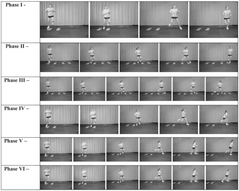
Reduced ability to activate the hip stabilizers may allow increased lateral trunk positions that can incite increased KAM (48). Decreased core control and muscular synergism, along with decreased control of inertial loads from the hip stabilizers, may affect performance in power activities and may also increase the incidence of injury secondary to lack of control of the center of mass, especially in female athletes (23,49,50). Targeted trunk and hip control neuromuscular training increases standing hip abduction strength in female athletes (29). Figure 3 presents a neuromuscular training progression that can be instituted with athletes to target deficits in trunk and hip control (29,30). Progressive exercise phases are used to facilitate incremental advancements that are designed to improve the athletes’ ability to control the increased inertial loads during dynamic activities. The progressive increases in intensity to given exercise techniques facilitate the adaptations that prepare the athlete for end-stage progressions that will incorporate lateral trunk perturbations. These perturbations require the athlete to decelerate and control the trunk in the frontal plane to successfully execute the prescribed technique. Similar exercise progressions have demonstrated that increased hip abduction strength improves the ability of female athletes to control the body center of mass and lower extremity alignment, which results in a decreased KAM during sports activities (7,11).
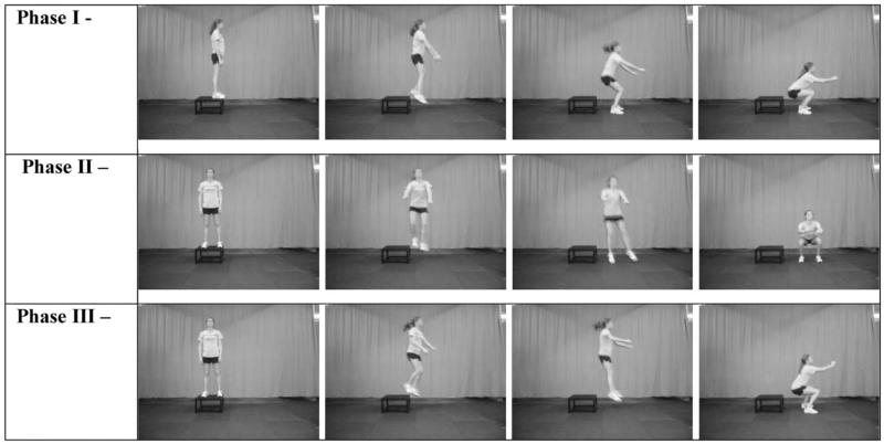
Figure 3.
Lateral Hop progression. Phase I: Two barrier lateral hop, hop opposite leg hold. Athlete faces forward and hops laterally over the barriers and into a hold position at each end. The hold position should be held stable for 3 seconds. The same movement is repeated back in the opposite direction. The maneuver is performed in both directions on both limbs. Phase II: Four barrier lateral hop, hop opposite leg hold with alternating legs. Athlete faces forward and hops laterally over the first 2 barriers and over the final 2 barriers with one hop and holds position at each end. The hold position should be held stable for 3 seconds. The same movement is repeated back in the opposite direction. The maneuver is performed in both directions on both limbs. Phase III: Four barrier dynamic lateral hopping drill. Athlete faces forward and hops laterally over the first 2 barriers and over the final 2 with one hop and immediately changes direction to repeat the movement back to starting position. The maneuver is performed on both limbs. Phase IV: Two barrier lateral hop, hop 90° opposite leg hold. Athlete faces forward and hops laterally over the first 2 barriers and over the final 2 barriers with one hop and immediately hops into a maximum effort 90° hop and hold. The hold position should be held stable for 3 seconds. The maneuver is performed in both directions on both limbs. Phase V: Two barrier lateral hop, hop 90° opposite, crossover hop hold. Athlete faces forward and hops laterally over the first 2 barriers and over the final 2 barriers with one hop and immediately hops into a maximum effort 90° hop, crossover hop, and hold. The hold position should be held stable for 3 seconds. The maneuver is performed on both limbs. Phase VI: Two barrier lateral hop, hop 90° opposite to sprint. Athlete faces forward and hops laterally over the first 2 barriers and takes the final 2 barriers with one hop and immediately hops into a maximum effort 90° hop, and sprint maneuver. The maneuver is performed on both limbs.
Excessive Knee Valgus Motion Contributes to High Knee Abduction Moments
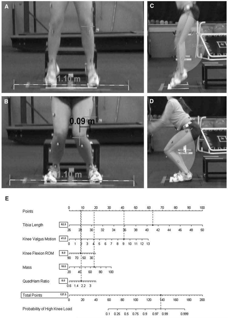
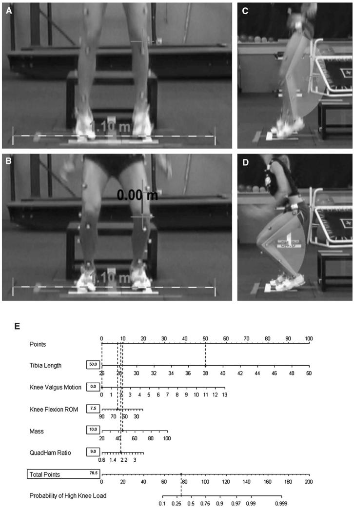
Neuromuscular control deficits are defined as muscle strength, power, or activation patterns that lead to abnormal joint alignments and increased KAM (19). Female athletes demonstrate neuromuscular control deficits that increase lower extremity joint loads during sports activities (10,12,39). One neuromuscular deficit, which can be termed “ligament dominance,” is defined as an imbalance between the neuromuscular and ligamentous control of dynamic knee joint stability (19). This imbalance in control of dynamic knee joint stability is demonstrated by an inability to control lower extremity frontal plane motion during landing and cutting (10,12,39). Figure 4 provides an example of a representative subject with excessive knee valgus motion during the drop vertical jump that contributes to her increased risk of high KAM landing mechanics. Figure 4E presents the completed algorithm for the representative subject (tibia length: 41.0 cm; knee valgus motion: 9.0 cm; knee flexion ROM: 59.8°; mass: 67.5 kg; QuadHam ratio: 1.90). Based on her demonstrated measurements, this subject would have a 98% (137.5 points) chance to demonstrate high KAM during the drop vertical jump. Her actual KAM measure for the presented drop vertical jump, which was quantified simultaneously with 3D motion analysis, was 44.1 N m of KAM. Figure 5 provides an example of a representative subject with optimal neuromuscular control that limits her potential to demonstrate high KAM risk factor and ultimately reduces her risk of noncontact ACL injury. Based on her demonstrated measurements (tibia length: 38 cm; knee valgus motion: 0.0 cm; knee flexion ROM: 63.8°; mass: 44.9 kg; QuadHam ratio: 1.89), this subject would have a 30% (76.5 points) chance to demonstrate high KAM during the drop vertical jump using the ACL injury prediction algorithm (Figure 5E). Her actual KAM measure for the presented drop vertical jump, quantified simultaneously with 3D motion analysis, was 7.6 N m of knee abduction load, indicating a likely reduced risk on noncontact ACL injury.
Figure 4.
Example of representative subject with excessive knee valgus during the drop vertical jump that contributes to her increased risk to demonstrate high KAM landing mechanics. A and B) The calibrated displacement measure between the 2 marked knee coordinates is representative of knee valgus motion during the drop vertical jump. C and D). The displacement of knee flexion is calculated as the differences in knee flexion angles at the frame before initial contact and maximum knee flexion and is representative of knee flexion ROM. E) Completed algorithm for the representative subject (tibia length: 41.0 cm; knee valgus motion: 9.0 cm; knee flexion ROM: 59.8°; mass: 67.5 kg; QuadHam ratio: 1.90). Based on her demonstrated measurements, this subject would have a 98% (137.5 points) chance to demonstrate high KAM during the drop vertical jump. Her actual KAM measure for the presented drop vertical jump that was quantified simultaneously with three-dimensional motion analysis was 44.1 N m of knee abduction load.
Figure 5.
Example of representative subject with optimal neuromuscular control during the drop vertical jump that limits her potential to demonstrate high KAM risk factor. A and B) The calibrated displacement measure between the 2 marked knee coordinates is representative of knee valgus motion during the drop vertical jump. C and D) The displacement of knee flexion is calculated as the differences in knee flexion angles at the frame before initial contact and maximum knee flexion and is representative of knee flexion ROM. E) Completed algorithm for the representative subject (tibia length: 38 cm; knee valgus motion: 0.0 cm; knee flexion ROM: 63.8°; mass: 44.9 kg; QuadHam ratio: 1.89). Based on her demonstrated measurements, this subject would have a 30% (76.5 points) chance to demonstrate high KAM during the drop vertical jump. Her actual KAM measure for the presented drop vertical jump that was quantified simultaneously with three-dimensional motion analysis was 7.6 N m of knee abduction load.
Targeted Training for Excessive Knee Valgus Motion Risk Factor
Although prior research demonstrates the benefit of injury prevention training among a wide variety of athletes, it appears that those athletes who demonstrate neuromuscular imbalances, as evidenced through poor frontal plane knee control and increased knee valgus motion, may benefit the most from training (34,39). Wall jumps are an example of an initial exercise that could be used to target excessive knee valgus motion (Figure 6). This low-to-moderate intensity jump also allows clinicians to begin analysis of the athlete’s level of frontal plane knee control by instructing the athletes to keep their knees apart when landing and which produces decreased ACL load in knee flexion angles used in this jump (27). Another useful exercise to target the high KAM driven by knee valgus motion is the tuck jump (Figure 7). The tuck jump is an exercise that is on the opposite end of the intensity spectrum from the wall jump and requires a high effort level from the athlete. Initially, the athlete will likely place a majority of their cognitive efforts on the performance of this difficult maneuver. Clinicians can readily identify an athlete who may demonstrate abnormal levels of frontal plane knee displacement during jumping and landing because the athlete usually devotes minimal attention to technique on the first few repetitions. In addition, tuck jumps can be used to assess progressive improvements in lower extremity biomechanics. The broad jump and hold (Figure 8) allows clinicians to assess the athlete’s propensity to demonstrate jumping mechanics termed as “active valgus,” a position of hip adduction and knee abduction that is the result of muscular contraction rather than the inability to control ground reaction forces. Active valgus occurs during taking off from a jump, rather than at landing, and should be corrected during neuromuscular training.
Figure 6.

Wall Jumps The athlete stands erect with her arms semi-extended overhead. This vertical jump requires minimal knee flexion. The gastrocnemius muscles should create the vertical height. The arms should extend fully at the top of the jump. Use this jump as a warm-up and coaching exercise because this relatively low intensity movement can reveal abnormal knee motion in athletes with poor frontal plane knee control.
Figure 7.
Tuck Jump progression. Phase I: Single Tuck Jump Soft Landing. The athlete starts in the athletic position with her feet shoulder width apart. The athlete initiates a vertical jump with a slight crouch downward while she extends her arms behind her. The athlete then swings her arms forward as she simultaneously jumps straight up and pulls her knees up as high as possible. At the highest point of the jump, the athlete should be positioned in the air with her thighs parallel to the ground. On landing, the athlete should land softly, using a toe-to-midfoot rocker landing. The athlete should not continue this jump if she cannot control the high landing force or keep her knees aligned during landing. Phase II: Double Tuck Jump. Similar to the single tuck jump described above but with an additional jump performed immediately after the first jump. The athlete should focus on maintaining good form and minimizing time on the ground between jumps. Phase III: Repeated Tuck Jump. The athlete starts in the athletic position with her feet shoulder width apart. The athlete initiates a vertical jump with a slight crouch downward while she extends her arms behind her. The athlete then swings her arms forward as she simultaneously jumps straight up and pulls her knees up as high as possible. At the highest point of the jump, the athlete should be positioned in the air with her thighs parallel to the ground. When landing, the athlete should immediately begin the next tuck jump. The athlete’s goal should be to perform the tuck jump exercise with near perfect technique for 10 seconds. Phase IV: Side-to-Side Tuck Jumps. The athlete starts in the athletic position with her feet shoulder width apart. The athlete initiates a vertical jump over a barrier with a slight crouch downward while she extends her arms behind her. The athlete then swings her arms forward as she simultaneously jumps straight up and pulls her knees up as high as possible. At the highest point of the jump, the athlete should be positioned in the air with her thighs parallel to the ground. When landing, the athlete should immediately begin the next tuck jump back to the other side of the barrier. Phase V: Side-to-Side Reaction Barrier Tuck Jumps. The athlete starts in the athletic position with her feet shoulder width apart. The athlete initiates a vertical jump over a barrier with a slight crouch downward while she extends her arms behind her. The athlete then swings her arms forward as she simultaneously jumps straight up and pulls her knees up as high as possible. At the highest point of the jump, the athlete should be positioned in the air with her thighs parallel to the ground. When landing, the athlete should immediately begin the next tuck jump. When prompted, the athlete should jump to the other side of the barrier without breaking rhythm. Encourage the athlete to land softly, using a toe-to-midfoot rocker landing. The athletes should not continue this jump if they cannot control the high landing force or if they use a knock-kneed landing.
Figure 8.

Broad Jump and Hold: The athlete prepares for this jump in the athletic position with her arms extended behind her at the shoulder. She begins by swinging her arms forward and jumping forward and vertically at approximately a 45° angle to achieve maximum horizontal distance. The athlete must stick the landing with her knees flexed to approximately 90° in an exaggerated athletic position. The athlete may not be able to stick the landing during a maximum effort jump in the early phases. In this situation, have the athlete perform a submaximal broad jump in which she can stick the landing with her toes straight ahead and without inward motion of her knees. As her technique improves, encourage her to add distance to her jumps but not at the expense of perfect technique.
The final progressions of the plyometric and movement training targeted toward reduced knee valgus motion should use unanticipated cutting movements during training. Knee abduction moment loads on the knee can double during unanticipated cutting maneuvers similar to those observed during actual sport (3). Figure 9 shows an athlete who demonstrated excessive dynamic knee valgus positions during agility and unanticipated cutting drills. Extensive verbal and visual feedback (via video tape) can be used to help the athlete with high KAM arising from inordinate knee valgus motion to correct unsafe biomechanics during these movements (44). Neuromuscular training that incorporates techniques to focus on unanticipated cutting maneuvers effectively reduces KAM in female athletes (41). By teaching the athlete to use movement techniques that produce lower KAM during unanticipated cutting maneuvers that inherently invoke high KAM loads on the joint, they may ultimately transfer the mechanics that reduce the risk of injury onto the field during competitive play (3,17).
Figure 9.
Unanticipated Cutting. Examples of dynamic valgus positions that athletes may demonstrate during agility and unanticipated cutting techniques. Clinicians should provide active feedback to the athlete to encourage them to perform reactive training with limited knee valgus positions.
Reduced Knee Flexion Contributes to High Knee Abduction Moments
In addition to limiting lower extremity frontal plane motion and KAM, a reduction in sports-related ACL injury rates in women may be achieved via improved sagittal plane biomechanics, especially increasing knee flexion during dynamic activities (4,43). A sagittal position of the knee close to full extension when landing or cutting is commonly observed in video analysis of ACL injuries in female athletes (4,43). In addition, a prospective study has shown that female athletes who subsequently sustained ACL injuries demonstrated significantly less (10.5°) knee flexion during a drop vertical jump than those who did not sustain injury (17). Current reports also indicate that increased sagittal plane moments and decreased knee flexion range of motion influence the propensity to demonstrate high KAM landing mechanics (37,39). Figure 10 provides an example of a representative subject whose knee valgus motion is exacerbated with small knee flexion ROM during the drop vertical jump that contributed to her high likelihood for a high KAM landing. In evaluation of her completed algorithm, the representative subject (tibia length: 42 cm; knee valgus motion: 4.6 cm; knee flexion ROM: 38.3°; mass: 65.3 kg; QuadHam ratio: 1.51) would have a 94% (123.0 points) chance to demonstrate high KAM during the drop vertical jump. Her actual KAM measure for the presented drop vertical jump that was quantified simultaneously with 3D motion analysis was 77.6 N m of KAM which is over 3 times the cut-score of 21.7 used to classify athletes as high risk for noncontact ACL injury (37,38). Thus, the current subject’s data indicate that control of knee valgus motion is critical when landing with knee angles close to full extension. Beyond her high KAM, the present landing mechanics may amplify direct ACL loads from forceful quadriceps contraction with her dynamic valgus positioning at small knee flexion angle (27).
Figure 10.
Example of representative subject with a combination of excessive knee valgus motion and small knee flexion ROM during the drop vertical jump that contribute to her increased risk for high KAM landing mechanics. A and B) The calibrated displacement measure between the 2 marked knee coordinates is representative of knee valgus motion during the drop vertical jump. C and D) The displacement of knee flexion is calculated as the differences in knee flexion angles at the frame before initial contact and maximum knee flexion and is representative of knee flexion ROM. E) Completed algorithm for the representative subject (tibia length: 42 cm; knee valgus motion: 4.6 cm; knee flexion ROM: 38.3°; mass: 65.3 kg; QuadHam ratio: 1.51). Based on her demonstrated measurements, this subject would have a 94% (123.0 points) chance to demonstrate high KAM during the drop vertical jump. Her actual KAM measure for the presented drop vertical jump that was quantified simultaneously with three-dimensional motion analysis was 77.6 N m of knee abduction load.
Targeted Training for Reduced Knee Flexion Range of Motion Risk Factor
To decrease the tendency toward utilization of small knee flexion ROM during dynamic sports-related tasks, exercises are employed that emphasize early (pre and initial contact) co-contraction of the knee flexor muscles (37,39). If the hamstrings are adequately activated at the proper time, they can increase knee flexion and decrease ACL loading. However, at low knee flexion angles, the hamstrings have little ability to protect against ACL loads (26,28,45). In addition, at angles greater than 45°, quadriceps recruitment can aid in the resistance of anterior tibial translation, providing an agonistic role to protect the ACL (1,8). Therefore, it is important to teach increased knee flexion ROM to reduce KAM and allow the hamstrings to provide protective muscular force to the ACL (35). To influence deep knee flexion, the box butt touch exercise (Figure 11) is used, in which a box is placed behind the athlete and the athlete starts with feet shoulder width apart and performs a squat down to the height of the box, softly touches the box without resting, and then ascends up to initial starting position. Once appropriate trunk and lower extremity control is achieved during the pseudodynamic maneuver, the athlete can progress to the box drop off-deep hold series of exercises (Figure 12).
Figure 11.
Box Butt Touch. A box is placed behind the athlete and the athlete starts with feet shoulder width apart and performs a squat down to the height of the box, softly touches the box without resting, and then ascends up to initial starting position.
Figure 12.
Box Drop Deep Hold. Phase I: Forward—Athlete drops down from a box landing with both feet simultaneously in the deep hold position. Phase II: Lateral—Athlete drops down laterally from a box landing with both feet simultaneously in the deep hold position. Phase III: Rotational—Athlete rotates 90° while dropping down from a box landing with both feet simultaneously in the deep hold position.
Squat jumps (Figure 13) can also be used to target improved knee flexion range of motion because their execution requires the athlete to go into deep knee flexion angles, past 90°. In addition, the squat jump can help teach the athlete to initiate landing in a more flexed knee position, which decreases the quadriceps ability to load the ACL and improves the ability of the hamstrings to offset anterior shear forces because of their line of pull (1,8,26,28,45). Utilization of the described exercises to influence increased knee flexion ROM during landing may reduce the potential high KAM and ACL injury stemming from an overextended landing position (4,43).
Figure 13.

Squat Jumps. The athlete begins in the athletic position with her feet flat on the mat pointing straight ahead. The athlete drops into deep knee, hip, and ankle flexion, touches the floor (or as close to her heels as possible) and then takes off into a maximum vertical jump. The athlete then jumps straight up vertically and reaches as high as possible. On landing, she immediately returns to starting position and repeats the initial jump. Repeat for the allotted time or until her technique begins to deteriorate. Teach the athlete to jump straight up vertically, reaching as high overhead as possible. Encourage her to land in the same spot on the floor and maintain upright posture when regaining the deep squat position. Do not allow the athlete to bend forward at the waist to reach the floor. The athlete should keep her eyes up, feet and knees pointed straight ahead, and have her arms to the outside of her legs.
Side-to-Side Differences in Landing Mechanics Contribute to Differential High Knee Abduction Moments
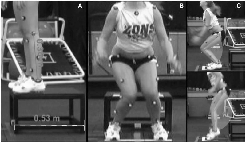
Side-to-side imbalances in muscular strength, flexibility, and coordination have been shown to be important predictors of increased injury risk (2,17,24). In addition, female athletes may generate lower hamstrings torques on the nondominant than in the dominant leg (20). Specific to ACL injury risk prediction, adolescent female athletes demonstrate significant side-to-side differences in maximum knee valgus angle compared with men during a box drop vertical jump (10). Half of the parameters used in a highly sensitive and specific regression model to predict increased KAM and ACL injury were side-to-side differences in lower extremity kinematics and kinetics (17). Leg-to-leg differences in KAM were also observed in injured, but not uninjured, women. Side-to-side KAM difference was 6.4 times greater in ACL-injured vs. the uninjured women (17). Female athletes tend to demonstrate side-to-side differences that are visibly evident for maximum knee valgus angle during a box drop vertical jump (Figure 14) (10). Overreliance on a single limb can put greater stress and torques on the knee increasing KAM and, in turn, increasing the risk for noncontact ACL injury on that limb (17).
Figure 14.
A, B, and C) provide an example of a representative subject’s landing mechanics. Note in image (B), there are visually evident side-to-side differences in the frontal plane landing biomechanics. Clinicians should evaluate side-to side differences in frontal plane mechanics, and the largest knee valgus motion measurements should be employed to maximize the utility of the proposed ACL injury prediction algorithm.
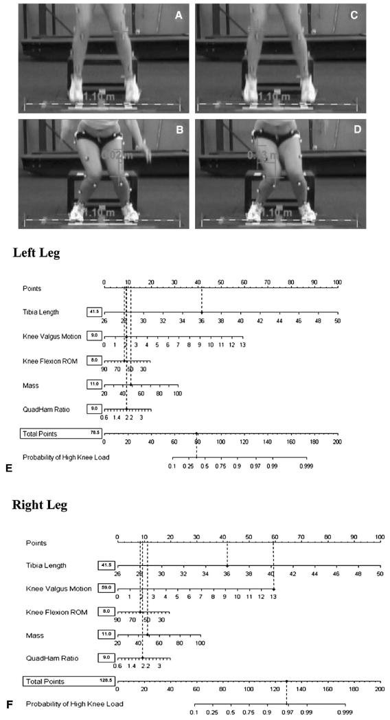
Figure 15 presents a subject with side-to-side differences in landing biomechanics that the ACL injury prediction algorithm also delineates with side-side differences in prediction of risk for high KAM. Based on measurements in her left leg (Figures 15A,B), this subject would have a 35% (78.5 points; Figure 15E) chance to demonstrate high KAM during the drop vertical jump. Her actual KAM measure for the presented drop vertical jump that was quantified simultaneously with 3D motion analysis was 15.4 N m of knee abduction load on her left leg. However, when using the frontal plane motion from the right leg (Figure 15C,D; 13 cm) in the ACL injury risk prediction algorithm, this subject would have a 96% (128.5 points; Figure 15F) chance to demonstrate high KAM during the drop vertical jump. Her actual KAM measure for the presented drop vertical jump that was quantified simultaneously with 3D motion analysis was 29.2 N m of knee abduction load on her right leg. The provided example of this important observation indicates that the ACL injury prediction algorithm is both sensitive and specific to high KAM, even between limbs in a single subject. Accordingly, clinicians should evaluate side-to-side differences in frontal plane mechanics, and the largest knee valgus motion measurements should be employed to maximize the utility of the proposed ACL injury prediction algorithm (17).
Figure 15.
Example of representative subject with side-to-side differences in landing biomechanics, which the ACL injury prediction algorithm also delineates with differences in prediction of risk for high KAM. A and B) Subject demonstrates 2 cm of knee valgus motion on her left limb during the drop vertical jump. C and D) Subject demonstrates 13 cm of knee valgus motion on her right limb during same trial of the drop vertical jump. E) Completed algorithm for the representative subject left leg (tibia length: 36 cm; knee valgus motion: 2.0 cm; knee flexion ROM: 58.0°; mass: 49.0 kg; QuadHam ratio: 2.0). Based on her demonstrated measurements in her left leg, this subject would have a 35% (78.5 points) chance to demonstrate high KAM during the drop vertical jump. Her actual KAM measure for the presented drop vertical jump that was quantified simultaneously with three-dimensional (3D) motion analysis was 15.4 N m of knee abduction load. F) When using the measures from the right leg frontal plane motion (13 cm) in the ACL injury risk prediction algorithm, the subject also shows side-to-side differences in prediction of high KAM. Based on her demonstrated measurements in her right leg, this subject would have a 96% (128.5 points) chance to demonstrate high KAM during the drop vertical jump. Her actual KAM measure for the presented drop vertical jump that was quantified simultaneously with 3D motion analysis was 29.2 N m of knee abduction load.
Targeted Training for Side-to-Side Difference Risk Factor
Before teaching dynamic movements focused to correct side-to-side imbalances, athletes should first be taught proper athletic position (Figure 16). The athletic position is a functionally stable position with the knees comfortably flexed, shoulders back, eyes up, feet approximately shoulder-width apart, and the body mass balanced evenly over the balls of the both feet. The athletic “ready position” should be the starting and finishing position for several of the training exercises and is focused to teach symmetry in weight distribution between limbs.
Figure 16.
Athletic position. The athletic position is a functionally stable position with the knees comfortably flexed, shoulders back, eyes up, feet approximately shoulder-width apart, and the body mass balanced over the balls of the feet. The knees should be over the balls of the feet, and chest should be over the knees. This is the athlete-ready position and is the starting and finishing position for most of the training exercises. During some of the exercises, the finishing position is exaggerated with deeper knee flexion to emphasize the correction of certain biomechanical deficiencies.
The majority of the initial dynamic movement exercises should involve both legs to safely introduce the athlete to plyometric training movements such as those portrayed in Figures 6 and 7 (6). Early training emphasis should be on balanced athletic positioning (Figure 16) that can help create dynamic control of the athlete’s center of mass (33,41,42). Once bilateral symmetry is gained during a bipedal task, clinicians can incorporate single limb balance exercises on unstable surfaces (Figure 17).
Figure 17.

Single Leg Balance: The balance drills are performed on a balance device that provides an unstable surface. Initially, the athlete should master drills on each device with a 2-leg stance with feet shoulder width apart, in athletic position (Figure 16). As the athlete improves, the training drills can incorporate ball catches and single leg balance drills. Encourage the athlete to maintain deep knee flexion when performing all balance drills.
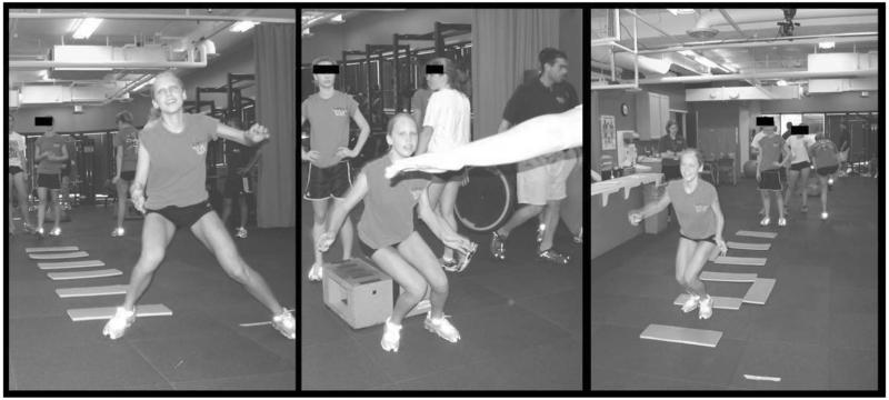
During the tuck jump exercise, some female athletes may unload their weaker limb (unloaded limb positioned anterior), as is visually evidenced by uneven foot placement at landing (Figure 18A) and asymmetrical limb alignment during flight of jumping (Figure 18B). To target limb-to-limb deficits, the single leg hop and hold (Figure 19) and single leg X hop progression are used (Figure 20). Single limb hopping maneuvers should be initiated with submaximal effort during the single limb hop and hold exercise so that they can experience the level of difficulty. Once the initial landing dissipation strategy is mastered, the distance of the broad hop can be progressively increased as the athlete improves her ability to stick and hold the final landing. The athlete should be instructed to keep her visual focus away from her feet to help prevent too much forward lean at the waist. Clinicians should provide real-time feedback to encourage the athlete to gain equal lower extremity biomechanics and ability on both limbs during these exercises to facilitate side-to-side sports-related symmetry.
Figure 18.
Tuck jump showing side-to-side differences in landing mechanics. A) Example of athlete demonstrating the tuck jump with staggered foot placement during landing. Clinicians should start the athlete in the desired foot position and encourage them to land in the same footprint when landing. B) Example of athlete performing the tuck jump with staggered limb alignment during flight. Clinicians should encourage the athlete to achieve symmetrical thigh placement at the top of the jump to decrease this symmetry deficit.
Figure 19.

Single-leg hop and hold. The starting position for this jump is a semicrouched position on a single leg. Her arms should be fully extended behind her at the shoulder. She initiates the jump by swinging the arms forward while simultaneously extending at the hip and knee. The jump should carry the athlete up at an approximately 45° angle and attain maximum distance for a single-leg landing. Athletes are instructed to land on the jumping leg with deep knee flexion (to 90°). The landing should be held for a minimum of 3 seconds. Coach this jump with care to protect the athlete from injury. Start her with a submaximal effort on the single leg hop and hold so that she can experience the level of difficulty. Continue to increase the distance of the broad hop as the athlete improves her ability to stick and hold the final landing. Have the athlete keep her visual focus away from her feet, to help prevent too much forward lean at the waist.
Figure 20.

Single-Leg X Hop. The athlete begins facing a quadrant pattern standing on a single limb with their support knee slightly bent. She will hop diagonally, landing in the opposite quadrant, maintaining forward stance, and holding the deep knee flexion landing for 3 seconds. The athlete then hops laterally into the side quadrant again holding the landing. Next, the athlete will hop diagonally backwards holding the landing. Finally, she hops laterally into the initial quadrant holding the landing. Athletes should repeat this figure 8 pattern for the required number of sets. Encourage the athlete to maintain balance during each landing, keeping their eyes up and focus away from their feet. Single-Leg X Hop (Reaction). Athlete performs the single leg X hop as described above with the exception that each landing must be held in a stable stance until the athlete receives an unanticipated cue from clinicians to hop to the next quadrant.
Targeted Training for Increased Quadriceps to Hamstring Ratio Risk Factor
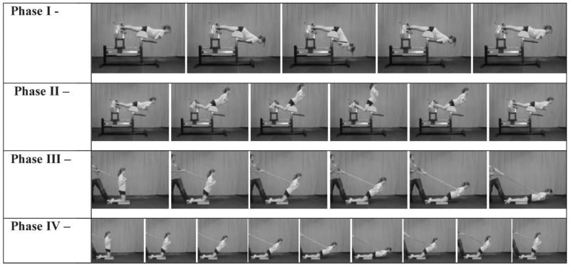
Female athletes who demonstrate the combination of decreased relative hamstrings and high relative quadriceps strength may be at increased risk for noncontact ACL injury (31). Targeted neuromuscular interventions that increase relative hamstrings muscle strength and recruitment may decrease injury risk and potentially increase performance in this population (20,31,33,40,41). Adequate co-contraction of the knee flexors may help balance active contraction of the quadriceps that can compress the joint and assist in the control of high KAM during deceleration tasks. To influence increased relative hamstring strength and recruitment, the single leg Romanian Deadlift progression (Figure 21) can be employed. In addition, the pelvic bridge progression (Figure 22) can influence synergistic posterior chain recruitment, especially from the gluteal musculature (maximus and medius) that can improve net hamstring recruitment and torque at the knee. End-stage exercise progression targeted to improved hamstring strength and recruitment should incorporate exercises from the Russian hamstring progression (Figure 23). These exercises may help improve dynamic knee stability during multiplanar movements via increased hamstring strength and coactivation to resist anterior tibial translation and KAM, which result from unequal opposition to forceful quadriceps contraction (9,20,46). As indicated in the current ACL injury risk prediction algorithm, increased relative strength and recruitment of the posterior chain musculature achieved through targeted neuromuscular training exercise may provide a mechanism for successful reduction of high KAM and noncontact ACL injury risk in female athletes (15,17,20,31,33,40).
Figure 21.
Single-Leg Romanian Deadlift (RDL) Progression. Phase I: Single Leg RDL. The athlete prepares for this exercise by standing on one leg with a slight bend in their knee. While keeping her shoulders back, the athlete should slowly flex only at the hip and lower her trunk toward the ground, reaching her hands to the floor. As she lowers her trunk, the athlete should extend the non-stance leg behind her out and as high as possible. She should try to minimize movement at the knee of her stance leg while maintaining her balance. Phase II: Single-Leg Airex RDL. The athlete prepares for this exercise by standing on the Airex pad with one leg with a slight bend in her knee. While keeping her shoulders back, the athlete should slowly flex only at the hip and lower her trunk toward the ground, reaching her hands to the floor. As she lowers her trunk, the athlete should extend the non-stance leg behind her out and as high as possible. She should try to minimize movement at the knee of her stance leg while maintaining her balance. Phase III: Single-Leg BOSU (Flat) RDL. The athlete prepares for this exercise by standing on the flat side of the BOSU with one foot in the center and a slight bend in her knee. While keeping her shoulders back, the athlete should slowly flex only at the hip and lower her trunk toward the ground, reaching her hands to the floor. As she lowers her trunk, the athlete should extend the non-stance leg behind her out and as high as possible. She should try to minimize movement at the knee of her stance leg while maintaining her balance. Also instruct the athlete to keep the BOSU as level and stable as possible. Phase III: Single-Leg RDL with dumbbell. The athlete prepares for this exercise by standing on one leg with a slight bend in their knee while holding dumbbells in each hand. While keeping her shoulders back, the athlete should slowly flex only at the hip and lower her trunk toward the ground, reaching her hands to the floor. As she lowers her trunk, the athlete should extend the non-stance leg behind her out and as high as possible. She should try to minimize movement at the knee of her stance leg while maintaining her balance.
Figure 22.
Pelvic Bridge progression. Phase I: Pelvic bridge 2 legs on ground. The athlete lays supine with her hip and knees flexed and her feet planted on the ground. The athlete then extends her hips and elevates her trunk off the ground to execute a pelvic bridge. This position should be held for 3 seconds before repeating the next repetition and is used to establish movement pattern with hips through and glute recruitment. Phase II: BOSU (Flat) Single-Leg Pelvic Bridge. The athlete lays supine with her hip and knees flexed and a single foot planted on the flat side of the BOSU and the contralateral leg fully extended. The athlete then extends her hips and elevates her trunk off the ground to execute a pelvic bridge. This position should be held for 3 seconds before repeating the next repetition. Phase III: BOSU (Flat) Single-Leg Pelvic Bridge with abduction and rotation. The athlete lays supine with her hip and knees flexed and a single foot planted on the flat side of the BOSU and the contralateral leg fully extended. The athlete then extends her hips and elevates her trunk off the ground to execute a pelvic bridge. This position should be held while the non-stance leg is abducted and externally rotated to further challenge strength, balance, and trunk control. Phase IV: Single-Leg Pelvic Bridge on Bench. The athlete supports a supine position with her shoulders positioned across a weight bench or plyometric box and her hip and knees flexed with a single foot planted on the flat side of a weight bench or chair. The athlete then extends her hips and elevates her trunk away from the ground to execute a pelvic bridge and thrust her non-stance leg in the air toward the ceiling. This position should be held for 3 seconds before repeating the next repetition.
Figure 23.
Russian Hamstring Progression. Phase I: Trunk Extension Glute Ham Raise. Athlete secures their feet in the chair. The athlete lowers herself down by flexing their trunk from a fully extended position to a point where her hip is flexed to nearly 90°. The torso should be perpendicular to the ground at the middle portion of the exercise. During the lifting phase, the athlete is instructed to use their glutes and trunk extensors to lift themselves back into the starting position again with their entire body parallel to the ground. Phase II: Glute Ham Raise. Athlete secures their feet in the chair. The athlete lowers herself down by extending their knee from a 90° to a 180° angle. The torso and lower extremity should be parallel to the ground (i.e., lying flat) at the end of the down portion of the exercise. During the lifting phase, the athlete is instructed to use their hamstrings and glutes to lift themselves back into the starting position where their thighs and torso are at a 90° angle to their calves (i.e., a kneeling position). Phase III: Eccentric Only Assisted Russian Hamstring Curl. The athlete begins in a kneeling position with a partner providing foot support and torso support (with band assistance). The athlete extends slowly at the knee to lower their torso toward the ground. Once touching the BOSU near the floor with their chest, the instructors provide substantial assistance to help the athletes maintain their trunk extension and return to the original position by flexing at the knee. The coach should provide enough assistance so that the exercise can be performed with relative ease during the concentric (raising) portion of the movement. Phase IV: Assisted Russian Hamstring Curl. The athlete begins in a kneeling position with a partner providing foot support and torso support (with band assistance). The athlete extends at the knee to lower their torso toward the ground. Once touching the BOSU near the floor with their chest, the athlete maintains her trunk extension and returns to the original position by flexing at the knee. The coach should provide enough assistance so that the exercise can be performed without flexing at the hip.
Practical Applications
To achieve the objective of reducing noncontact injury risk in female athletes, identification and treatment of those who preferentially demonstrate high KAM landing mechanics appear salient. The provided ACL injury risk prediction algorithm provides an integrative approach to guide targeted dynamic neuromuscular analysis training to specifically address and correct neuromuscular deficits that increase high KAM risk within the algorithm. Targeted correction of high KAM risk factors is important for both optimal biomechanics of athletic movements that maximize sport performance and ultimately the reduction of knee injury incidence in female athletes.
Although this article attempts to delineate proper training techniques that are specifically targeted for biomechanical deficits, the practitioner should understand the synergistic nature of the suggested training modes. Several of the individual components of the aforementioned training may have positive effects outside of the discrete changes that are referenced. Although it is beyond the scope of this paper to elucidate all the effects of the recommended training, we propose a method by which the practitioner can improve and refine a training program intended to target identified deficits in individual athletes. As coaches and practitioners apply their own knowledge and understanding of how each exercise works both discretely and in concord with the proposed integrative approach, we would expect even further positive adaptive responses from their athletes. Future research focused to determine the injury risk reductions using the proposed methods is warranted.
Acknowledgments
The authors would like to acknowledge funding support from National Institutes of Health/NIAMS Grants R01-AR049735, R01-AR05563 and R01-AR056259. The authors would like to thank Boone County Kentucky, School District, especially the School Superintendent Randy Poe, for participation in this study. We would also like to thank Mike Blevins, Ed Massey, Dr. Brian Blavatt, and the athletes of Boone County public school district for their participation in this study. The authors would also like to acknowledge the Cincinnati Children’s Sports Medicine Biodynamics Center Team who have contributed intellectually and physically to the presented research outcomes.
References
- 1.Andriacchi TP, Andersson GBJ, Fermier RW, Stern D, Galante JO. Study of lower-limb mechanics during stair-climbing. J Bone Joint Surg Am. 1980;62A:749–757. [PubMed] [Google Scholar]
- 2.Baumhauer J, Alosa D, Renstrom A, Trevino S, Beynnon B. A prospective study of ankle injury risk factors. Am J Sports Med. 1995;23:564–570. doi: 10.1177/036354659502300508. [DOI] [PubMed] [Google Scholar]
- 3.Besier TF, Lloyd DG, Ackland TR, Cochrane JL. Anticipatory effects on knee joint loading during running and cutting maneuvers. Med Sci Sports Exerc. 2001;33:1176–1181. doi: 10.1097/00005768-200107000-00015. [DOI] [PubMed] [Google Scholar]
- 4.Boden BP, Dean GS, Feagin JA, Garrett WE. Mechanisms of anterior cruciate ligament injury. Orthopedics. 2000;23:573–578. doi: 10.3928/0147-7447-20000601-15. [DOI] [PubMed] [Google Scholar]
- 5.Brent JL, Myer GD, Ford KR, Hewett TE. A longitudinal examination of hip abduction strength in adolescent males and females. Med Sci Sport Exerc. 2008;40:731. [Google Scholar]
- 6.Chmielewski TL, Myer GD, Kauffman D, Tillman S. Plyometric exercise in the rehabilitation of athletes: Physiological responses and clinical application. J Orthop Sports Phys Ther. 2006;36:308–319. doi: 10.2519/jospt.2006.2013. [DOI] [PubMed] [Google Scholar]
- 7.Claiborne TL, Armstrong CW, Gandhi V, Pincivero DM. Relationship between hip and knee strength and knee valgus during a single leg squat. J Appl Biomech. 2006;22:41–50. doi: 10.1123/jab.22.1.41. [DOI] [PubMed] [Google Scholar]
- 8.Daniel DM, Malcom LL, Losse G, Stone ML, Sachs R, Burks R. Instrumented measurement of anterior laxity of the knee. J Bone Joint Surg Am. 1985;67A:720–726. [PubMed] [Google Scholar]
- 9.Draganich LF, Vahey JW. An in vitro study of anterior cruciate ligament strain induced by quadriceps and hamstrings forces. J Orthop Res. 1990;8:57–63. doi: 10.1002/jor.1100080107. [DOI] [PubMed] [Google Scholar]
- 10.Ford KR, Myer GD, Hewett TE. Valgus knee motion during landing in high school female and male basketball players. Med Sci Sports Exerc. 2003;35:1745–1750. doi: 10.1249/01.MSS.0000089346.85744.D9. [DOI] [PubMed] [Google Scholar]
- 11.Ford KR, Myer GD, Hewett TE. Increased trunk motion in female athletes compared to males during single leg landing. Med Sci Sports Exerc. 2007;39:S70. [Google Scholar]
- 12.Ford KR, Myer GD, Toms HE, Hewett TE. Gender differences in the kinematics of unanticipated cutting in young athletes. Med Sci Sports. 2005;37:124–129. [PubMed] [Google Scholar]
- 13.Hewett TE, Biro FM, McLean SG, Van den Bogert AJ. Identifying Female Athletes at High Risk for ACL Injury. Cincinnati Children’s Hospital, National Institutes of Health; Bethesda, MD: 2003. [Google Scholar]
- 14.Hewett TE, Ford KR, Myer GD. Anterior cruciate ligament injuries in female athletes: Part 2, a meta-analysis of neuromuscular interventions aimed at injury prevention. Am J Sports Med. 2006;34:490–498. doi: 10.1177/0363546505282619. [DOI] [PubMed] [Google Scholar]
- 15.Hewett TE, Lindenfeld TN, Riccobene JV, Noyes FR. The effect of neuromuscular training on the incidence of knee injury in female athletes. A prospective study. Am J Sports Med. 1999;27:699–706. doi: 10.1177/03635465990270060301. [DOI] [PubMed] [Google Scholar]
- 16.Hewett TE, Myer GD, Ford KR. Decrease in neuromuscular control about the knee with maturation in female athletes. J Bone Joint Surg Am. 2004;86-A:1601–1608. doi: 10.2106/00004623-200408000-00001. [DOI] [PubMed] [Google Scholar]
- 17.Hewett TE, Myer GD, Ford KR, Heidt RS, Jr, Colosimo AJ, McLean SG, van den Bogert AJ, Paterno MV, Succop P. Biomechanical measures of neuromuscular control and valgus loading of the knee predict anterior cruciate ligament injury risk in female athletes: A prospective study. Am J Sports Med. 2005;33:492–501. doi: 10.1177/0363546504269591. [DOI] [PubMed] [Google Scholar]
- 18.Hewett TE, Myer GD, Ford KR, Slauterbeck JR. Preparticipation physical exam using a box drop vertical jump test in young athletes: The effects of puberty and sex. Clin J Sport Med. 2006;16:298–304. doi: 10.1097/00042752-200607000-00003. [DOI] [PubMed] [Google Scholar]
- 19.Hewett TE, Paterno MV, Myer GD. Strategies for enhancing proprioception and neuromuscular control of the knee. Clin Orthop Relat Res. 2002;402:76–94. doi: 10.1097/00003086-200209000-00008. [DOI] [PubMed] [Google Scholar]
- 20.Hewett TE, Stroupe AL, Nance TA, Noyes FR. Plyometric training in female athletes. Decreased impact forces and increased hamstring torques. Am J Sports Med. 1996;24:765–773. doi: 10.1177/036354659602400611. [DOI] [PubMed] [Google Scholar]
- 21.Hodges PW, Richardson CA. Contraction of the abdominal muscles associated with movement of the lower limb. Phys Ther. 1997;77:132–142. doi: 10.1093/ptj/77.2.132. discussion 142-144. [DOI] [PubMed] [Google Scholar]
- 22.Hodges PW, Richardson CA. Feedforward contraction of transversus abdominis is not influenced by the direction of arm movement. Exp Brain Res. 1997;114:362–370. doi: 10.1007/pl00005644. [DOI] [PubMed] [Google Scholar]
- 23.Ireland ML. The female ACL: Why is it more prone to injury? Orthop Clin North Am. 2002;33:637–651. doi: 10.1016/s0030-5898(02)00028-7. [DOI] [PubMed] [Google Scholar]
- 24.Knapik JJ, Bauman CL, Jones BH, Harris JM, Vaughan L. Preseason strength and flexibility imbalances associated with athletic injuries in female collegiate athletes. Am J Sports Med. 1991;19:76–81. doi: 10.1177/036354659101900113. [DOI] [PubMed] [Google Scholar]
- 25.Krosshaug T, Nakamae A, Boden BP, Engebretsen L, Smith G, Slauterbeck JR, Hewett TE, Bahr R. Mechanisms of anterior cruciate ligament injury in basketball: Video analysis of 39 cases. Am J Sports Med. 2007;35:359–367. doi: 10.1177/0363546506293899. [DOI] [PubMed] [Google Scholar]
- 26.Lloyd DG, Buchanan TS. Strategies of muscular support of varus and valgus isometric loads at the human knee. J Biomech. 2001;34:1257–1267. doi: 10.1016/s0021-9290(01)00095-1. [DOI] [PubMed] [Google Scholar]
- 27.Markolf KL, Burchfield DM, Shapiro MM, Shepard MF, Finerman GA, Slauterbeck JL. Combined knee loading states that generate high anterior cruciate ligament forces. J Orthop Res. 1995;13:930–935. doi: 10.1002/jor.1100130618. [DOI] [PubMed] [Google Scholar]
- 28.More RC, Karras BT, Neiman F, Fritschy D, Woo SL-Y, Daniel DM. Hamstrings-an anterior cruciate ligament protagonist: An in vitro study. Am J Sports Med. 1993;21:231–237. doi: 10.1177/036354659302100212. [DOI] [PubMed] [Google Scholar]
- 29.Myer GD, Brent JL, Ford KR, Hewett TE. A pilot study to determine the effect of trunk and hip focused neuromuscular training on hip and knee isokinetic strength. Br J Sports Med. 2008;42:614–619. doi: 10.1136/bjsm.2007.046086. [DOI] [PMC free article] [PubMed] [Google Scholar]
- 30.Myer GD, Chu DA, Brent JL, Hewett TE. Trunk and hip control neuromuscular training for the prevention of knee joint injury. Clin Sports Med. 2008;27:425–448. doi: 10.1016/j.csm.2008.02.006. ix. [DOI] [PMC free article] [PubMed] [Google Scholar]
- 31.Myer GD, Ford KR, Barber Foss KD, Liu C, Nick TG, Hewett TE. The relationship of hamstrings and quadriceps strength to anterior cruciate ligament injury in female athletes. Clin J Sport Med. 2009;19:3–8. doi: 10.1097/JSM.0b013e318190bddb. [DOI] [PMC free article] [PubMed] [Google Scholar]
- 32.Myer GD, Ford KR, Brent JL, Hewett TE. The effects of plyometric versus dynamic balance training on landing force and center of pressure stabilization in female athletes. Br J Sports Med. 2005;39:397. [Google Scholar]
- 33.Myer GD, Ford KR, Brent JL, Hewett TE. The effects of plyometric versus dynamic balance training on power, balance and landing force in female athletes. J Strength Cond Res. 2006;20:345–353. doi: 10.1519/R-17955.1. [DOI] [PubMed] [Google Scholar]
- 34.Myer GD, Ford KR, Brent JL, Hewett TE. Differential neuromuscular training effects on ACL injury risk factors in “high-risk” versus “low-risk” athletes. BMC Musculoskelet Disord. 2007;8:1–39. doi: 10.1186/1471-2474-8-39. [DOI] [PMC free article] [PubMed] [Google Scholar]
- 35.Myer GD, Ford KR, Hewett TE. Rationale and clinical techniques for anterior cruciate ligament injury prevention among female athletes. J Athl Train. 2004;39:352–364. [PMC free article] [PubMed] [Google Scholar]
- 36.Myer GD, Ford KR, Hewett TE. New method to identify athletes at high risk of ACL injury using clinic-based measurements and freeware computer analysis. Br J Sports Med. 2011;45:238–244. doi: 10.1136/bjsm.2010.072843. [DOI] [PMC free article] [PubMed] [Google Scholar]
- 37.Myer GD, Ford KR, Khoury J, Succop P, Hewett TE. Clinical correlates to laboratory measures for use in non-contact anterior cruciate ligament injury risk prediction algorithm. Clin Biomech (Bristol, Avon) 2010;25:693–699. doi: 10.1016/j.clinbiomech.2010.04.016. [DOI] [PMC free article] [PubMed] [Google Scholar]
- 38.Myer GD, Ford KR, Khoury J, Succop P, Hewett TE. Development and validation of a clinic-based prediction tool to identify female athletes at high risk for anterior cruciate ligament injury. Am J Sports Med. 2010;38:2025–2033. doi: 10.1177/0363546510370933. [DOI] [PMC free article] [PubMed] [Google Scholar]
- 39.Myer GD, Ford KR, Khoury J, Succop P, Hewett TE. Biomechanics laboratory-based prediction algorithm to identify female athletes with high knee loads that increase risk of ACL injury. Br J Sports Med. 2011;45:245–252. doi: 10.1136/bjsm.2009.069351. [DOI] [PMC free article] [PubMed] [Google Scholar]
- 40.Myer GD, Ford KR, McLean SG, Hewett TE. The effects of plyometric versus dynamic stabilization and balance training on lower extremity biomechanics. Am J Sports Med. 2006;34:490–498. doi: 10.1177/0363546505281241. [DOI] [PubMed] [Google Scholar]
- 41.Myer GD, Ford KR, Palumbo JP, Hewett TE. Neuromuscular training improves performance and lower-extremity biomechanics in female athletes. J Strength Cond Res. 2005;19:51–60. doi: 10.1519/13643.1. [DOI] [PubMed] [Google Scholar]
- 42.Myklebust G, Engebretsen L, Braekken IH, Skjolberg A, Olsen OE, Bahr R. Prevention of anterior cruciate ligament injuries in female team handball players: A prospective intervention study over three seasons. Clin J Sport Med. 2003;13:71–78. doi: 10.1097/00042752-200303000-00002. [DOI] [PubMed] [Google Scholar]
- 43.Olsen OE, Myklebust G, Engebretsen L, Bahr R. Injury mechanisms for anterior cruciate ligament injuries in team handball: A systematic video analysis. Am J Sports Med. 2004;32:1002–1012. doi: 10.1177/0363546503261724. [DOI] [PubMed] [Google Scholar]
- 44.Onate JA, Guskiewicz KM, Marshall SW, Giuliani C, Yu B, Garrett WE. Instruction of jump-landing technique using videotape feedback: altering lower extremity motion patterns. Am J Sports Med. 2005;33:831–842. doi: 10.1177/0363546504271499. [DOI] [PubMed] [Google Scholar]
- 45.Pandy MG, Shelburne KB. Dependence of cruciate-ligament loading on muscle forces and external load. J Biomech. 1997;30:1015–1024. doi: 10.1016/s0021-9290(97)00070-5. [DOI] [PubMed] [Google Scholar]
- 46.White KK, Lee SS, Cutuk A, Hargens AR, Pedowitz RA. EMG power spectra of intercollegiate athletes and anterior cruciate ligament injury risk in females. Med Sci Sports Exerc. 2003;35:371–376. doi: 10.1249/01.MSS.0000053703.65057.31. [DOI] [PubMed] [Google Scholar]
- 47.Wilson JD, Dougherty CP, Ireland ML, Davis IM. Core stability and its relationship to lower extremity function and injury. J Am Acad Orthop Surg. 2005;13:316–325. doi: 10.5435/00124635-200509000-00005. [DOI] [PubMed] [Google Scholar]
- 48.Winter DA. Biomechanics and Motor Control of Human Movement. John Wiley & Sons, Inc.; New York, NY: 2005. [Google Scholar]
- 49.Zatsiorsky VM. Science and Practice of Strength Training. Human Kinetics; Champaign, IL: 1995. [Google Scholar]
- 50.Zazulak BT, Hewett TE, Reeves NP, Goldberg B, Cholewicki J. The effects of core proprioception on knee injury: A prospective biomechanical-epidemiological study. Am J Sports Med. 2007;35:368–373. doi: 10.1177/0363546506297909. [DOI] [PubMed] [Google Scholar]


