Abstract
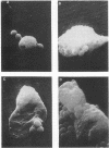
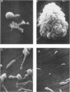
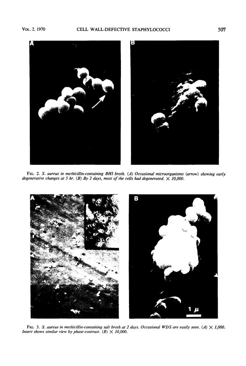
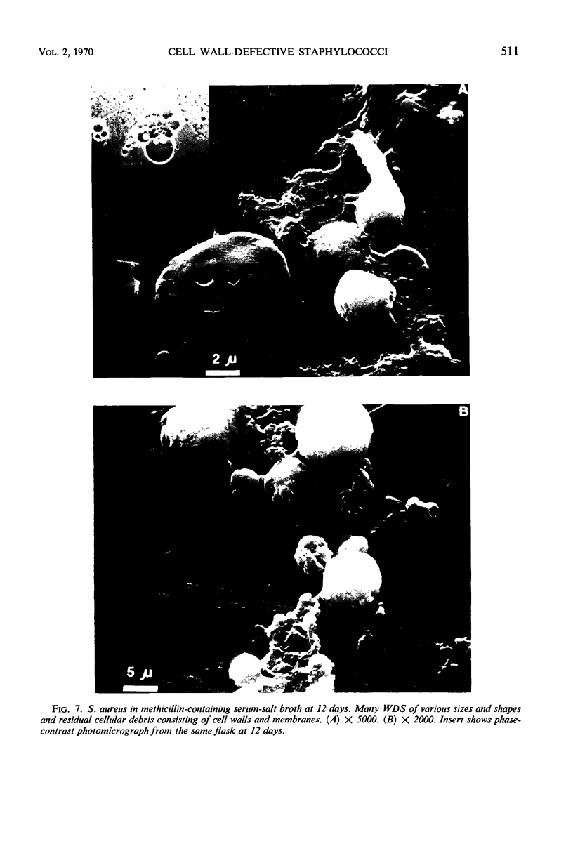
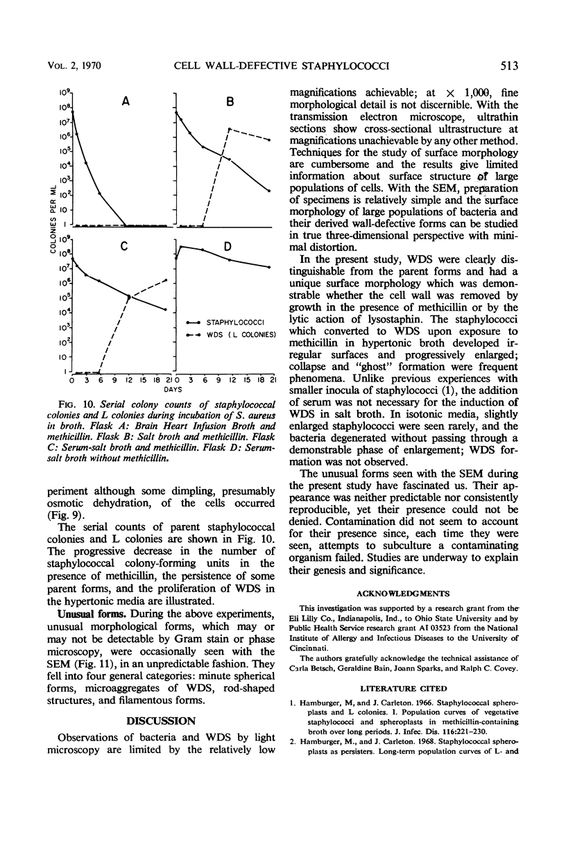
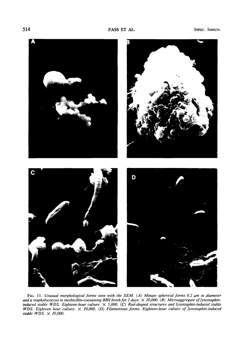
Staphylococci and their derived wall-defective forms (WDS) were studied with Gram stain, phase microscopy, and the scanning-beam electron microscope. Staphylococci were smooth, spherical, and relatively uniform in size. Stable WDS had corrugated surfaces and were larger; those prepared with lysostaphin were indistinguishable from those prepared with methicillin. During induction of WDS in methicillin-containing hypertonic broth, WDS were first observed after 7 hr of incubation and progressively, thereafter, increased in number. They were larger than the stable WDS and varied more in size and shape. Microscopically, “wisps” were seen to consist of WDS, persistent parent staphylococci, and residual cell membranes.
Full text
PDF





Images in this article
Selected References
These references are in PubMed. This may not be the complete list of references from this article.
- Hamburger M., Carleton J. Staphylococcal spheroplasts and L colonies. I. Population curves of vegetative staphylococci and spheroplasts in methicillin-containing broth over long periods. J Infect Dis. 1966 Apr;116(2):221–230. doi: 10.1093/infdis/116.2.221. [DOI] [PubMed] [Google Scholar]
- Klainer A. S., Betsch C. J. Scanning-beam electron microscopy of selected microorganisms. J Infect Dis. 1970 Mar;121(3):339–343. doi: 10.1093/infdis/121.3.339. [DOI] [PubMed] [Google Scholar]
- Watanakunakorn C., Browder H. P. Effects of lysostaphin and its two active components on stable wall-defective forms of Staphylococcus aureus. J Infect Dis. 1970 Feb;121(2):124–128. doi: 10.1093/infdis/121.2.124. [DOI] [PubMed] [Google Scholar]
- Watanakunakorn C., Goldberg L. M., Carleton J., Hamburger M. Staphylococcal spheroplasts and L-colonies. 3. Induction by lysostaphin. J Infect Dis. 1969 Jan;119(1):67–74. doi: 10.1093/infdis/119.1.67. [DOI] [PubMed] [Google Scholar]


