
Figure 1.
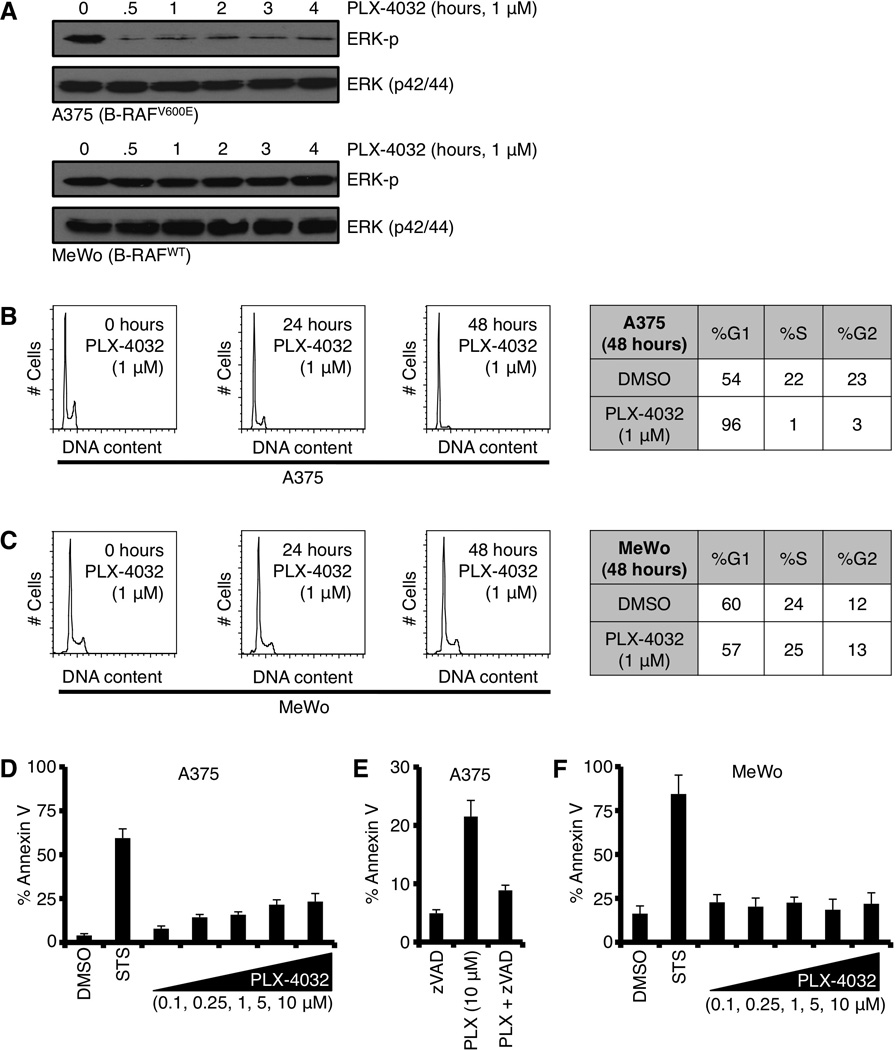
The majority of B-RAFV600E positive cells respond to PLX-4032 treatment by reducing phosphorylated ERK and G1 cell cycle arrest, but only a minority of cells undergoes apoptosis. (A) A375 (B-RAFV600E) and MeWo (B-RAFWT) were treated with PLX-4032 (1 µM) for indicated time points before western blot for phosphorylated ERK (ERK-p) and total ERK1/2 (p42/44). (B–C) A375 and MeWo were treated with PLX-4032 (1 µM) for indicated time points and analyzed for cell cycle distribution. Representative percentages are shown in charts. (D) A375 was treated with PLX-4032 (0.1, 0.25, 1, 5, 10 µM) for 72 hours before AnnexinV staining and flow cytometry. Staurosporine (50 nM, STS) is shown as a positive control. (E) A375 cells were pre-treated with zVAD-fmk (50 µM) for 1 hour before treatment with PLX-4032 (10 µM) for 48 hours, and analyzed by AnnexinV and flow cytometry. (F) MeWo was treated as in D. Staurosporine (50 nM, STS) is shown as a positive control. All data are representative of at least triplicate experiments, and reported as ± S.D., as required.