
Figure 2.
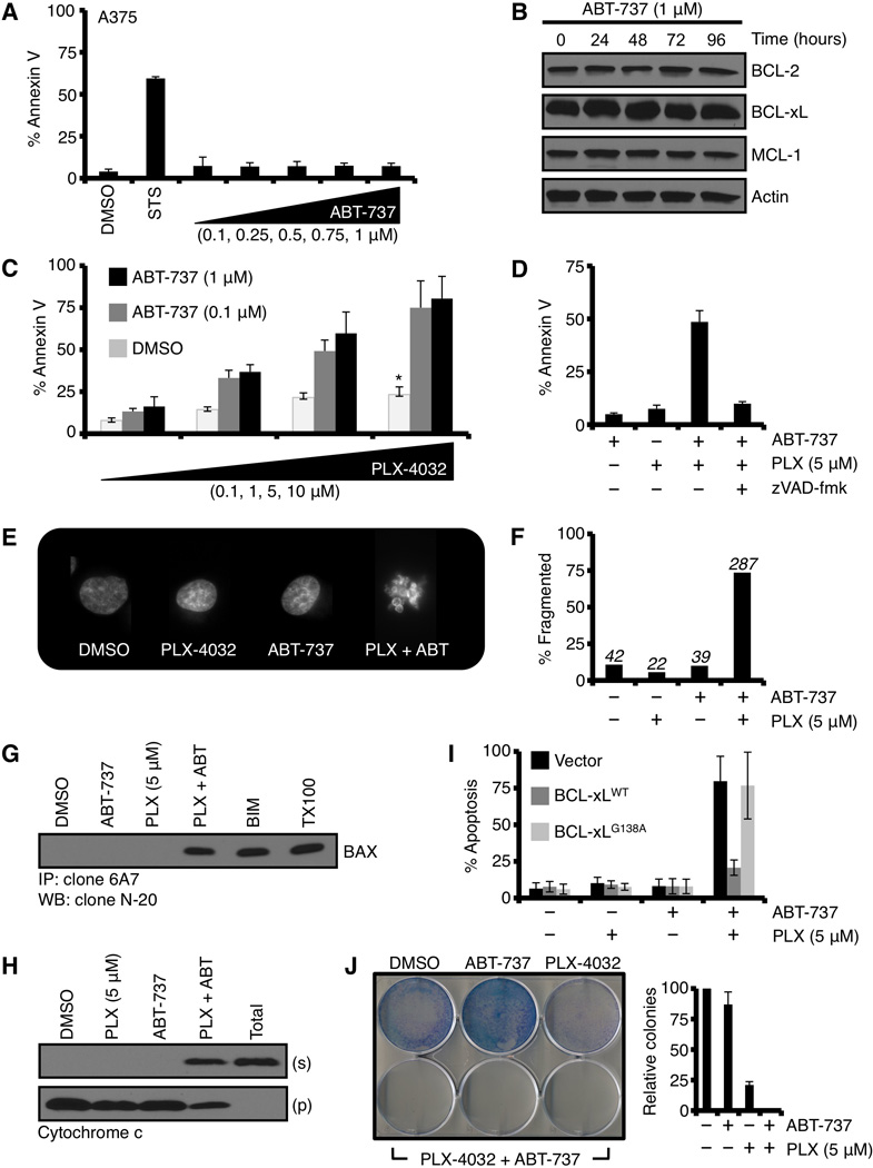
Collateral inhibition of B-RAFV600E signaling and anti-apoptotic BCL-2 protein function reveals marked commitment to the mitochondrial pathway of apoptosis and loss of clonogenic survival. (A) A375 was treated with ABT-737 (0.1, 0.25, 0.5, 0.75, 1 µM) for 48 hours before AnnexinV staining and flow cytometry. Staurosporine (50 nM, STS) is shown as a positive control. (B) A375 was treated with ABT-737 (1 µM) for indicated time points before western blotting for BCL-2, BCL-xL, and MCL-1. Actin is shown as a loading control. (C) A375 cells were pre-treated with ABT-737 (0.1, 1.0 µM) for 1 hour before treatment with PLX-4032 (0.1, 1, 5, 10 µM) for 48 hours, and analyzed by AnnexinV and flow cytometry. *p value = < 0.005. (D) A375 cells were pre-treated with ABT-737 (1.0 µM) and zVAD-fmk (50 µM) for 1 hour before treatment with PLX-4032 (5 µM) for 48 hours, and analyzed by AnnexinV and flow cytometry. (E) A375 cells were pre-treated with ABT-737 (1.0 µM) for 1 hour before treatment with PLX-4032 (5 µM) for 48 hours. The cells were stained with Hoechst 33342 (20 µM) and imaged by fluorescent microscopy. (F) The data obtained in E were quantified. ~ 400 cells per condition were evaluated. (G) A375 cells were pre-treated with ABT-737 (1.0 µM) for 1 hour before treatment with PLX-4032 (5 µM) for 48 hours. The cells were lysed in CHAPS buffer, and the lysates were subjected to anti-BAX immunoprecipitation using clone 6A7 and analyzed by western blot for BAX. BIM-transfected cells and TritonX-100 (0.25%, TX100) are positive controls for 6A7 positive BAX. (H) A375 cells were pre-treated with ABT-737 (1.0 µM) for 1 hour before treatment with PLX-4032 (5 µM) for 24 hours. The cells were then subjected to fractionation for cytosol and heavy membranes (i.e., mitochondria), and these fractions were subjected to western blot for cytochrome c. Supernatants (s) and pellets (p) are shown. “Total” is a detergent (0.25% TX100) solubilized sample. (I) A375 were transiently transfected with empty vector, pcDNA3.1-BCL-xLWT, or pcDNA3.1-BCL-xLG138A. The next day, cells were pre-treated with ABT-737 (1.0 µM) for 1 hour before treatment with PLX-4032 (5 µM) for 48 hours, and analyzed by AnnexinV and flow cytometry. (J) A375 were pre-treated with ABT-737 (1.0 µM) for 1 hour before treatment with PLX-4032 (5 µM) for 48 hours, the media was replaced, and the cells were cultured for an additional 10 days. The resulting colonies were stained (left panel) and quantified (right panel). The PLX-4032 + ABT-737 combination is shown in triplicate. All data are representative of at least triplicate experiments, and reported as ± S.D., as required.