
Figure 3.
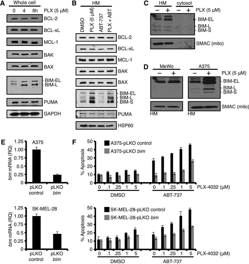
PLX-4032 induced inhibition of B-RAFV600E promotes marked expression and rapid mitochondrial accumulation of BIM; however, this pro-apoptotic signal remains silenced. (A) A375 was treated with PLX-4032 (5 µM) for indicated time points before western blot for the indicated BCL-2 family members. GAPDH is shown as a loading control. (B) A375 cells were pre-treated with ABT-737 (1.0 µM) for 1 hour before treatment with PLX-4032 (5 µM) for 24 hours. The heavy membrane fractions (“HM”, i.e., mitochondria) were then analyzed for the indicated BCL-2 family members. HSP60 is shown as a loading control. (C) A375 cells were treated with PLX-4032 (5 µM) for 24 hours, and fractionated into cytosol and heavy membranes. These fractions were subjected to western blot for BIM. SMAC and actin are shown as mitochondrial and cytosolic fractionation controls. (D) A375 and MeWo were treated with PLX-4032 (5 µM) for 24 hours, the HM fractions were isolated and subjected to western blot analysis for BIM. SMAC is shown as a mitochondrial loading control. (E) A375 and SK-MEL-28 cells stably expressing bim shRNA (or the control vector, pLKO) were quantified for bim knock-down by qPCR. Expression was normalized with β-actin and gapdh. (F) The cells in E were treated with PLX-4032 (0, 0.1, 0.25, 1, 5 µM) ± ABT-737 (0.5 µM) for 48 hours before AnnexinV staining and flow cytometry. All data are representative of at least triplicate experiments, and reported as ± S.D., as required.