
Figure 4.
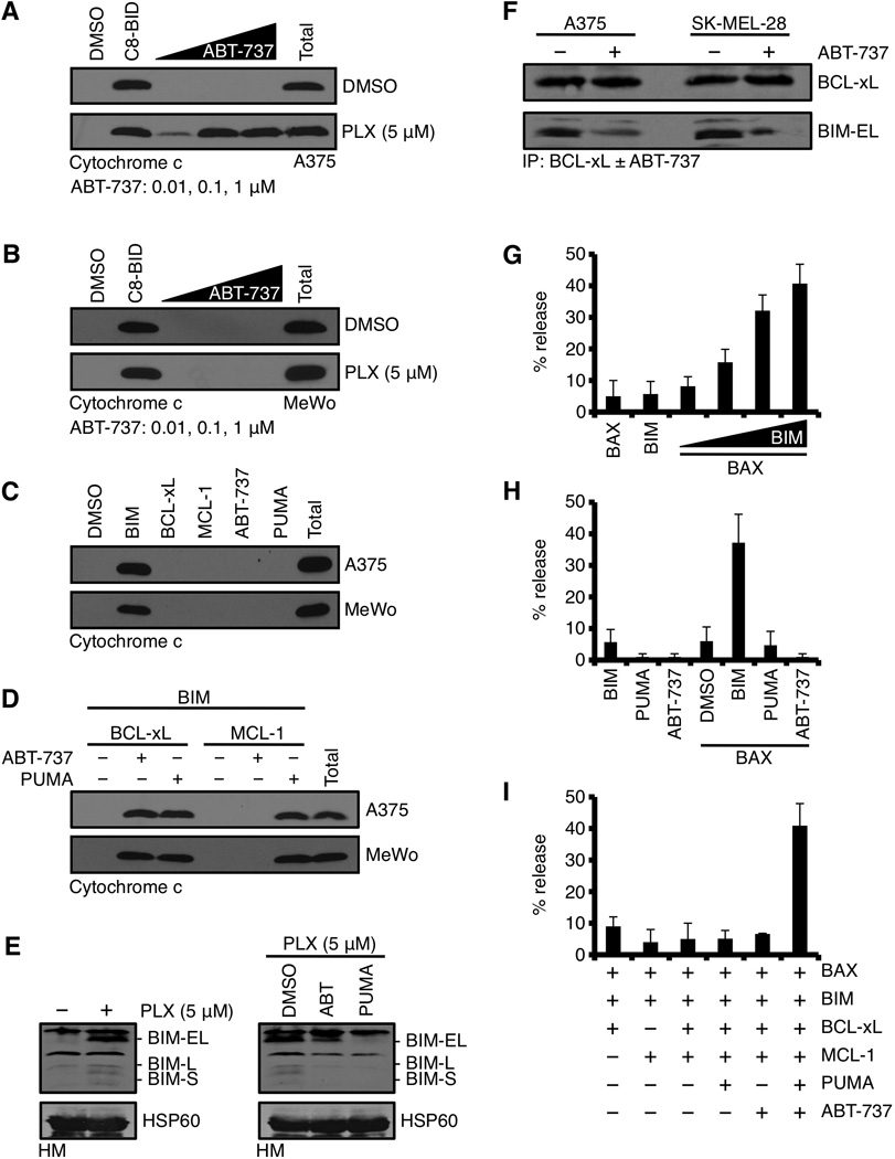
Despite the presence of numerous pro-apoptotic BH3-only proteins, ABT-737 is required to reveal PLX-4032 induced BIM-dependent mitochondrial outer membrane permeabilization. (A) A375 were treated with DMSO or PLX-4032 (5 µM) for 24 hours, the HM fractions isolated, treated with ABT-737 (0.01, 0.1, 1 µM) for 30 minutes at 37°C, centrifuged, and the supernatants were subjected to western blot for cytochrome c. Caspase-8 cleaved BID (C8-BID, 10 nM) is a positive control for physiological cytochrome c release; “Total” is a detergent solubilized sample and is the maximal cytochrome c per sample. (B) The same assay as in A, but with MeWo. (C) HM fractions from untreated A375 or MeWo were incubated with the BIM BH3 domain peptide (5 µM), BCL-xL (50 nM), MCL-1 (50 nM), ABT-737 (1 µM), or PUMA (1 µM) for 30 minutes at 37°C, centrifuged, and the supernatants were subjected to western blot for cytochrome c. (D) HM fractions from untreated A375 or MeWo were incubated with the indicated combinations of BCL-2 family reagents and analyzed as in C. (E) A375 were treated with PLX-4032 (5 µM) for 24 hours, the HM fractions were isolated and subjected to western blot analysis for BIM (left panel). Isolated mitochondria were treated with DMSO, ABT-737 (100 nM), PUMA (100 nM) for 1 hour at 37°C, centrifuged, and the pellets were subjected to western blot for BIM (right panel). HSP60 is shown as a mitochondrial loading control. (F) A375 and SK-MEL-28 were treated with PLX-4032 (5 µM) for 24 hours, the HM fractions were isolated, lysed, and subjected to immunoprecipitation with an anti-BCL-xL antibody (1 µg) ± ABT-737 (100 nM). BCL-xL and BIM were detected by western blot. (G) LUVs were treated with combinations of BAX (100 nM) and the BIM BH3 (5 µM alone, or 0.1, 0.5, 1, 5 µM) for 30 minutes at 37°C. 100% release was determined by a detergent treated sample. (H) LUVs were treated with combinations of BIM BH3 (5 µM), PUMA (1 µM), ABT-737 (1 µM), and BAX (100 nM) for 30 minutes at 37°C. (I) LUVs were treated with combinations of reagents described in E, including BCL-xL (50 nM) and MCL-1 (50 nM) for 30 minutes at 37°C. All data are representative of at least triplicate experiments, and reported as ± S.D., as required.