Abstract
An L-rhamnose-based hydrogelator self-assembles to form nanofibrils, which, contrasting to the properties of monomeric L-rhamnose, suppress the antibody response of mice to phycoerythrin (PE), a fluorescent protein antigen. As the first example of the supramolecular assemblies of a saccharide to suppress immunity, this work illustrates a new approach of immunomodulation.
Recent vaccine design mainly relies on purified antigen-based acellular approaches due to their low toxicity compared with whole pathogen-based approaches. However, purified antigens require a helper (adjuvant) because of their inherently poor immunogenicity. Currently, there are only two FDA-approved vaccine adjuvants: long-standing aluminium salts (collectively termed alum) and the recently approved monophosphoryl lipid A (MPL).1 Despite alum’s effectiveness to induce Th2-biased immune responses, there are unmet needs for adjuvants that are safer, have more precise molecular control, and induce Th1 or Th17 T cell responses, which has stimulated the development of synthetic immunomodulatory materials.2 While most studies have focused on fabricating polymer-based nanoparticles that are preferentially internalized by antigen presenting cells (APCs),3 liposomes/micelles or peptide-based nanofibrils resulting from molecular self-assembly are emerging as promising alternatives. One promising approach is to covalently incorporate antigenic epitopes into the structural components (lipids or peptides) of the adjuvants.4 Another option is to physically encapsulate purified antigens or antigen-coding DNA into liposomes or nanofibrils.5 The initial explorations of both approaches have successfully demonstrated dramatically enhanced immune responses, thus validating usage of self-assembly of small molecules for immunomodulation.
On the other hand, recruitment of pre-existing “natural” antibodies has become an important and integrated part of vaccine design. Because the human immune system produces a large amount of natural antibodies against certain small organic molecules including α-Gal epitope (Gal-α(1,3)-Gal-β(1,4)- GlcNAc/Glc), constituting about 1% of human serum IgG,6 the covalent linkage of antigens or antigenic epitopes to α-Gal would facilitate formation of immune complexes with natural antibodies to induce complement deposition, opsonization, and phagocytosis of antigens by APCs.7 However, the complicated anomeric stereochemistry of α-Gal makes it a rather prohibitive target of synthesis. Thus, it is still impractical to covalently conjugate α-Gal with antigenic epitopes. Two independent groups recently discovered even higher human natural antibody levels against L-rhamnose via glycan array screening.8 Although it is generally considered D-configuration monomeric carbohydrate units are unable to elicit immunological response, the observation on the immunogenicity of L-rhamonse8c, 8d clearly suggests that the stereochemistry of monosaccharides plays important role in understanding immune response of carbohydrates. The relative facile synthesis of L-rhamnose offers a unique opportunity to explore the incorporation of natural antigen saccharides into supramolecular assemblies for modulating immune responses because the incorporation of simple saccharides in supramolecular assemblies is able to achieve new functions.9
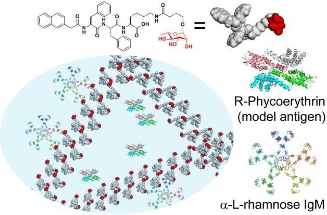
Encouraged by the development of self-assembling small glycopeptides10 and study of L-rhamnose,8c, 8d we designed and synthesized a conjugate (1) of a self-assembling motif11 and L-rhamnose (Scheme 1) to examine its immunomodulatory properties. Our results show that 1 self-assembles in water to form supramolecular nanofibrils and to result in a weak hydrogel, which allows the encapsulation of a fluorescent model antigen, R-phycoerythrin12 (PE, Scheme 1). Intraperitoneal (i.p.) injection of PE encapsulated by the nanofibrils of 1 into the mice being pre-injected with purified human IgM (containing circulating natural α-L-rhamnose antibodies), induces a reduction in IgG against PE in the murine model. As a control, free L-rhamnose or nanofibrils without L-rhamnose are unable to reduce anti-PE IgG. This immunosuppression by the L-rhamnose hydrogel is independent of complement component 3 (Figure 2) and absent in intraplantar (i.pl.) vaccination of PE. These results not only indicate that the murine peritoneal cavity result in a different mode of immunization, but also highlight different immunological behaviours of monomeric L-rhamnose and the supramolecular assemblies of L-rhamnose. Thus, the finding of this work ultimately may lead to the development of new immunosuppressing materials by taking advantage of molecular self-assembly of saccharides.
Scheme 1.
Illustration of the interactions of L-rhamnose in molecular nanofibrils with natural antibodies (α-L-rhamnose IgM) in mediating the suppression of antibody responses against a model foreign antigen (R-phycoerythrin) in mice.
Fig. 2.
ELISA readouts of absorbances at 405 nm for total Immunoglobulin G (IgG) levels against PE in serum of mice 28 days after intraperitoneal immunization by the indicated samples (NS: P > 0.05; **: 0.05<P≤ 0.01; ***: P ≤ 0.001).
To obtain supramolecular nanofibrils that not only encapsulate antigens, but also interact with human natural antibodies, we designed and synthesized 1 by introducing L-rhamnose to a self-assembly motif (NapFFK).10c, 13 Using the procedure reported by Sarkar and co-authors,8d we synthesized 2, a L-rhamnopyranoside with carboxylic acid group. N-(3- dimethylaminopropyl)-N’-ethylcarbodiimide hydrochloride (EDC) and N-hydroxysuccinimide (NHS) activate 2 to react with the εNH2 group of the lysine residue in NapFFK. Finally, Zemplén deacetylation gives 1 with a total yield of 79%. Similarly, the amide bond formation between butyric acid and NapFFK by offers 5, the self-assembling molecule without L-rhamnose (as a control), with a yield of 84%. In addition, Zemplén deacetylation of 2 gives 4 in a quantitative yield, a L-rhamnose derivative that acts as another control.
After the synthesis, we tested the gelation properties of 1 and 5. At the centration of 0.4% (w/v) in DPBS buffer (pH=7.4),† the molecules of 1 self-assemble to form nanofibrils with the diameters of 7±2 nm. The entanglement of the nanofibrils forms a relatively dense network (Fig. 1A), which results in a weak hydrogel (inset of Fig. 1A). Rheology measurement confirms that the hydrogel is rather liquid-like, as supported by that both its storage and loss moduli are around 1 Pa (Fig. S1). Molecules of 5, the control without L-rhamnose, exhibit a much better gelation property in the same buffer, and form a stable hydrogel at the concentration of 0.4 w/v% (inset of Fig. 1B). Rheology analysis of the hydrogel of 5 reveals that the storage modulus to be around 1400 Pa and the critical strain around 20.0% (Fig. S1). According TEM, the nanofibrils of 5 have the diameters of 10±2 nm (Fig. 2B), which are slightly wider than those of the nanofibrils of 1. The enhanced gelation property of 5 likely originates from an improved balance of amphiphilicity due to the absence of the hydrophilic L-rhamnose in 5.
Fig. 1.
Transmission electron microscopy (TEM) images of the negatively stained self-assembled nanofibrils of (A) 1 and (B) 5 at the concentration of 0.4 % (w/v) (scale bar: 100 nm; inset: the optical images of the weak hydrogel of 1 and hydrogel of 5)
After obtaining the nanofibrils of 1 and 5, we examined their abilities for immunomodulation by encapsulating PE in the nanofibrils and injecting the hydrogels into mice. Due to the low levels of anti-rhamnose antibodies in mice (Fig. S2),8c we injected purified human IgM 12 hours prior to the immunization. Instead of observing an increased antibody response, as exhibited by monomeric L-rhamnose,8d we found a dramatic reduced antibody response to PE (Fig. 2) in the case of i.p. immunization of PE encapsulated by the nanofibrils of 1 in the mice pre-injected with purified human IgM (“PE with 1 + IgM”, Fig. 2). This reduction is absent in i.p. immunization of PE inside nanofibrils of 5 in the mice pre-injected with purified human IgM (“PE with 5 + IgM”, Fig. 2). These two results indicate that the presence of L-rhamnose is necessary for mediating the observed immunosuppression. Without being pre-injected with purified human IgM, the mice, after receiving i.p. immunization of PE inside nanofibrils of 1 (“PE with 1”, Fig. 2), barely show any reduction of immune responses, suggesting that IgM is also essential for the immunosuppression. More interestingly, being immunized with a mixture of 4 and PE, neither the mice with the pre-injection of IgM (“PE with 4 + IgM”, Fig. 2) nor the mice without the pre-injection (“PE with 4”, Fig. 2) exhibits a reduction of αPE levels, strongly suggesting that the monomeric L-rhamnose is unable to mediate the immunosuppression. This result not only agrees with previous reports about the immune response of L-rhamnose,8c, 8d but also confirms that molecular assemblies of L-rhamnose differ drastically from the properties of the monomeric L-rhamnose. Moreover, our result shows that there is little further immunosuppression in complement component 3 (C3) knock-out mice (“PE with 1 + IgM (C3−/−)”, Fig. 2). Since C3 is most abundant and immunologically most important component of the complement system that mediate complement deposition, opsonization, and some pathways of phagocytosis, the independence of C3 suggests that this immunosuppression process may be mediated by the Toso/IgM Fc receptor, which has a high expression in B cells relative to innate immune cells.14 The modest reduction of IgG responses to PE mixed with anti-PE antibodies likely stems of the fast clearance before proper antigen presentation (“PE + αPE”, Fig. 2). No detected signal of IgG or IgM against 5 in the serums from the mice receiving injections of 1 or 5 suggests that the self-assembly motif 5 is non-immunogenic. In terms of the anti-PE titering of IgG isotypes (i.e., IgGγ1, IgGγ2a, IgGγ2b and IgGγ3) in Fig. S3, the amount of IgGγ2a is largely dominant and this matches the IgG isotype response induced by IFN-gamma.15 Therefore, we speculate that this antibody response is Th1 biased. Interestingly, the lack of immunosuppression in the mice received i.pl. immunization of PE encapsulated by the nanofibrils of 1 (Fig. S4) indicates that the immunosuppression also depends on the distinct immune environment of peritoneal cavity, which is largely unexplored.16
In conclusion, this unique immunosuppression by L-rhamnose nanofibrils highlights the immunomodulating properties offered by the supramolecular glycopeptide assemblies of a simple saccharide. Although most work on immunomodulation by synthetic materials has focused on boosting the immune response, immunosuppression by synthetic materials should find its own clinical relevance to many immunological disorders where rampant immune activation is deleterious, such as inflammatory bowel disease (IBD) and allergy. The understanding of the mechanisms behind this work certainly will offer a new path, hopefully among many, for developing immunomodulatory materials.
Supplementary Material
Scheme 2.
Synthetic route of L-rhamnose-containing glycopeptide (1) and the corresponding controls (4, 5).
Footnotes
We chose the concentration 0.4 w/v% for 1 and 5, because 5 has limited solubility in DPBS buffer above 0.4 w/v% while 1 can still self-assemble to form nanofibrils.
We acknowledge the financial support provided by the National Institute of Health (CA142746, B. X.; AI039246 and AI078897, M. C. C.), innovator award from Kenneth Rainin Foundation (B. X.), and Chinese Council Scholarship (J. S.). We thank Brandeis EM facility for the help on EM experiment.
Electronic Supplementary Information (ESI) available: [Experimental procedures and supporting figures]. See DOI: 10.1039/c000000x/
Notes and references
- 1.(a) McKee AS, Munks MW, Marrack P. Immunity. 2007;27:687. doi: 10.1016/j.immuni.2007.11.003. [DOI] [PubMed] [Google Scholar]; (b) Reed SG, Orr MT, Fox CB. Nat. Med. 2013;19:1597. doi: 10.1038/nm.3409. [DOI] [PubMed] [Google Scholar]
- 2.Hubbell JA, Thomas SN, Swartz MA. Nature. 2009;462:449. doi: 10.1038/nature08604. [DOI] [PubMed] [Google Scholar]
- 3.(a) Bachmann MF, Jennings GT. Nat. Rev. Immunol. 2010;10:787. doi: 10.1038/nri2868. [DOI] [PubMed] [Google Scholar]; (b) Reddy ST, van der Vlies AJ, Simeoni E, Angeli V, Randolph GJ, O'Neil CP, Lee LK, Swartz MA, Hubbell JA. Nat. Biotech. 2007;25:1159. doi: 10.1038/nbt1332. [DOI] [PubMed] [Google Scholar]; (c) St. John AL, Chan CY, Staats HF, Leong KW, Abraham SN. Nat. Mater. 2012;11:250. doi: 10.1038/nmat3222. [DOI] [PMC free article] [PubMed] [Google Scholar]; (d) Nochi T, Yuki Y, Takahashi H, Sawada S-i, Mejima M, Kohda T, Harada N, Kong IG, Sato A, Kataoka N, Tokuhara D, Kurokawa S, Takahashi Y, Tsukada H, Kozaki S, Akiyoshi K, Kiyono H. Nat. Mater. 2010;9:572. doi: 10.1038/nmat2784. [DOI] [PubMed] [Google Scholar]
- 4.(a) Rudra JS, Tian YF, Jung JP, Collier JH. Proc. Natl. Acad. Sci. U.S.A. 2010;107:622. doi: 10.1073/pnas.0912124107. [DOI] [PMC free article] [PubMed] [Google Scholar]; (b) Huang Z-H, Shi L, Ma J-W, Sun Z-Y, Cai H, Chen Y-X, Zhao Y-F, Li Y-M. J. Am. Chem. Soc. 2012;134:8730. doi: 10.1021/ja211725s. [DOI] [PubMed] [Google Scholar]; (c) Sarkar S, Salyer ACD, Wall KA, Sucheck SJ. Bioconjugate Chem. 2013;24:363. doi: 10.1021/bc300422a. [DOI] [PMC free article] [PubMed] [Google Scholar]
- 5.(a) Liu H, Moynihan KD, Zheng Y, Szeto GL, Li AV, Huang B, Van Egeren DS, Park C, Irvine DJ. Nature. 2014;507:519. doi: 10.1038/nature12978. [DOI] [PMC free article] [PubMed] [Google Scholar]; (b) Tian Y, Wang H, Liu Y, Mao L, Chen W, Zhu Z, Liu W, Zheng W, Zhao Y, Kong D, Yang Z, Zhang W, Shao Y, Jiang X. Nano Lett. 2014;14:1439. doi: 10.1021/nl404560v. [DOI] [PubMed] [Google Scholar]; (c) Huang H, Shi J, Laskin J, Liu Z, McVey DS, Sun XS. Soft Matter. 2011;7:8905. [Google Scholar]
- 6.Macher BA, Galili U. Biochim. Biophys. Acta. 2008;1780:75. doi: 10.1016/j.bbagen.2007.11.003. [DOI] [PMC free article] [PubMed] [Google Scholar]
- 7.Fridman WH. FASEB J. 1991;5:2684. doi: 10.1096/fasebj.5.12.1916092. [DOI] [PubMed] [Google Scholar]
- 8.(a) Oyelaran O, McShane LM, Dodd L, Gildersleeve JC. J. Proteome Res. 2009;8:4301. doi: 10.1021/pr900515y. [DOI] [PMC free article] [PubMed] [Google Scholar]; (b) Huflejt ME, Vuskovic M, Vasiliu D, Xu H, Obukhova P, Shilova N, Tuzikov A, Galanina O, Arun B, Lu K, Bovin N. Mol. Immunol. 2009;46:3037. doi: 10.1016/j.molimm.2009.06.010. [DOI] [PubMed] [Google Scholar]; (c) Chen W, Gu L, Zhang W, Motari E, Cai L, Styslinger TJ, Wang PG. ACS Chem. Biol. 2010;6:185. doi: 10.1021/cb100318z. [DOI] [PubMed] [Google Scholar]; (d) Sarkar S, Lombardo SA, Herner DN, Talan RS, Wall KA, Sucheck SJ. J. Am. Chem. Soc. 2010;132:17236. doi: 10.1021/ja107029z. [DOI] [PubMed] [Google Scholar]
- 9.Du XW, Zhou J, Guvench O, Sangiorgi FO, Li XM, Zhou N, Xu B. Bioconjugate Chem. 2014;25:1031. doi: 10.1021/bc500187m. [DOI] [PMC free article] [PubMed] [Google Scholar]
- 10.(a) Yang Z, Liang G, Ma M, Abbah AS, Lu WW, Xu B. Chem. Commun. 2007:843. doi: 10.1039/b616563j. [DOI] [PubMed] [Google Scholar]; (b) Wang WJ, Wang HM, Ren CH, Wang JY, Tan M, Shen J, Yang ZM, Wang PG, Wang L. Carbohydr. Res. 2011;346:1013. doi: 10.1016/j.carres.2011.03.031. [DOI] [PubMed] [Google Scholar]; (c) Zhao F, Weitzel CS, Gao Y, Browdy HM, Shi J, Lin H-C, Lovett ST, Xu B. Nanoscale. 2011;3:2859. doi: 10.1039/c1nr10333d. [DOI] [PMC free article] [PubMed] [Google Scholar]; (d) Ochi R, Kurotani K, Ikeda M, Kiyonaka S, Hamachi I. Chem. Commun. 2013;49:2115. doi: 10.1039/c2cc37908b. [DOI] [PubMed] [Google Scholar]
- 11.Zhang Y, Kuang Y, Gao Y, Xu B. Langmuir. 2010;27:529. doi: 10.1021/la1020324. [DOI] [PMC free article] [PubMed] [Google Scholar]
- 12.(a) Maruyama M, Lam K-P, Rajewsky K. Nature. 2000;407:636. doi: 10.1038/35036600. [DOI] [PubMed] [Google Scholar]; (b) Xiang Z, Cutler AJ, Brownlie RJ, Fairfax K, Lawlor KE, Severinson E, Walker EU, Manz RA, Tarlinton DM, Smith KGC. Nat Immunol. 2007;8:419. doi: 10.1038/ni1440. [DOI] [PubMed] [Google Scholar]; (c) Denzel A, Maus UA, Gomez MR, Moll C, Niedermeier M, Winter C, Maus R, Hollingshead S, Briles DE, Kunz-Schughart LA, Talke Y, Mack M. Nat Immunol. 2008;9:733. doi: 10.1038/ni.1621. [DOI] [PubMed] [Google Scholar]; (d) Pape KA, Taylor JJ, Maul RW, Gearhart PJ, Jenkins MK. Science. 2011;331:1203. doi: 10.1126/science.1201730. [DOI] [PMC free article] [PubMed] [Google Scholar]
- 13.Gao Y, Kuang Y, Guo Z-F, Guo Z, Krauss IJ, Xu B. J. Am. Chem. Soc. 2009;131:13576. doi: 10.1021/ja904411z. [DOI] [PubMed] [Google Scholar]
- 14.(a) Kubagawa H, Oka S, Kubagawa Y, Torii I, Takayama E, Kang D-W, Gartland GL, Bertoli LF, Mori H, Takatsu H, Kitamura T, Ohno H, Wang J-Y. J. Exp. Med. 2009;206:2779. doi: 10.1084/jem.20091107. [DOI] [PMC free article] [PubMed] [Google Scholar]; (b) Honjo K, Kubagawa Y, Kubagawa H. Proc. Natl. Acad. Sci. U.S.A. 2013;110:E2540. doi: 10.1073/pnas.1304904110. [DOI] [PMC free article] [PubMed] [Google Scholar]
- 15.Snapper C, Paul W. Science. 1987;236:944. doi: 10.1126/science.3107127. [DOI] [PubMed] [Google Scholar]
- 16.Rangel-Moreno J, Moyron-Quiroz JE, Carragher DM, Kusser K, Hartson L, Moquin A, Randall TD. Immunity. 2009;30:731. doi: 10.1016/j.immuni.2009.03.014. [DOI] [PMC free article] [PubMed] [Google Scholar]
Associated Data
This section collects any data citations, data availability statements, or supplementary materials included in this article.


